Abstract
The coordinated migration of bilateral cardiomyocytes and the formation of the cardiac cone are essential for heart tube formation. We investigated gene regulatory mechanisms involved in myocardial migration, and regulation of the timing of cardiac cone formation in zebrafish embryos. Through screening of zebrafish treated with ethylnitrosourea, we isolated a mutant with a hypomorphic allele of mil (s1pr2)/edg5, called s1pr2as10 (as10). Mutant embryos with this allele expressed less mil/edg5 mRNA and exhibited cardia bifida prior to 28 hours post-fertilization. Although the bilateral hearts of the mutants gradually fused together, the resulting formation of two atria and one tightly-packed ventricle failed to support normal blood circulation. Interestingly, cardia bifida of s1pr2as10 embryos could be rescued and normal circulation could be restored by incubating the embryos at low temperature (22.5°C). Rescue was also observed in gata5 and bon cardia bifida morphants raised at 22.5°C. The use of DNA microarrays, digital gene expression analyses, loss-of-function, as well as mRNA and protein rescue experiments, revealed that low temperature mitigates cardia bifida by regulating the expression of genes encoding components of the extracellular matrix (fibronectin 1, tenascin-c, tenascin-w). Furthermore, the addition of N-acetyl cysteine (NAC), a reactive oxygen species (ROS) scavenger, significantly decreased the effect of low temperature on mitigating cardia bifida in s1pr2as10 embryos. Our study reveals that temperature coordinates the development of the heart tube and somitogenesis, and that extracellular matrix genes (fibronectin 1, tenascin-c and tenascin-w) are involved.
Introduction
During zebrafish cardiac development, heart progenitors are located near the ventral lateral margins at 5 hours post-fertilization (hpf). By 12 hpf, both myocardial and endocardial progenitors appear bilaterally under the future hindbrain region. Bilateral cardiomyocytes then migrate toward the midline and fuse to form a cone-shaped heart at the 21-somite stage (ss). Next, the heart cone begins to elongate, and forms a single heart tube at 24 hpf [1]–[3].
Previous studies have demonstrated that mutations in several genes cause defective migration of myocardial precursors, including genes involved in myocardium formation [4], [5], endoderm formation [5]–[7], extracellular matrix composition [8], and sphingosine-1-phosphate signaling [9]–[11]. Studies on fibronectin mutants of mice and chick embryos incubated with fibronectin antibodies demonstrated that fibronectin is the major extracellular matrix component that directs myocardial migration to the midline [12], [13]. In zebrafish, fibronectin has been shown to be required for the proper formation of adherens junctions between myocardial precursor cells; this in turn maintains epithelial integrity, which is essential for myocardial migration [8]. Increased levels of fibronectin 1 (fn1) mRNA were previously observed in hand2 mutants; a genetic reduction of fn1 gene dosage in hand2 mutants partially rescued the cardia bifida phenotype, thereby implicating fibronectin in the development of this disorder [14].
The mil/edg5 gene encodes a G protein-coupled receptor which binds to sphingosine-1-phosphate in the lateral plate mesoderm [10], while toh/spns2 encodes the sphingosine-1-phosphate transporter in the yolk syncytial layer, required for transport of sphingosine-1-phosphate to the mesoderm [9], [11]. Mutations in either gene have been reported to cause cardia bifida. Injection of fibronectin into the midline of mil 14-ss embryos could partially rescue the cardia bifida, and caused partial fusion of bilateral cardiomyocytes [15]. Decreased levels of fibronectin were also detected at the midline in embryos injected with edg5-morpholino antisense oligomers (MO) [11]. The findings of these studies indicate that the cardia bifida exhibited by mil mutant embryos can be partially attributed to decreased cell-fibronectin interactions. However, both the regulatory mechanisms that control genes involved in myocardial migration, and the developmental processes required for heart tube formation, remain unclear.
In this study, we screened zebrafish treated with ethylnitrosourea (ENU), and isolated a hypomorphic allele of mil (s1pr2)/edg5, designated as s1pr2as10 (as10). Embryos with the s1pr2as10 allele exhibited decreased mil/edg5 expression, and this was associated with cardia bifida at 24 hpf. Interestingly, the cardia bifida phenotype of s1pr2as10 embryos could be rescued (restoring normal circulation) by incubating the embryos at a low temperature (22.5°C). A similar rescue was observed in gata5 and bon cardia bifida morphants raised at 22.5°C. DNA microarray, MO knockdown, and rescue experiments with mRNA or protein revealed that low temperature mitigates cardia bifida by regulating the expression of extracellular matrix genes, particularly in the anterior lateral plate mesoderm, midline, and pharyngeal arch regions.
Materials and Methods
Ethics Statement
All animal procedures were approved by the Animal Use and Care Committee of Academia Sinica (Protocol # RFiZOOHS2010065).
Zebrafish Maintenance and Staging, and Low Temperature and N-acetyl Cysteine (NAC) Treatment
Adult zebrafish strains, including AB, SJD, Tg(−7.5bmp4:GFP)as1, Tg(cmlc2:EGFP, cmlc2:H2AFZmCherry)cy13, s1pr2as10 (as10), and mil (s1pr2m93/+)/edg5, were raised under standard conditions, as previously described [16]. To perform the rescue experiments, embryos were incubated at 22.5°C from the one-cell zygote stage onwards. Two staging systems for zebrafish embryos were used in this study, the somite stage (ss), which is determined by counting the somite number, and the conventional temporal stage, which is determined by hours post-fertilization (hpf). We also followed the previously described morphological criteria for staging [17]. One-cell zygotes of as10 were incubated at 22.5°C and treated with 50 or 150 µM N-acetyl cysteine (NAC) (Merck) from the tailbud stage onwards. NAC-treated embryos were continuously incubated at 22.5°C and allowed to develop to the 26 ss stage; the position of migrating cardiomyocytes was examined at this stage.
Mutagenesis and Isolation of s1pr2as10 Mutants
Random mutagenesis was conducted using Tg(−7.5bmp4:GFP)as1 transgenic fish, as previously described [18]. ENU-treated Tg(−7.5bmp4:GFP)as1 males were outcrossed to untreated homozygous Tg(−7.5bmp4:GFP)as1 females to generate a larger stock of F1 founders, and mutant screening was performed using the early pressure protocol to generate parthenogenic homozygous F2 embryos [18].
Positional Cloning for s1pr2as10
s1pr2as10 heterozygous mutant fish in the AB background were outcrossed to the wild-type SJD background to generate mapping stocks. Simple sequence length polymorphism (SSLP) markers were used to perform the linkage analyses. For the low-resolution mapping analysis, s1pr2as10 was mapped to chromosome 3 between markers z3725 (9 recombinants out of 71 meioses, 9/71) and Z39291 (2/78). To construct a high-resolution map around the s1pr2as10 locus, 900 s1pr2as10 homozygous mutant embryos and custom-designed SSLP markers were used. The minimum recombinants included markers CU57-40022 (1/900) and CR38-83127 (2/900). The sequences of the primer pair for CU57-40022 were F-TCATATCGATTGACCACCCTAA and R-ATTATGGAGCAGCCGAGCTA. The sequences of the primer pair for CR38-83127 were F-GTCAGTGTCACTCCCCTGGT and R-TGTGACCCATTCTTTCATTTCTT.
Ligation-mediated Polymerase Chain Reaction (PCR)
Ligation-mediated PCR was performed to identify an insertion in intron 1 of the mil/edg5 gene. Genomic DNA isolated from s1pr2as10 mutants was digested with NlaIII, and ligated to an Nla linker prepared by annealing Nla oligo 1 (GTAATACGACTCACTATAGGGCTCCGCTTAAGGGACCATG) and Nla oligo 2 (GTCCCTTAAGCGGAG). The first PCR was conducted using the Nla linker-ligated genomic DNA of s1pr2as10 mutants as a template with primer 1 (GTAATACGACTCACTATAGGGC) and primer 2 (AGAGTGGTCATGTGGTGGTC). A nested PCR reaction was subsequently performed using a 1∶50 dilution of the first PCR product as a template with primer 1 (AGGGCTCCGCTTAAGGGAC) and primer 2 (CTGATAAGTAAATGAATGAACACA). The candidate PCR products were subsequently gel-eluted and cloned into the pGEM-T vector (Promega). Sequence analyses indicated that the insertion was derived from chromosome 11. To further confirm the presence of the insertion in intron 1 of the mil/edg5 gene, genomic DNA was isolated from wild-type, the wild-type siblings of s1pr2as10, and s1pr2as10 mutant embryos, and used as templates for PCR with primers either flanking or within the insertion (Figure S1B). The sequences of the three primers were as follows: F1-AAAACATCTGCACGCGCTTCTTC; R1-ATAAGAGTGGTCATGTGGTGG TC; and I1-GCGTGGCTCATTATACTTCAGGA.
Quantitative Real-time Reverse-transcription Polymerase Chain Reaction (qRT-PCR)
qRT-PCR was conducted using 2x SYBR green PCR master mix (Roche) with a Roche Light Cycler 480 II thermal cycler. The following genes were amplified using the indicated primers: edg5 (F- GCAGCAGTCACTCCGCAAAAG and R- ACGC CCAGCCCGAAGTCAC); fn1 (F- GACACGGCCCACAGAGACTAT and R- TGG GCGTGATTTTACAGGTG); tnc (F- TTGGAGAAGGCCGGTTGCTAAAAT and R- CAGGGTTCAGGCCAGTCAGGATG); tnw (F- CAGTGGGAGCAGCAGGCAG AC and R- GTATGGACGTTGTGGATTTCAGTA); β-actin (F-CGAGCAGGAGA TGGGAACC and R-GGGCAACGGAAACGCTCAT).
Morpholino Antisense Oligomer (MO)-mediated Knockdown
The sequences of MOs used in this study include: bon-MO [19] (GACTGCCATTGTGCTGCTGTCCTTC); edg5-MO [20] (AGACGGCAAGTAGTCATTCAGAGGG); edg5-5 mm (AGtCcGCAAcTAcTCATTCAcAGGG); fn1-MO [8] (TTTTTTCACAGGTGCGATTGAACAC); gata5-MO [21] (TGTTAAGATTTTTACCTATACTGGA); tnc-MO1 [22] (GAGAGGATCTCACAGGACACTCC); tnc−5mm (GAcAcGATgTCACAGcAgACTCC); tnc-MO2 [22] (TATATGGGCTCACCTGTAACCTGAG); tnw-MO1 (AATATTCCCTGCCACAGTAACTTTC); and tnw−5mm (AATAaTgCCTcCCACAcTAAgTTTC); tnw-MO2 (ACGTCTATTACTGCAAGCACCTGTT) (Gene Tools). MOs or mRNA synthesized in vitro were individually microinjected into one- or two-cell zygotes, using an IM-300 microinjector (NARISHIGE). Human fibronectin (1 mg/mL; Sigma) and Tenascin-C (0.1 mg/mL; Millipore) protein were individually microinjected into the midline region at 14–16 hpf (10–14 ss).
Whole-mount in Situ Hybridization, Histological Analysis, and Photography
Embryos treated with 0.003% phenylthiocarbamide were subjected to whole mount in-situ hybridization, using digoxigenin- or fluorescein-labeled antisense RNA probes and either alkaline phosphatase-conjugated anti-digoxigenin or anti-fluorescein antibodies, as previously described. Various templates derived from the pGEM-T vector were linearized, and the following antisense RNA probes were generated, using the restriction sites and promoters in parentheses: amhc (Nco I/sp6), cmlc2 (Nco I/sp6), foxa1 (Sal I/T7), fn1 (Nco I/sp6), tnc (Apa I/sp6), and tnw (Sac II/sp6). Paraffin sectioning (5 µm) and haematoxylin and eosin staining were performed using standard procedures. All images were taken using a Zeiss AxioCam HRC camera mounted on a Zeiss Imager M1 microscope.
Immunohistochemistry
Immunohistochemistry was performed as previously described [23]. In brief, embryos were fixed in 2% paraformaldehyde at 4°C overnight. Fixed embryos were embedded in 4% low-melting point agarose and sectioned with a vibratome (Leica VT1000M) to sections of 200 µm. The sections were washed with 1% Triton X-100 in PBS (PBSTx) at room temperature, before being incubated in blocking solution (10% lamb serum in PBSTx). Sections were then incubated with an anti-fibronectin antibody (Sigma) at a dilution of 1∶100 for 1.5 days at 4°C. Alexa Fluor 488 goat anti-rabbit IgG (Invitrogen) was used as a secondary antibody, at a dilution of 1∶200. The sections were washed with blocking solution and PBSTx at room temperature. After staining, sections were examined with a Leica Confocal Microscope (TCS SP5 MP).
DNA Microarray, Gene Ontology (GO), and Pathway Analyses
Total RNA was extracted from 22-ss wild-type and s1pr2as10 mutant embryos raised at either 28.5 or 22.5°C using an RNeasy Plant Mini Kit (Qiagen), and 10 µg of RNA was treated with RNase-free DNase I. The RNA quality was verified with a Bioanalyzer 2100 (Agilent). DNA microarray analysis was performed by NimbleGen using Zebrafish Gene Expression 12×135K arrays (NimbleGen) containing 38,489 genes. The 1,844 genes that exhibited a 1.3-fold or greater change in expression between embryos raised at 28.5 and 22.5°C for either s1pr2as10 homozygous mutants or wild-type were clustered into 5 groups using GeneSpring GX software (Agilent). The same strategy was applied to the 264 genes that exhibited a 1.5-fold or greater change in expression. The identified genes were subjected to gene ontology (GO) analysis using a Batch-Genes tool based on a zebrafish or human database (GOEAST, http://omicslab.genetics.ac.cn/GOEAST/tools.php), for which the p values were smaller than 0.1 [24]. Pathway analysis was performed on these genes using the website of the Database for Annotation, Visualization and Integrated Discovery (DAVID, http://david.abcc.ncifcrf.gov/), for which p values were smaller than 0.1 [25], [26].
Digital Gene Expression (DGE) Analysis
Total RNA was extracted from 22-ss s1pr2as10 mutant embryos raised at 28.5 or 22.5°C using an RNeasy Plant Mini Kit (Qiagen), and 4 µg was treated with RNase-free DNase I. The RNA quality was verified with a Bioanalyzer 2100 (Agilent). DGE analysis was performed by BGI through next-generation sequencing (NGS). In brief, Oligo(dT) beads were used to enrich mRNA from total RNA, and the mRNA was then reverse transcribed into double-stranded cDNA, using reverse transcriptase and DNA polymerase. The cDNA was digested with NlaIII and ligated to Illumina adaptor 1. The cDNA was then cut at 17 bp downstream of the CATG site using MmeI, and its 3′ end was ligated to Illumina adaptor 2. PCR was performed using primers GX1 and GX2, which target adaptors 1 and 2, respectively. The resulting 95-bp fragments were isolated by 6% TBE polyacrylamide gel electrophoresis (PAGE), and the DNA was purified and subjected to Illumina sequencing using a Genome Analyzer II (Illumina).
Results
Phenotypic Characterization of the s1pr2as10 Mutation
In order to screen for mutants with heart morphogenetic defects, we conducted a ethylnitrosourea mutagenesis screen of the Tg(−7.5bmp4:GFP)as1 transgenic zebrafish line, which expresses GFP in the heart after 24 hpf. Through this screen, we isolated the s1pr2as10 mutant line (Figure 1B), which is a recessive embryonic lethal mutation with several phenotypes, including pericardial edema (Figure 1 B’), small eyes, and tail blisters (Figure 1 B’). Blood circulation was not established during the embryonic and larval periods. We subsequently crossed s1pr2as10 into a Tg(cmlc2:EGFP, cmlc2:H2AFZmCherry)cy13 background to generate higher fluorescence intensity in the bilateral cardiomyocytes. In contrast to wild-type (WT) myocardial precursors, which migrated toward the midline and fused to form a single heart tube by 24 hpf (Figure 1C), myocardial precursors in the s1pr2as10 mutants remained in the bilateral lateral plate mesoderm (LPM) at 24 hpf (Figure 1D). Although the s1pr2as10 bilateral myocardial precursors eventually reached the midline, they formed a three-chambered heart with two atria and one tightly-packed ventricle from 48 hpf (Figure 1F and 1H); this is in contrast to WT hearts, which contain a single atrium and ventricle (Figure 1E and 1G). These results indicate that the newly identified s1pr2as10 mutation delays the fusion of bilateral myocardiocytes, leading to the formation of non-functional three-chambered hearts.
Figure 1. The phenotype of the s1pr2as10 mutant.
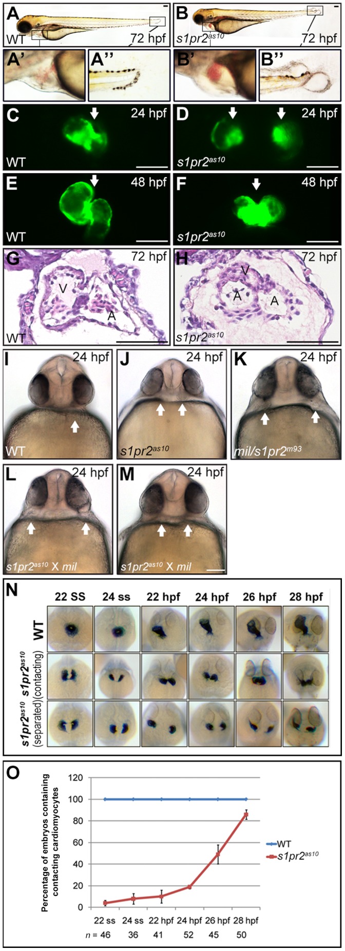
(A-H) Lateral views of wild-type (WT) (A, C, E and G) and s1pr2as10 mutants (B, D, F and H). Pericardial edema (B’), small eyes, and tail blisters (B’) were observed in 72-hpf s1pr2as10 mutant embryos (B). Two separate hearts (white arrows, D) were detected at 24 hpf, whereas a fused heart (white arrow, F) was observed at 48 hpf in s1pr2as10 mutant embryos. Paraffin sectioning with haematoxylin and eosin staining revealed the presence of two atria and one ventricle in s1pr2as10 mutant embryos at 72 hpf (H). (I-M) Ventral views of wild-type (WT) (I), s1pr2as10 (J), mil (K), and the intercross mutant progeny of s1pr2as10 and mil mutants (L and M) at 24 hpf are shown. The arrows indicate the positions of the hearts. (N) Cardiomyocytes were labeled via cmlc2 staining. Two representative s1pr2as10 mutant embryos are shown. Both contacting and separated cardiomyocytes were detected in the s1pr2as10 mutant embryos from the 22 ss to 28 hpf. (O) Percentages of WT and s1pr2as10 mutants containing contacting myocardia at different developmental stages. Scale bar = 100 µm. The error bars indicate the standard error.
Genetic Mapping of the s1pr2as10 Mutant Gene
By performing positional cloning with simple sequence length polymorphism markers, we established that s1pr2as10 is located on chromosome 3 near the mil/edg5 locus (Figure S1A). Sequencing of the mil/edg5 coding regions within cDNA or genomic DNA of s1pr2as10 mutant embryos did not predict any changes in the amino acid sequence (data not shown). Next, we examined the non-coding regions of the mil/edg5 locus via ligation-mediated polymerase chain reaction (PCR) and identified a large insertion (more than 1,000 base pairs from linkage group 11) in the first intron of mil/edg5, near exon 1 (data not shown). To confirm this insertion, we performed PCR with three primers, including F1 and R1 outside the insertion and I1 within the insertion (Figure S1B and S1C). A 426-bp DNA fragment amplified by primers I1 and R1 was detected only in the s1pr2as10 heterozygote and homozygote mutant embryos. To examine the impact of the insertion on mil/edg5 gene expression, we performed quantitative real-time reverse-transcription polymerase chain reaction (qRT-PCR) to compare the expression levels of mil/edg5 between the s1pr2as10 mutants and their WT siblings during and after cardiogenesis. Significantly reduced mil/edg5 mRNA levels were detected in the s1pr2as10 mutants prior to 96 hpf (Figure S1D). These results strongly suggest that the s1pr2as10 cardiac defect is caused by hypomorphic mil/edg5 gene expression and that s1pr2as10 may represent a new allele of mil/s1pr2m93. Complementation experiments further demonstrated that s1pr2as10 failed to complement a mil mutant at 24 hpf (Figure 1L and 1M vs. 1K). In summary, s1pr2as10 is a hypomorphic allele of mil that is caused by an insertion into intron 1.
s1pr2as10 Mutants Exhibit a Weaker Phenotype than mil Mutants
Next, we carefully compared the cardiac phenotypes of s1pr2as10 and mil mutations. We observed that the average distance between bilateral hearts at 24 hpf in s1pr2as10 mutant embryos was shorter than that of mil mutants (Figure 1J and 1K). In addition, while the bilateral hearts persisted until at least 48 hpf in mil mutants, the myocardia fused by 48 hpf in s1pr2as10 mutants (Figure 1F). Both of these observations indicate that the cardia bifida of s1pr2as10 mutants is weaker than that of mil mutants. We subsequently conducted time-lapse analyses of cmlc2-stained embryos to investigate the timing of bilateral heart fusion in the s1pr2as10 mutant (Figure 1N). At 24 hpf, the majority of s1pr2as10 mutant embryos contained two clusters of laterally positioned myocardial precursors, whereas by 28 hpf, over 80% of mutant embryos contained myocardial precursors in contact with one another (Figure 1O). To demonstrate that the s1pr2as10 mutant phenotype could be attributed to the low levels of mil/edg5 mRNA, we injected different doses of edg5-MO or edg5-5mm MO into Tg(cmlc2:EGFP, cmlc2:H2AFZmCherry)cy13 transgenic fish at the one-cell zygote stage. To quantitatively assess the severity of cardiac abnormalities, we grouped the cardiac phenotypes into three classes; Class I represented a normal heart tube, Class II represented cardiomyocytes either in close proximity or in contact, and Class III represented the most severe cardia bifida (Figure S2A). At 24 hpf, dose-dependent cardia bifida was detected in embryos injected with 1 to 16 ng of edg5-MO, but not in embryos injected with edg5-5mm (Figure S2B). Injection of s1pr2as10 mutant embryos with 30 pg mil/edg5 mRNA resulted in a partial rescue of cardia bifida at 24 hpf, as compared to mutant embryos injected with LacZ (Figure S3). Overall, these data indicate that the hypomorphic migration defect of myocardial precursors in s1pr2as10 mutant embryos was a result of decreased levels of mil/edg5 mRNA.
Raising s1pr2as10 Embryos at low Temperature Rescued the Cardiac Phenotype
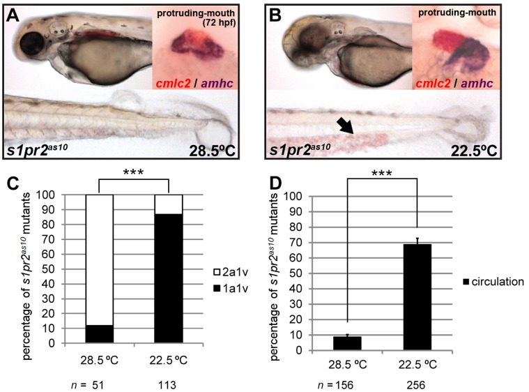
The hearts of s1pr2as10 mutant embryos raised at a standard temperature (28.5°C) contained two atria and one tightly-packed ventricle, as visualized by cmlc2 and amhc double-staining; most of these embryos lacked blood circulation at 72 hpf (Figure 2A). However, we found that the s1pr2as10 mutants raised at a lower temperature (22.5°C) presented with a less severe phenotype (Figure 2B). More than 85% of the s1pr2as10 mutant embryos raised at 22.5°C had hearts with one atrium and one ventricle at 72 hpf, as compared to only 12% of the embryos raised at 28.5°C (Figure 2C). In addition, normal blood circulation was detected at 72 hpf in 69% of the s1pr2as10 mutant embryos raised at 22.5°C, but in only 9% of those raised at 28.5°C (Figure 2D). Rescued s1pr2as10 homozygous mutants grew to adulthood. A high proportion of the offspring of rescued mutants were also rescued when raised at 22.5°C (Table S1). However, the tail blister phenotype of the s1pr2as10 mutant embryos could not be rescued by development at 22.5°C (Figure 2B). This result indicates that incubation at low temperature specifically affects myocardial migration, and that s1pr2as10 is not simply a temperature-sensitive allele of mil/edg5. We subsequently performed temperature shift experiments, by incubating s1pr2as10 mutants from different stages at 28.5°C or 22.5°C. We found that the critical time window in which low temperature could rescue cardia bifida in s1pr2as10 mutants was from ∼16 to the 22 somite stage (ss) (Figure S4).
Figure 2. Development at low temperature rescues the s1pr2as10 cardiac phenotype.
(A) s1pr2as10 mutants raised at 28.5°C contained hearts with two atria (dark purple, amhc staining) and one ventricle (red, cmlc2 staining), and presented with tail blisters at 72 hpf. (B) s1pr2as10 mutant embryos raised at 22.5°C contained hearts with a single atrium and ventricle, and exhibited blood circulation at 72 hpf (arrow). (C) Percentages of s1pr2as10 mutant embryos raised at either 28.5 or 22.5°C containing two atria and one ventricle (2a1v) or one atrium and one ventricle (1a1v) at 72 hpf. (D) Percentages of s1pr2as10 mutant embryos raised at either 28.5 or 22.5°C displaying blood circulation at 72 hpf. The error bars indicate the standard error. Statistical significance was determined using Student’s t-test. *** indicates p<0.001.
Low Temperature Shifts the Timing of Cardiac Cone Formation
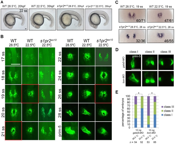
To examine why the cardiac defects of the s1pr2as10 mutants were rescued by low temperature, we conducted time-lapse experiments to observe bilateral myocardial precursor migration at different somite stages. In Tg(cmlc2:EGFP, cmlc2:H2AFZmCherry)cy13 transgenic embryos raised at 28.5°C, the bilateral myocardial precursors fused at the midline and became cone-shaped at the 21-ss (Figure 3B, red boxes in WT at 28.5°C). Surprisingly, the timing of cardiac cone formation was shifted to the 19 ss when the embryos were raised at the lower temperature (Figure 3B, red boxes in WT at 22.5°C). Although the overall growth rate at 22.5°C was about 2-fold slower than that at 28.5°C [27], low temperature does not seem to cause any morphological defects in WT and s1pr2as10 embryos (Figure 3A). Similarly, the bilateral cardiomyocytes in the s1pr2as10 mutant embryos fused at the 26 ss at low temperature (Figure 3B, red boxes in s1pr2as10 22.5°C); this result was in contrast to the delayed fusion (28 hpf) observed at 28.5°C (Figure 1O). In situ hybridization using a cmlc2 probe revealed that 77% WT and 84% s1pr2as10 mutant embryos raised at low temperature formed a fused cardiac cone at the 19 ss and 26 ss, respectively (Figure 3C). In contrast, the majority of WT and s1pr2as10 mutant embryos raised at standard temperature presented with horseshoe-shaped hearts or bilateral hearts at 19 ss and 26 ss, respectively (Figure 3C). However, cardia bifida of mil mutant embryos cannot be rescued by low temperature treatment (Figure S5A). Since defects in endoderm formation also result in cardia bifida [7], [28], we induced cardia bifida by injecting different doses of gata5 MO or bon MO. We found that the cardia bifida phenotypes of embryos injected with 10 ng of gata5 MO or bon MO can be significantly rescued by being raised at low temperature (Figure 3D and 3E) while cardia bifida defects in embryos injected with 20 ng of gata5 MO were not rescued by low temperature treatment (Figure S5B). Although the rescue effect was not statistically significant, a reduced percentage of cardia bifida phenotype was notable in embryos injected with 20 ng of bon MO. (Figure S5B). In addition to cardia bifida, cells of the anterior gut tissues also failed to migrate to the midline in gata5/fau mutants [28]. We found that the distance between the two lateral anterior gut tubes was also significantly reduced in embryos injected with 10 ng gata5 MO and incubated at low temperature, as compared to those incubated at standard temperature (145 µm vs. 160 µm) at the prim-25 stage (Figure S6). Thus, low temperature alters the timing of cardiac cone formation in WT, s1pr2as10 mutants (which exhibit hypomorphic migration defects) and other cardia bifida morphants, resulting in a rescue of the cardia bifida phenotype that is independent of the genetic background.
Figure 3. Low temperature shifts the timing of formation of the cardiac cone.
(A) Lateral views of 22-ss wild-type (WT) and s1pr2as10 mutant embryos raised at 28.5 or 22.5°C. (B) Time-lapse analyses of the medial migration of bilateral cardiomyocytes during the ∼17–30-ss in Tg(cmlc2:EGFP, cmlc2:H2AFZmCherry)cy13 transgenic fish (WT) or s1pr2as10 mutant embryos raised at 28.5 or 22.5°C. Five WT or s1pr2as10 mutant embryos were analyzed for each stage. The images outlined in red indicate the stage at which cardiac cone formation took place under each condition. (C) In situ hybridization of 19-ss wild-type (WT) or 26-ss s1pr2as10 mutant embryos raised at 28.5°C or 22.5°C with a cmlc2 RNA probe. The number of embryos displaying cmlc2 staining/the total number of embryos analyzed is shown for each panel. (D) Different degrees of myocardial migration defects were observed in gata5 and bon morphants at 24 hpf. Class I (single heart tube), Class II (cardiomyocytes in close proximity but not in contact), and Class III (two separate hearts) defects are shown. (E) Percentages of each class of myocardial migration defects in 10 ng gata5-MO or bon-MO-injected embryos raised at 28.5 or 22.5°C. Scale bar = 100 µm. Statistical significance was determined using Student’s t-test. * indicates p<0.05.
Gene Expression Profiles of Zebrafish Embryos Raised at 28.5 and 22.5°C
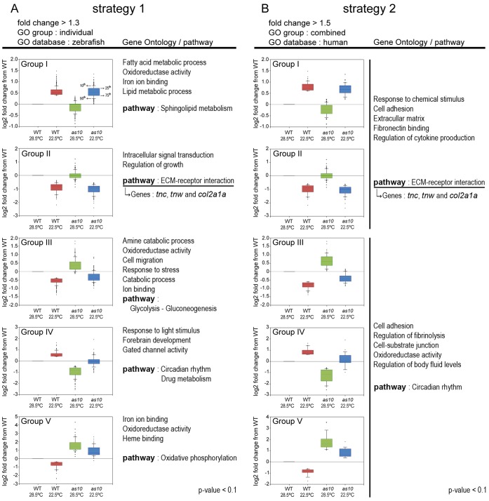
Previous reports have demonstrated that changes in temperature affect developmental processes and gene expression in zebrafish embryos [29], [30]. We performed DNA microarray (Figure 4) and digital gene expression (DGE) (Table S2) analyses to further investigate how low temperature mitigates cardia bifida. By combining these two analyses, we identified several candidate gene groups and demonstrated that genes encoding extracellular matrix (ECM) proteins are important for preventing cardia bifida. DNA microarray and qRT-PCR revealed that mil/edg5 levels were not affected by raising WT or s1pr2as10 embryos at different temperatures (28.5°C or 22.5°C) (Figure S7).
Figure 4. Gene ontology (GO) and pathway analyses for differentially expressed genes identified by DNA microarray analysis.
GO and pathway analyses were conducted on different groups of genes using two strategies. (A) Genes with a greater than 1.3-fold change in expression between embryos raised at 28.5 and 22.5°C were used in strategy 1. GO analysis was performed using the zebrafish database. (B) Genes with a greater than 1.5-fold change in expression between embryos raised at 28.5 and 22.5°C were used in strategy 2. GO analysis was performed using the human database. The GO and pathway analyses were performed on genes from both Groups I and II or on genes from Groups III, IV and V. Box and Whisker plots for each group indicate the median (line within the box), 25th and 75th percentiles (top and bottom of the box), and 10th and 90th percentiles (Whiskers/error bars). The P-value from GO and pathway analyses are smaller than 0.1.
Low Temperature Mitigates Cardia Bifida by Altering ECM Gene Expression
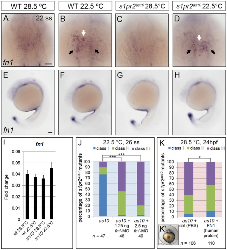
Based on the DNA microarray and DGE analyses, we established that several genes involved in the ECM were affected by low temperature; these genes included tenascin-c (tnc), tenascin-w (tnw), and collagen type II, alpha-1a identified through the DNA microarray analysis (Figure 4), and fibronectin 1 (fn1), collagen, type XI alpha 2 and sid4 through the DGE analysis (Table S2). We first focused on fn1, which was previously found to be important for myocardial migration [8]. An increased level of fn1 was initially identified via DGE in s1pr2as10 mutant embryos raised at 22.5°C (Table S2). Whole mount in situ hybridization and immunocytochemistry revealed increased expression of fn1 mRNA and protein in the midline regions and bilateral anterior LPM of both 22-ss WT and s1pr2as10 mutant embryos raised at 22.5°C (Figure 5B and 5D and Figure S8). However, levels of fn1 at the tail bud or yolk regions were not affected by incubation at low temperature (Figure 5E–5H). qRT-PCR revealed an approximately 1.3-fold increase of fn1 mRNA in s1pr2as10 mutant embryos raised at 22.5°C, as compared to those raised at 28.5°C. However, this temperature-mediated up-regulation was not statistically significant (Figure 5I). The failure to detect a significant change in WT and s1pr2as10 mutant embryos raised at low temperature may be due to the inability of temperature to affect fn1 expression at the tail bud and yolk regions (Figure 5E–5H).
Figure 5. Expression of fibronectin 1 is affected by temperature.
(A-H) In situ hybridization against fibronectin 1 (fn1) in 22-ss wild-type (WT) (A, B, E, F) and s1pr2as10 (as10) mutant (C, D, G, H) embryos raised at 28.5 or 22.5°C. Expression of fn1 mRNA increased at the midline region (white arrows in B and D) and bilateral LPM (black arrows in B and D) of both WT and s1pr2as10 mutant embryos raised at 22.5°C, as compared to those raised at 28.5°C (A and C). Expression of fn1 at the tail bud or yolk regions was not affected (E-H). (I) qRT-PCR revealed a trend towards increased expression of fn1 mRNA in s1pr2as10 mutant embryos raised at 22.5°C. The error bars indicate the standard error. (J) Knockdown of fn1 expression in s1pr2as10 mutants increased the percentage of 26-ss embryos raised at 22.5°C with the Class III cardia bifida phenotype. (K) Injection of human fibronectin protein into s1pr2as10 mutant embryos raised at 28.5°C partially rescued the cardia bifida phenotypes at 24 hpf. (K’) The red dye indicates the protein injection region at 14–16 hpf. Scale bars = 100 µm. Statistical significance was determined using Student’s t-test. * indicates p<0.05, *** indicates p<0.001.
As incubation at low temperature raised fn1 expression and rescued the cardia bifida phenotype in s1pr2as10, we hypothesized that fnl up-regulation may be required for the rescue of heart development. To test this hypothesis, we injected fn1-MO into s1pr2as10 mutants raised at 22.5°C. As shown in Figure 5J, 26-ss mutant embryos injected with 1.25 or 2.5 ng fn1-MO displayed severe cardia bifida, even after being raised at the low temperature. In addition, we injected human fibronectin protein into the midline region (Figure 5K’) of 14–16 hpf s1pr2as10 mutant embryos raised at 28.5°C, to determine whether elevated levels of fibronectin can rescue the cardia bifida phenotype. We found that the percentage of mutant embryos with class III severe cardia bifida was reduced from 60% to 42% (Figure 5K). Together, these results indicate that incubation at low temperature mitigates cardia bifida in s1pr2as10 mutants by promoting fn1 expression in the midline and bilateral anterior LPM regions.
Two Tenascin Genes, Tenascin-C and Tenascin-W, Play Important Roles in myocardial migration in embryos raised at low temperature
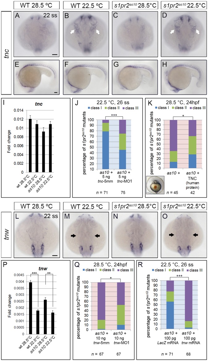
In addition to fn1, expression levels of two other extracellular matrix genes, tenascin-C (tnc) and tenascin-W (tnw), were affected by incubation at low temperature. The tnc gene can produce four different isoforms through differential splicing. By using RNA probes against exons common to all tnc isoforms for in situ hybridization, we detected increased expression of tnc at the pharyngeal arches and in the cells between the brain and eyes of 22-ss WT and s1pr2as10 mutant embryos raised at 22.5°C as compared to those raised at 28.5°C (Figure 6B and 6D vs. 6A and 6C). However, expression of tnc in the somite region was not affected by the temperature of incubation (Figure 6E–6H). Accordingly, the use of qRT-PCR to measure tnc mRNA did not uncover significant differences between WT and s1pr2as10 mutant embryos raised at 22.5°C as compared to those raised at 28.5°C (Figure 6I). We subsequently used a previously published tnc-MO1 that can prevent the translation of all tnc isoforms [22] and designed a tnc−5mm MO based on the tnc-MO1 sequence for use as controls. Additionally, we used the previously published tnc-MO2, which blocks splicing of tnc exon 1 [22]. Mutant s1pr2as10 embryos incubated at 22.5°C and injected with either tnc-MO1 or tnc-MO2 presented with more severe cardia bifida phenotypes, as compared to tnc−5 mm injected embryos (Figure 6J and Figure S9A). We also injected human TNC protein into the midline region of s1pr2as10 mutants, and found the percentage of embryos with class III severe cardia bifida defects was reduced from 64% to 33% (Figure 6K). These results indicate that low temperature rescues s1pr2as10 cardiac phenotype partially due to increased expression of tnc.
Figure 6. Expression of two tenascin genes is affected by temperature.
In situ hybridization against tenascin-c (tnc) (A-H) and tenascin-w (tnw) (L-O) in 22-ss wild-type (WT) and s1pr2as10 (as10) mutant embryos raised at 28.5°C or 22.5°C. Expression of tnc increased in the pharyngeal arches (white arrows in B and D) and the cells between the brain and eyes of embryos raised at 22.5°C, but was unaffected in the trunk somite region (E-H). (I) qRT-PCR revealed a trend towards increased expression of tnc mRNA in s1pr2as10 mutant embryos raised at 22.5°C. (J) Knockdown of tnc expression with tnc-MO1 in s1pr2as10 mutants increased the percentage of 26-ss embryos raised at 22.5°C with the Class III cardia bifida phenotype. (K) Injection of s1pr2as10 mutant embryos raised at 28.5°C with human TNC protein partially rescued cardia bifida phenotypes at 24 hpf. (L-O) Decreased tnw expression in all tissues, including scattered epidermal cells in the head (black arrows in M and O) was observed in both 22-ss WT and s1pr2as10 mutants raised at 22.5°C. (P) qRT-PCR was used to confirm that expression of tnw mRNA is significantly reduced in 22-ss WT and s1pr2as10 mutant embryos raised at 22.5°C. (Q) Knockdown of tnw with tnw-MO1 in s1pr2as10 mutant embryos raised at 28.5°C partially rescued cardia bifida phenotypes at 24 hpf. (R) Injection of s1pr2as10 mutant embryos raised at 22.5°C with 100 pg tnw mRNA increased the percentage of 26-ss embryos with the Class III cardia bifida phenotype. Scale bars = 100 µm. The error bars indicate the standard error. Statistical significance was determined using Student’s t-test. * indicates p<0.05, ** indicates p<0.01, *** indicates p<0.001.
DNA microarray analysis also revealed decreased expression of another tenascin gene, tnw, in 22-ss WT and s1pr2as10 mutant embryos raised at 22.5°C. Whole-mount in situ hybridization and qRT-PCR confirmed that tnw mRNA expression was reduced in all tissues, including the LPM, of both 22-ss WT and s1pr2as10 mutant embryos raised at 22.5°C (Figure 6M, 6O and 6P). We subsequently designed morpholinos (tnw-MO1 and tnw-MO2) against the 5′ untranslated region (UTR) of tnw to prevent its translation; we also designed a tnw−5 mm based on the tnw-MO1 sequence as a control. In order to test the efficiency and specificity of tnw-MOs, we fused EGFP in frame with the tnw 5′UTR under the control of the CMV enhancer/promoter, and co-injected this construct with tnw-MO1, tnw-MO2, or tnw−5 mm into one-cell zygotes. As shown in Figure S9D and E, we detected EGFP expression in the majority of embryos co-injected with tnw−5 mm and CMV-tnwUTR-EGFP plasmid. In contrast, the majority of embryos (85%) co-injected with tnw-MO1 and CMV-tnwUTR-EGFP did not express GFP. However, 30% of embryos co-injected with tnw-MO2 and CMV-tnwUTR-EGFP expressed GFP, indicating that tnw-MO1 is more efficient at blocking EGFP translation. This may be a consequence of tnw-MO2 targeting a region further upstream from that of tnw-MO1.
We then injected either tnw-MO1 or tnw-MO2 into s1pr2as10 mutants raised at 28.5°C, and observed partial rescue of the cardia bifida phenotype at 24 hpf (Figure 6Q and Figure S9B). Injection of tnw-MO1 resulted in greater rescue of the cardia bifida phenotype than with tnw-MO2; this may be attributed to the reduced efficiency of tnw-MO2 in blocking translation. Furthermore, some of the rescued embryos contained a single heart tube and displayed normal circulation (data not shown). Interestingly, injection of tnw mRNA at the one-cell stage caused a highly severe cardia bifida phenotype in s1pr2as10 mutants, which was observed even at 22.5°C (Figure 6R).
We also injected fn1-MO, tnc-MO1, or tnw mRNA into wild type embryos to investigate their effects on myocardial migration. We found that the percentage of tnc-MO1-injected morphants (26%) which developed a horseshoe-shaped heart at 22ss was similar to that of control embryos (27%) (Figure S10A and S10B). The percentage of tnw mRNA-injected embryos with a horseshoe-shaped heart at 22ss was greater (38%) than that of control, and 12% of these embryos also exhibited cardia bifida (Figure S10D). The percentage of embryos with horseshoe-shaped hearts at 22 ss was increased further by fn1-MO1-injection (69%), and 9% of these embryos exhibited cardia bifida (9%) (Figure S10C); these results indicate that fibronectin plays a more important role in regulating myocardial migration than tenascin-C or tenascin-W. However, the majority (93%) of embryos injected with a mixture of tnc-MO1, fn1-MO, and tnw mRNA exhibited cardia bifida at 22 ss (Figure S10E); this result further demonstrates that tenascin-C and fibronectin positively regulate, while tenascin-W negatively regulates, myocardial migration in zebrafish embryos.
Together, these data indicate that tnc and tnw play opposing roles in zebrafish myocardial migration, and that low temperature mitigates cardia bifida by altering the expression of ECM genes: fn1 and tnc are up-regulated, and tnw is down-regulated.
Reactive Oxygen Species (ROS) Mediate Mitigation of Cardia Bifida by Low Temperature
Cold exposure was previously shown to increase expression of peroxisome proliferator-activated receptors (PPARs) and uncoupling proteins (UCPs) in the zebrafish brain, to prevent oxidative damage and to maintain metabolic balance and homeostasis [31]. We investigated whether ROS mediates mitigation of the cardia bifida phenotype in s1pr2as10 mutants by low temperature treatment. To test this hypothesis, we treated s1pr2as10 mutant embryos with either 50 or 150 µM of N-acetyl cysteine (NAC), a ROS scavenger, at the tailbud stage, and incubated the embryos at 22.5°C. As shown in Figure 7, we detected a dose-dependent of NAC on the percentage of s1pr2as10 mutant embryos with a class I single heart tube at 26 ss when incubated at low temperature; class I heart tubes were reduced by NAC to 46–32% as compared to 67–73% in untreated s1pr2as10 mutant embryos. This result suggests that ROS may mediate mitigation of cardia bifida by low temperature.
Figure 7. Reactive oxygen species (ROS) mediate mitigation of cardia bifida in zebrafish embryos incubated at low temperature.
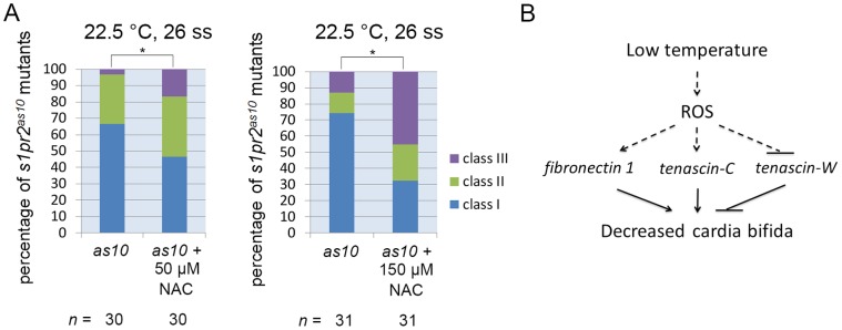
(A) s1pr2as10 mutant embryos raised at 22.5°C were treated with 50 or 150 µM N-acetyl cysteine (NAC) at the tailbud stage, and were then incubated at 22.5°C. Different degrees of myocardial migration defects were observed at the 26 ss. Class I (single heart tube), Class II (cardiomyocytes in close proximity but not in contact), and Class III (two separate hearts). (B) A proposed model showing how low temperature mitigates cardia bifida in zebrafish embryos. Statistical significance was determined using Student’s t-test. * indicates p<0.05.
Discussion
Previous reports have demonstrated that changes in temperature affect various aspects of development and gene expression [29], [30]. In this study, we have demonstrated that temperature also affects the timing of cardiac cone formation. We have specifically demonstrated that low temperature (22.5°C) mitigates cardia bifida in both s1pr2as10 mutant and gata5 and bon morphant embryos. DNA microarray and digital gene expression analyses revealed that low temperature regulates expression of several extracellular matrix (ECM) genes to control myocardial migration.
An increase in fn1 expression at the midline region and bilateral anterior lateral plate mesoderm (LPM) was identified in both 22-ss WT and s1pr2as10 mutant embryos raised at 22.5°C (Figure 5B, and 5D and Figure S8). Injection of s1pr2as10 mutant embryos with fn1-MO abolished the rescue of cardia bifida by low temperature (Figure 5J), while injection with human fibronectin protein decreased the percentage of s1pr2as10 mutant embryos raised at 28.5°C with class III severe cardia bifica at 24 hpf (Figure 5K). During myocardial precursor migration, fibronectin secreted by the endoderm and/or endocardial precursors is deposited at the midline and laterally around the myocardial precursors, to maintain the integrity of myocardial epithelia, and mediate interactions between the endoderm and migrating myocardial precursors [8], [15]. It has been demonstrated that knockdown of mil/edg5 results in cardia bifida and decreased levels of fibronectin [11]. Furthermore, injection of the midline with fibronectin partially rescued the cardia bifida phenotype in mil mutant embryos [15]. All of these findings suggest that both mil/edg5 and low temperature play a positive role in regulating myocardial migration. Proper deposition of extracellular matrix proteins is also important for normal cardiac function after formation of the four-chambered heart, and excess collagen and fibronectin deposition results in cardiac fibrosis in cardiac hypertrophy. Reactive oxygen species (ROS) have been shown to up-regulate expression of profibrotic genes, such as col2a1, col1a1, and fn1, thereby resulting in direct extracellular matrix deposition in a transgenic mouse model, and resulting in cardiac hypertrophy [32].
Tenascins are a family of large multimeric extracellular matrix glycoproteins involved in cell adhesion, migration and proliferation [33], [34]. Tenascin-C has been shown to either promote or inhibit cell adhesion and migration, depending on the cell type. Addition of tenascin-C stimulated the migration of human adult dermal fibroblasts cultured within matrices comprised of fibronectin [35]. Smooth muscle cells cultured on tenascin-C substrate exhibited increased expression of αv-integrin and elevated phosphorylation of focal adhesion kinase (FAK), followed by enhanced migration [36]. Tenascin-C also promoted bovine retinal endothelial cell migration by enhancement of FAK phosphorylation [37]. On the other hand, high levels of tenascin-C produced by glioma tumour tissues actively hindered T-cell migration, resulting in an accumulation of T cells in the peritumoral region [38]. Although the function of tenascin-W and its gene regulation has not been well-characterized, it has been shown to confer anti-adhesive properties [39]. Addition of soluble tenascin-W was reported to inhibit the formation of lamellipodia with stress fibers and focal adhesion complexes in a murine myoblast cell line (C2C12) cultured on fibronectin. Various factors, including inflammatory cytokines, growth factors, oxidative stress, and hypoxia, have been shown to induce tnc expression [40]. In neonatal rat cardiomyocytes, mechanical strain increased tnc expression by activating nuclear factor-κB through ROS [41]. Here, we observed up-regulation of tnc and down-regulation of tnw in embryos raised at low temperature (Figures 5 and 6). MO knockdown and mRNA or protein rescue confirmed the opposing roles of tenascin-C and tenascin-W in mitigating cardia bifida in zebrafish embryos. Moreover, we found that the addition of N-acetyl cysteine (NAC), a ROS scavenger, decreased mitigation of cardia bifida in s1pr2as10 mutant embryos raised at 22.5°C (Figure 7A).
Based on our results and previously published studies, we propose a model describing how low temperature mitigates cardia bifida in zebrafish embryos (Figure 7B). Low temperature may enhance ROS production in cranial neural crest cells, endocardial and myocardial cells, and endoderm (in a similar manner to ROS induction in zebrafish brain upon cold exposure [31]), as these tissues are more vulnerable to oxidative stress [42]–[47]. The presence of ROS in the endoderm and anterior LPM may then up-regulate fn1 expression, resulting in the secretion of fibronectin in the midline and bilateral anterior LPM (Figure 5 and Figure S8). Meanwhile, ROS production in the cranial neural crest derived-bilateral pharyngeal arches may activate tnc expression, thereby promoting secretion of tenascin-C near migrating bilateral cardiomyocytes (Figure 6). Increased levels of secreted tenascin-C may enhance integrin expression and promote FAK phosphorylation in migrating bilateral cardiomyocytes, thereby facilitating their interaction with fibronectin, formation of focal adhesion complexes, actin polymerization, and protrusion [37], [48]–[50]. Increased fibronectin around migrating cardiomyocytes can further promote the integrity of myocardial epithelia, which is a prerequisite of myocardial migration [8]. Since tenascin-W is known to inhibit cell adhesion to fibronectin, ROS may also down-regulate tnw expression, consequently preventing secretion of tenascin-W from scattered epidermal cells in the head region, and thereby allowing migrating cardiomyocytes to interact with fibronectin [51]. Furthermore, a combinatorial effect of up-regulation of tnc and fn1, and down-regulation of tnw are required to mitigate cardia bifida of s1pr2as10 mutant embryos and gata5 and bon cardia bifida morphants upon low temperature treatment.
In conclusion, our identification of the s1pr2as10 mutation serendipitously led to our finding that incubation at low temperature mitigates cardia bifida, a phenomena caused by up-regulation of fn1 and tnc and down-regulation of tnw. In addition, ROS mediates mitigation of cardia bifida upon low temperature treatment. Future studies may be expected to delineate the mechanisms underlying regulation of fn1, tnc and tnw expression by ROS under low temperatures.
Supporting Information
Genetic mapping of the s1pr2as10 mutant gene. (A) Meiotic map of the s1pr2as10 locus. The numbers above the line indicate the number of meiotic recombination events out of the total number of meioses. (B) An enlarged view of the mil/edg5 locus shows the insertion in intron 1. Three primers (F1, R1, and I1) were used to confirm the insertion. (C) A 182-bp DNA fragment amplified using primers F1 and R1 was detected in wild-type (WT) and s1pr2as10 heterozygous mutants, whereas a 426-bp DNA fragment amplified using primers I1 and R1 was detected in s1pr2as10 heterozygous and homozygous mutant embryos. (D) Expression of mil/edg5 mRNA was reduced in s1pr2as10 mutants prior to 96 hpf, as revealed by qRT-PCR. WT (+, W); s1pr2as10 mutant (−, M). The error bars indicate the standard error.
(TIF)
Dosage curve of an edg5 -morpholino antisense oligomer (MO) and its association with cardia bifida. (A) Tg(cmlc2:EGFP, cmlc2:H2AFZmCherry)cy13 embryos injected with different doses of edg5-MO and edg5-5 mm exhibited varying degrees of myocardial migration defects, ranging from Class I (a single heart tube) to Class II (cardiomyocytes either in close proximity or in contact) and Class III (separated cardiomyocytes) at 24 hpf. Scale bar = 100 µm. (B) Percentages of Class I, II and III myocardial migration defects in Tg(cmlc2:EGFP, cmlc2:H2AFZmCherry)cy13 embryos injected with 1–16 ng of edg5-MO at 24 hpf.
(TIF)
Injection of s1pr2as10 mutants with mil / edg5 mRNA partially rescued the cardia bifida phenotype. Injection of s1pr2as10 mutant embryos with mil/edg5 mRNA, but not LacZ mRNA, decreased the percentage of embryos with the Class III cardia bifida phenotype. Statistical significance was determined using Student’s t-test. * indicates p<0.05.
(TIF)
The 16 ss to the 22 ss is the critical time interval for rescue of the s1pr2as10 mutant heart at 22.5°C. s1pr2as10 mutants raised at 28.5°C (red line) or 22.5°C (blue line) at different stages were rescued to differing levels, as determined by the percentage of embryos containing one atrium and one ventricle at the protruding-mouth stage.
(TIF)
Low temperature treatment cannot rescue the cardia bifida phenotype of mil mutants and 20 ng gata5 MO-injected embryos. (A) milm93 mutant embryos were incubated at 28.5°C or 22.5°C. Different degrees of myocardial migration defects were observed from prim 5-prim 15. (B) Percentages of each class of myocardial migration defect in 20 ng gata5-MO or bon-MO-injected embryos raised at 28.5 or 22.5°C at the 26 ss. Myocardial migration defects were observed at prim-5. Class I (single heart tube), Class II (cardiomyocytes in close proximity but not in contact), and Class III (two separate hearts).
(TIF)
Anterior gut migration defects in gata5 morphants can be rescued by low temperature treatment. Embryos injected with 10 ng gata5 MO were incubated at 28.5°C (A) or 22.5°C (B). Embryos were harvested at prim-25 and stained with foxa1 RNA probe. (C). The distance between two lateral anterior gut tubes was significantly different between morphants incubated at 28.5°C or 22.5°C. Scale bars = 100 μm. The error bars indicate the standard error. Statistical significance was determined using Student’s t-test. *** indicates p <0.001.
(TIF)
mil/edg5 levels were similar in embryos raised at 28.5°C or 22.5°C. qRT-PCR measurement of mil/edg5 levels in 22-ss WT or s1pr2as10 mutant embryos raised at 28.5 or 22.5°C. The error bars indicate the standard error.
(TIF)
Low temperature increases fibronectin 1 expression in the midline region. Immunohistochemistry was used to demonstrate increased fibronectin 1 expression at the midline region (white arrows) of 22-ss wild type (WT) and s1pr2as10 mutant embryos raised at 22.5°C. Scale bars = 100 µm.
(TIF)
Evaluation of the roles of tnc and tnw in the mitigation of cardia bifida, and of the efficiency and specificity of tnw MOs. (A) Knockdown of tnc with tnc-MO2 in s1pr2as10 mutants increased the percentages of 26-ss embryos raised at 22.5°C with the Class II and Class III cardia bifida phenotype. (B) Knockdown of tnw with tnw-MO2 in s1pr2as10 mutant embryos raised at 28.5°C partially rescued cardia bifida phenotypes at 24 hpf. Class I (a single heart tube) to Class II (cardiomyocytes either in close proximity or in contact) and Class III (separated cardiomyocytes). Statistical significance was determined using Student’s t-test. * indicates p <0.05, ** indicates p <0.01. (C) Diagram indicating the relative binding positions of two tnw-MOs in the 5’untranslated region of tnw mRNA. (D) Green fluorescence can be detected in embryos co-injected with CMV-tnwUTR-EGFP and tnw-5mm at 24 hpf. Green fluorescence was not observed in the majority of embryos co-injected with CMV-tnwUTR-EGFP and tnw-MO1, while green fluorescence was detected in 30% of embryos co-injected with CMV-tnwUTR-EGFP and tnw-MO2. (E) Percentage of embryos expressing EGFP following co-injection of CMV-tnwUTR-EGFP with tnw-5mmMO, tnw-MO1 or tnw-MO2. The error bars indicate the standard error.
(TIF)
Knockdown of fn1 or tnc and overexpression of tnw results in cardia bifida in wild type embryos. Wild type embryos at the one-cell zygote stage were injected with 5 ng tnc-MO1 (B), 2.5 ng fn1-MO (C), 100 pg tnw mRNA (D), or a mixture of tnc-MO1, fn1-MO, and tnw mRNA (E), and incubated at 28.5°C. Un-injected embryos were used as a control (A). Embryos were harvested at the 22 ss and stained with cmlc2.
(TIF)
Circulation defect of s1pr2as10 mutant embryos from different genotypes could be partially rescued when raised at 22.5°C. The statistics of the offspring derived from different genotypes of s1pr2as10 mutant raised at 28.5°C or 22.5°C. Genotype labeled with +/− or −/− indicated the heterozygous or homozygous s1pr2as10 mutants respectively. For example, among 213 embryos from heterozygous mutants intercross (+/− x +/−) raised at 28.5°C, 162 embryos showed normal wild-type phenotype (a), 46 embryos contained tail blisters phenotype and established no blood circulation (b), and 5 embryos contained tail blisters with normal circulation (c). Mendel ratio was calculated by number of embryos with tail blister phenotype divided by number of total embryos. Rescue of tail blister with circulation phenotype was observed in offspring derived from different genotypes of s1pr2as10 mutant raised at 22.5°C. Rescue percentage of tail blister with circulation phenotype was calculated by number of embryos showing tail blister with circulation phenotype divided by total number of embryos showing tail blister phenotype.
(DOC)
Up- and down-regulated genes in s1pr2as10 mutant at 22.5°C as determined by digital gene expression (DGE) analysis. Double-stranded cDNA from 22 ss-s1pr2as10 mutant embryos raised at 28.5°C and 22.5°C were synthesized for next generation sequencing. The Up- and down-regulated genes at 22.5°C were subsequently analyzed by pathway analysis according to Kyoto Encyclopedia of Genes and Genomes (KEGG) database. Several groups of genes were selected for further analysis.
(DOC)
Acknowledgments
The authors thank Dr. Atsuo Kawahara for providing the various plasmids of the sphingosine-1-phosphate synthesis pathway. We thank Dr. Jr-Kai Yu and Dr. Yi-Hsien Su for helpful discussion. We thank Dr. Yi-Wen Liu and Ms. Chih-Wei Chou for their technical assistance. We thank Dr. Mei-Chun Tseng and Ms. Ping-Yu Lin in the Mass facility, Institute of Chemistry, Academia Sinica, for their assistance with mass spectrometry. We thank the members of the Core Facility of the Institute of Cellular and Organismic Biology, Academia Sinica, for their assistance with DNA sequencing. We thank the Taiwan Zebrafish Core Facility at Academia Sinica (TZCAS) for providing the Tg(cmlc2:EGFP, cmlc2:H2AFZmCherry)cy13 fish line and the Zebrafish International Resource Center (ZIRC) for providing the mil/s1pr2m93/+ fish line.
Funding Statement
This work was supported by Academia Sinica [AS-99-TP-B08 to S.P.L.H.] and the National Science Council [NSC 99-2628-B-001-003-MY3 to SH]. The funders had no role in study design, data collection and analysis, decision to publish, or preparation of the manuscript.
References
- 1. Tu S, Chi NC (2012) Zebrafish models in cardiac development and congenital heart birth defects. Differentiation 84: 4–16. [DOI] [PMC free article] [PubMed] [Google Scholar]
- 2. Miura GI, Yelon D (2011) A guide to analysis of cardiac phenotypes in the zebrafish embryo. Methods Cell Biol 101: 161–180. [DOI] [PMC free article] [PubMed] [Google Scholar]
- 3. Bakkers J (2011) Zebrafish as a model to study cardiac development and human cardiac disease. Cardiovasc Res 91: 279–288. [DOI] [PMC free article] [PubMed] [Google Scholar]
- 4. Yelon D, Ticho B, Halpern ME, Ruvinsky I, Ho RK, et al. (2000) The bHLH transcription factor hand2 plays parallel roles in zebrafish heart and pectoral fin development. Development 127: 2573–2582. [DOI] [PubMed] [Google Scholar]
- 5. Reiter JF, Alexander J, Rodaway A, Yelon D, Patient R, et al. (1999) Gata5 is required for the development of the heart and endoderm in zebrafish. Genes Dev 13: 2983–2995. [DOI] [PMC free article] [PubMed] [Google Scholar]
- 6. Alexander J, Rothenberg M, Henry GL, Stainier DY (1999) casanova plays an early and essential role in endoderm formation in zebrafish. Dev Biol 215: 343–357. [DOI] [PubMed] [Google Scholar]
- 7. Kikuchi Y, Trinh LA, Reiter JF, Alexander J, Yelon D, et al. (2000) The zebrafish bonnie and clyde gene encodes a Mix family homeodomain protein that regulates the generation of endodermal precursors. Genes Dev 14: 1279–1289. [PMC free article] [PubMed] [Google Scholar]
- 8. Trinh LA, Stainier DY (2004) Fibronectin regulates epithelial organization during myocardial migration in zebrafish. Dev Cell 6: 371–382. [DOI] [PubMed] [Google Scholar]
- 9. Kawahara A, Nishi T, Hisano Y, Fukui H, Yamaguchi A, et al. (2009) The sphingolipid transporter spns2 functions in migration of zebrafish myocardial precursors. Science 323: 524–527. [DOI] [PubMed] [Google Scholar]
- 10. Kupperman E, An S, Osborne N, Waldron S, Stainier DY (2000) A sphingosine-1-phosphate receptor regulates cell migration during vertebrate heart development. Nature 406: 192–195. [DOI] [PubMed] [Google Scholar]
- 11. Osborne N, Brand-Arzamendi K, Ober EA, Jin SW, Verkade H, et al. (2008) The spinster homolog, two of hearts, is required for sphingosine 1-phosphate signaling in zebrafish. Curr Biol 18: 1882–1888. [DOI] [PMC free article] [PubMed] [Google Scholar]
- 12. George EL, Georges-Labouesse EN, Patel-King RS, Rayburn H, Hynes RO (1993) Defects in mesoderm, neural tube and vascular development in mouse embryos lacking fibronectin. Development 119: 1079–1091. [DOI] [PubMed] [Google Scholar]
- 13. Linask KK, Lash JW (1988) A role for fibronectin in the migration of avian precardiac cells. I. Dose-dependent effects of fibronectin antibody. Dev Biol 129: 315–323. [DOI] [PubMed] [Google Scholar]
- 14. Garavito-Aguilar ZV, Riley HE, Yelon D (2010) Hand2 ensures an appropriate environment for cardiac fusion by limiting Fibronectin function. Development 137: 3215–3220. [DOI] [PMC free article] [PubMed] [Google Scholar]
- 15. Matsui T, Raya A, Callol-Massot C, Kawakami Y, Oishi I, et al. (2007) miles-apart-Mediated regulation of cell-fibronectin interaction and myocardial migration in zebrafish. Nat Clin Pract Cardiovasc Med 4 Suppl 1S77–82. [DOI] [PubMed] [Google Scholar]
- 16. Shentu H, Wen HJ, Her GM, Huang CJ, Wu JL, et al. (2003) Proximal upstream region of zebrafish bone morphogenetic protein 4 promoter directs heart expression of green fluorescent protein. Genesis 37: 103–112. [DOI] [PubMed] [Google Scholar]
- 17. Kimmel CB, Ballard WW, Kimmel SR, Ullmann B, Schilling TF (1995) Stages of embryonic development of the zebrafish. Dev Dyn 203: 253–310. [DOI] [PubMed] [Google Scholar]
- 18. Solnica-Krezel L, Schier AF, Driever W (1994) Efficient recovery of ENU-induced mutations from the zebrafish germline. Genetics 136: 1401–1420. [DOI] [PMC free article] [PubMed] [Google Scholar]
- 19. Trinh LA, Meyer D, Stainier DY (2003) The Mix family homeodomain gene bonnie and clyde functions with other components of the Nodal signaling pathway to regulate neural patterning in zebrafish. Development 130: 4989–4998. [DOI] [PubMed] [Google Scholar]
- 20. Serluca FC (2008) Development of the proepicardial organ in the zebrafish. Dev Biol 315: 18–27. [DOI] [PubMed] [Google Scholar]
- 21. Trinh LA, Yelon D, Stainier DY (2005) Hand2 regulates epithelial formation during myocardial diferentiation. Curr Biol 15: 441–446. [DOI] [PubMed] [Google Scholar]
- 22. Schweitzer J, Becker T, Lefebvre J, Granato M, Schachner M, et al. (2005) Tenascin-C is involved in motor axon outgrowth in the trunk of developing zebrafish. Dev Dyn 234: 550–566. [DOI] [PubMed] [Google Scholar]
- 23. Koshida S, Kishimoto Y, Ustumi H, Shimizu T, Furutani-Seiki M, et al. (2005) Integrinalpha5-dependent fibronectin accumulation for maintenance of somite boundaries in zebrafish embryos. Dev Cell 8: 587–598. [DOI] [PubMed] [Google Scholar]
- 24. Zheng Q, Wang XJ (2008) GOEAST: a web-based software toolkit for Gene Ontology enrichment analysis. Nucleic Acids Res 36: W358–363. [DOI] [PMC free article] [PubMed] [Google Scholar]
- 25. Huang da W, Sherman BT, Lempicki RA (2009) Bioinformatics enrichment tools: paths toward the comprehensive functional analysis of large gene lists. Nucleic Acids Res 37: 1–13. [DOI] [PMC free article] [PubMed] [Google Scholar]
- 26.Huang da W, Sherman BT, Zheng X, Yang J, Imamichi T, et al.. (2009) Extracting biological meaning from large gene lists with DAVID. Curr Protoc Bioinformatics Chapter 13: Unit 13 11. [DOI] [PubMed]
- 27. Schroter C, Herrgen L, Cardona A, Brouhard GJ, Feldman B, et al. (2008) Dynamics of zebrafish somitogenesis. Dev Dyn 237: 545–553. [DOI] [PubMed] [Google Scholar]
- 28. Reiter JF, Kikuchi Y, Stainier DY (2001) Multiple roles for Gata5 in zebrafish endoderm formation. Development 128: 125–135. [DOI] [PubMed] [Google Scholar]
- 29. Kulkeaw K, Ishitani T, Kanemaru T, Fucharoen S, Sugiyama D (2010) Cold exposure down-regulates zebrafish hematopoiesis. Biochem Biophys Res Commun 394: 859–864. [DOI] [PubMed] [Google Scholar]
- 30. Lahiri K, Vallone D, Gondi SB, Santoriello C, Dickmeis T, et al. (2005) Temperature regulates transcription in the zebrafish circadian clock. PLoS Biol 3: e351. [DOI] [PMC free article] [PubMed] [Google Scholar]
- 31. Tseng YC, Chen RD, Lucassen M, Schmidt MM, Dringen R, et al. (2011) Exploring uncoupling proteins and antioxidant mechanisms under acute cold exposure in brains of fish. PLoS One 6: e18180. [DOI] [PMC free article] [PubMed] [Google Scholar]
- 32. Kumar S, Seqqat R, Chigurupati S, Kumar R, Baker KM, et al. (2011) Inhibition of nuclear factor kappaB regresses cardiac hypertrophy by modulating the expression of extracellular matrix and adhesion molecules. Free Radic Biol Med 50: 206–215. [DOI] [PubMed] [Google Scholar]
- 33.Chiquet-Ehrismann R, Tucker RP (2011) Tenascins and the importance of adhesion modulation. Cold Spring Harb Perspect Biol 3. [DOI] [PMC free article] [PubMed]
- 34. Jones FS, Jones PL (2000) The tenascin family of ECM glycoproteins: structure, function, and regulation during embryonic development and tissue remodeling. Dev Dyn 218: 235–259. [DOI] [PubMed] [Google Scholar]
- 35. Trebaul A, Chan EK, Midwood KS (2007) Regulation of fibroblast migration by tenascin-C. Biochem Soc Trans 35: 695–697. [DOI] [PubMed] [Google Scholar]
- 36. Ishigaki T, Imanaka-Yoshida K, Shimojo N, Matsushima S, Taki W, et al. (2011) Tenascin-C enhances crosstalk signaling of integrin alphavbeta3/PDGFR-beta complex by SRC recruitment promoting PDGF-induced proliferation and migration in smooth muscle cells. J Cell Physiol 226: 2617–2624. [DOI] [PubMed] [Google Scholar]
- 37. Zagzag D, Shiff B, Jallo GI, Greco MA, Blanco C, et al. (2002) Tenascin-C promotes microvascular cell migration and phosphorylation of focal adhesion kinase. Cancer Res 62: 2660–2668. [PubMed] [Google Scholar]
- 38. Huang JY, Cheng YJ, Lin YP, Lin HC, Su CC, et al. (2010) Extracellular matrix of glioblastoma inhibits polarization and transmigration of T cells: the role of tenascin-C in immune suppression. J Immunol 185: 1450–1459. [DOI] [PubMed] [Google Scholar]
- 39. Brellier F, Martina E, Chiquet M, Ferralli J, van der Heyden M, et al. (2012) The adhesion modulating properties of tenascin-W. Int J Biol Sci 8: 187–194. [DOI] [PMC free article] [PubMed] [Google Scholar]
- 40. Imanaka-Yoshida K, Hiroe M, Yoshida T (2004) Interaction between cell and extracellular matrix in heart disease: multiple roles of tenascin-C in tissue remodeling. Histol Histopathol 19: 517–525. [DOI] [PubMed] [Google Scholar]
- 41. Yamamoto K, Dang QN, Kennedy SP, Osathanondh R, Kelly RA, et al. (1999) Induction of tenascin-C in cardiac myocytes by mechanical deformation. Role of reactive oxygen species. J Biol Chem 274: 21840–21846. [DOI] [PubMed] [Google Scholar]
- 42. Davis WL, Crawford LA, Cooper OJ, Farmer GR, Thomas D, et al. (1990) Generation of radical oxygen species by neural crest cells treated in vitro with isotretinoin and 4-oxo-isotretinoin. J Craniofac Genet Dev Biol 10: 295–310. [PubMed] [Google Scholar]
- 43. Lum H, Roebuck KA (2001) Oxidant stress and endothelial cell dysfunction. Am J Physiol Cell Physiol 280: C719–741. [DOI] [PubMed] [Google Scholar]
- 44. Zhang X, Azhar G, Nagano K, Wei JY (2001) Differential vulnerability to oxidative stress in rat cardiac myocytes versus fibroblasts. J Am Coll Cardiol 38: 2055–2062. [DOI] [PubMed] [Google Scholar]
- 45. Giordano FJ (2005) Oxygen, oxidative stress, hypoxia, and heart failure. J Clin Invest 115: 500–508. [DOI] [PMC free article] [PubMed] [Google Scholar]
- 46. Wang X, Michaelis EK (2010) Selective neuronal vulnerability to oxidative stress in the brain. Front Aging Neurosci 2: 12. [DOI] [PMC free article] [PubMed] [Google Scholar]
- 47. Aw TY (1999) Molecular and cellular responses to oxidative stress and changes in oxidation-reduction imbalance in the intestine. Am J Clin Nutr 70: 557–565. [DOI] [PubMed] [Google Scholar]
- 48. Iyoda T, Fukai F (2012) Modulation of Tumor Cell Survival, Proliferation, and Differentiation by the Peptide Derived from Tenascin-C: Implication of beta1-Integrin Activation. Int J Cell Biol 2012: 647594. [DOI] [PMC free article] [PubMed] [Google Scholar]
- 49. Ridley AJ, Schwartz MA, Burridge K, Firtel RA, Ginsberg MH, et al. (2003) Cell migration: integrating signals from front to back. Science 302: 1704–1709. [DOI] [PubMed] [Google Scholar]
- 50. Huttenlocher A, Horwitz AR (2011) Integrins in cell migration. Cold Spring Harb Perspect Biol 3: a005074. [DOI] [PMC free article] [PubMed] [Google Scholar]
- 51. Weber P, Montag D, Schachner M, Bernhardt RR (1998) Zebrafish tenascin-W, a new member of the tenascin family. J Neurobiol 35: 1–16. [PubMed] [Google Scholar]
Associated Data
This section collects any data citations, data availability statements, or supplementary materials included in this article.
Supplementary Materials
Genetic mapping of the s1pr2as10 mutant gene. (A) Meiotic map of the s1pr2as10 locus. The numbers above the line indicate the number of meiotic recombination events out of the total number of meioses. (B) An enlarged view of the mil/edg5 locus shows the insertion in intron 1. Three primers (F1, R1, and I1) were used to confirm the insertion. (C) A 182-bp DNA fragment amplified using primers F1 and R1 was detected in wild-type (WT) and s1pr2as10 heterozygous mutants, whereas a 426-bp DNA fragment amplified using primers I1 and R1 was detected in s1pr2as10 heterozygous and homozygous mutant embryos. (D) Expression of mil/edg5 mRNA was reduced in s1pr2as10 mutants prior to 96 hpf, as revealed by qRT-PCR. WT (+, W); s1pr2as10 mutant (−, M). The error bars indicate the standard error.
(TIF)
Dosage curve of an edg5 -morpholino antisense oligomer (MO) and its association with cardia bifida. (A) Tg(cmlc2:EGFP, cmlc2:H2AFZmCherry)cy13 embryos injected with different doses of edg5-MO and edg5-5 mm exhibited varying degrees of myocardial migration defects, ranging from Class I (a single heart tube) to Class II (cardiomyocytes either in close proximity or in contact) and Class III (separated cardiomyocytes) at 24 hpf. Scale bar = 100 µm. (B) Percentages of Class I, II and III myocardial migration defects in Tg(cmlc2:EGFP, cmlc2:H2AFZmCherry)cy13 embryos injected with 1–16 ng of edg5-MO at 24 hpf.
(TIF)
Injection of s1pr2as10 mutants with mil / edg5 mRNA partially rescued the cardia bifida phenotype. Injection of s1pr2as10 mutant embryos with mil/edg5 mRNA, but not LacZ mRNA, decreased the percentage of embryos with the Class III cardia bifida phenotype. Statistical significance was determined using Student’s t-test. * indicates p<0.05.
(TIF)
The 16 ss to the 22 ss is the critical time interval for rescue of the s1pr2as10 mutant heart at 22.5°C. s1pr2as10 mutants raised at 28.5°C (red line) or 22.5°C (blue line) at different stages were rescued to differing levels, as determined by the percentage of embryos containing one atrium and one ventricle at the protruding-mouth stage.
(TIF)
Low temperature treatment cannot rescue the cardia bifida phenotype of mil mutants and 20 ng gata5 MO-injected embryos. (A) milm93 mutant embryos were incubated at 28.5°C or 22.5°C. Different degrees of myocardial migration defects were observed from prim 5-prim 15. (B) Percentages of each class of myocardial migration defect in 20 ng gata5-MO or bon-MO-injected embryos raised at 28.5 or 22.5°C at the 26 ss. Myocardial migration defects were observed at prim-5. Class I (single heart tube), Class II (cardiomyocytes in close proximity but not in contact), and Class III (two separate hearts).
(TIF)
Anterior gut migration defects in gata5 morphants can be rescued by low temperature treatment. Embryos injected with 10 ng gata5 MO were incubated at 28.5°C (A) or 22.5°C (B). Embryos were harvested at prim-25 and stained with foxa1 RNA probe. (C). The distance between two lateral anterior gut tubes was significantly different between morphants incubated at 28.5°C or 22.5°C. Scale bars = 100 μm. The error bars indicate the standard error. Statistical significance was determined using Student’s t-test. *** indicates p <0.001.
(TIF)
mil/edg5 levels were similar in embryos raised at 28.5°C or 22.5°C. qRT-PCR measurement of mil/edg5 levels in 22-ss WT or s1pr2as10 mutant embryos raised at 28.5 or 22.5°C. The error bars indicate the standard error.
(TIF)
Low temperature increases fibronectin 1 expression in the midline region. Immunohistochemistry was used to demonstrate increased fibronectin 1 expression at the midline region (white arrows) of 22-ss wild type (WT) and s1pr2as10 mutant embryos raised at 22.5°C. Scale bars = 100 µm.
(TIF)
Evaluation of the roles of tnc and tnw in the mitigation of cardia bifida, and of the efficiency and specificity of tnw MOs. (A) Knockdown of tnc with tnc-MO2 in s1pr2as10 mutants increased the percentages of 26-ss embryos raised at 22.5°C with the Class II and Class III cardia bifida phenotype. (B) Knockdown of tnw with tnw-MO2 in s1pr2as10 mutant embryos raised at 28.5°C partially rescued cardia bifida phenotypes at 24 hpf. Class I (a single heart tube) to Class II (cardiomyocytes either in close proximity or in contact) and Class III (separated cardiomyocytes). Statistical significance was determined using Student’s t-test. * indicates p <0.05, ** indicates p <0.01. (C) Diagram indicating the relative binding positions of two tnw-MOs in the 5’untranslated region of tnw mRNA. (D) Green fluorescence can be detected in embryos co-injected with CMV-tnwUTR-EGFP and tnw-5mm at 24 hpf. Green fluorescence was not observed in the majority of embryos co-injected with CMV-tnwUTR-EGFP and tnw-MO1, while green fluorescence was detected in 30% of embryos co-injected with CMV-tnwUTR-EGFP and tnw-MO2. (E) Percentage of embryos expressing EGFP following co-injection of CMV-tnwUTR-EGFP with tnw-5mmMO, tnw-MO1 or tnw-MO2. The error bars indicate the standard error.
(TIF)
Knockdown of fn1 or tnc and overexpression of tnw results in cardia bifida in wild type embryos. Wild type embryos at the one-cell zygote stage were injected with 5 ng tnc-MO1 (B), 2.5 ng fn1-MO (C), 100 pg tnw mRNA (D), or a mixture of tnc-MO1, fn1-MO, and tnw mRNA (E), and incubated at 28.5°C. Un-injected embryos were used as a control (A). Embryos were harvested at the 22 ss and stained with cmlc2.
(TIF)
Circulation defect of s1pr2as10 mutant embryos from different genotypes could be partially rescued when raised at 22.5°C. The statistics of the offspring derived from different genotypes of s1pr2as10 mutant raised at 28.5°C or 22.5°C. Genotype labeled with +/− or −/− indicated the heterozygous or homozygous s1pr2as10 mutants respectively. For example, among 213 embryos from heterozygous mutants intercross (+/− x +/−) raised at 28.5°C, 162 embryos showed normal wild-type phenotype (a), 46 embryos contained tail blisters phenotype and established no blood circulation (b), and 5 embryos contained tail blisters with normal circulation (c). Mendel ratio was calculated by number of embryos with tail blister phenotype divided by number of total embryos. Rescue of tail blister with circulation phenotype was observed in offspring derived from different genotypes of s1pr2as10 mutant raised at 22.5°C. Rescue percentage of tail blister with circulation phenotype was calculated by number of embryos showing tail blister with circulation phenotype divided by total number of embryos showing tail blister phenotype.
(DOC)
Up- and down-regulated genes in s1pr2as10 mutant at 22.5°C as determined by digital gene expression (DGE) analysis. Double-stranded cDNA from 22 ss-s1pr2as10 mutant embryos raised at 28.5°C and 22.5°C were synthesized for next generation sequencing. The Up- and down-regulated genes at 22.5°C were subsequently analyzed by pathway analysis according to Kyoto Encyclopedia of Genes and Genomes (KEGG) database. Several groups of genes were selected for further analysis.
(DOC)


