

Abstract
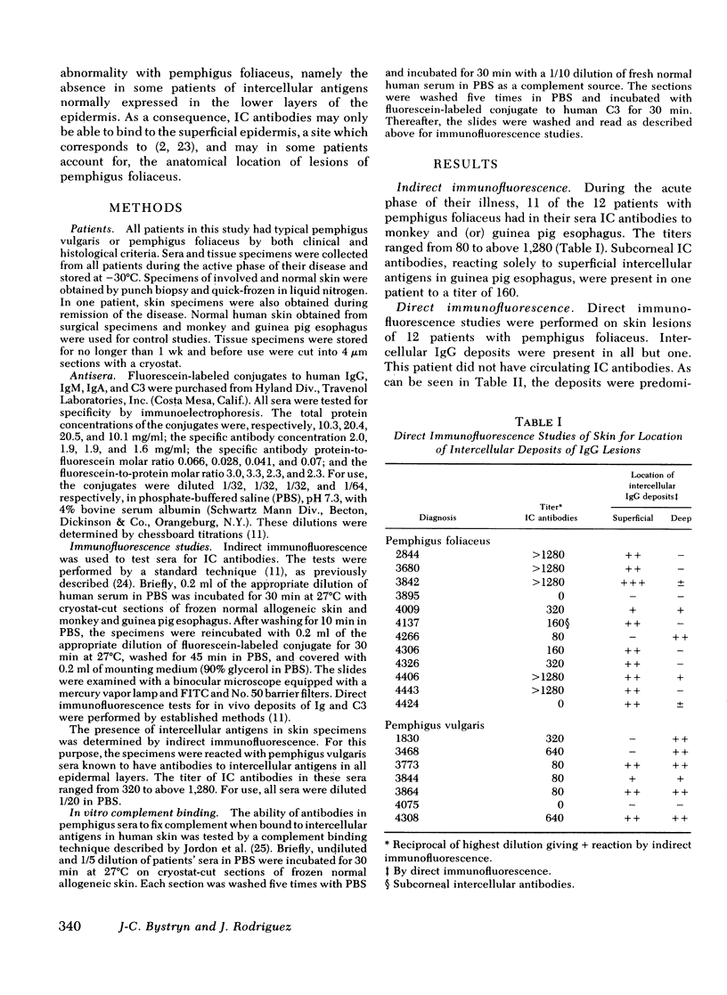
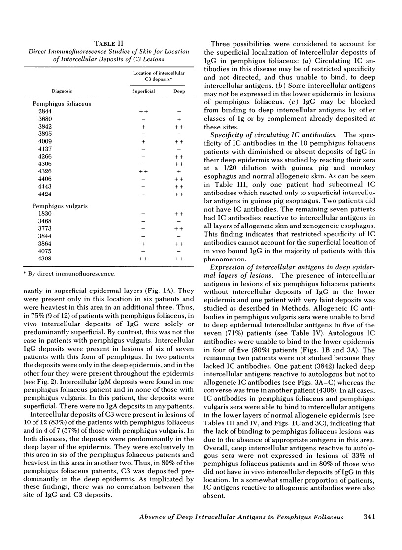
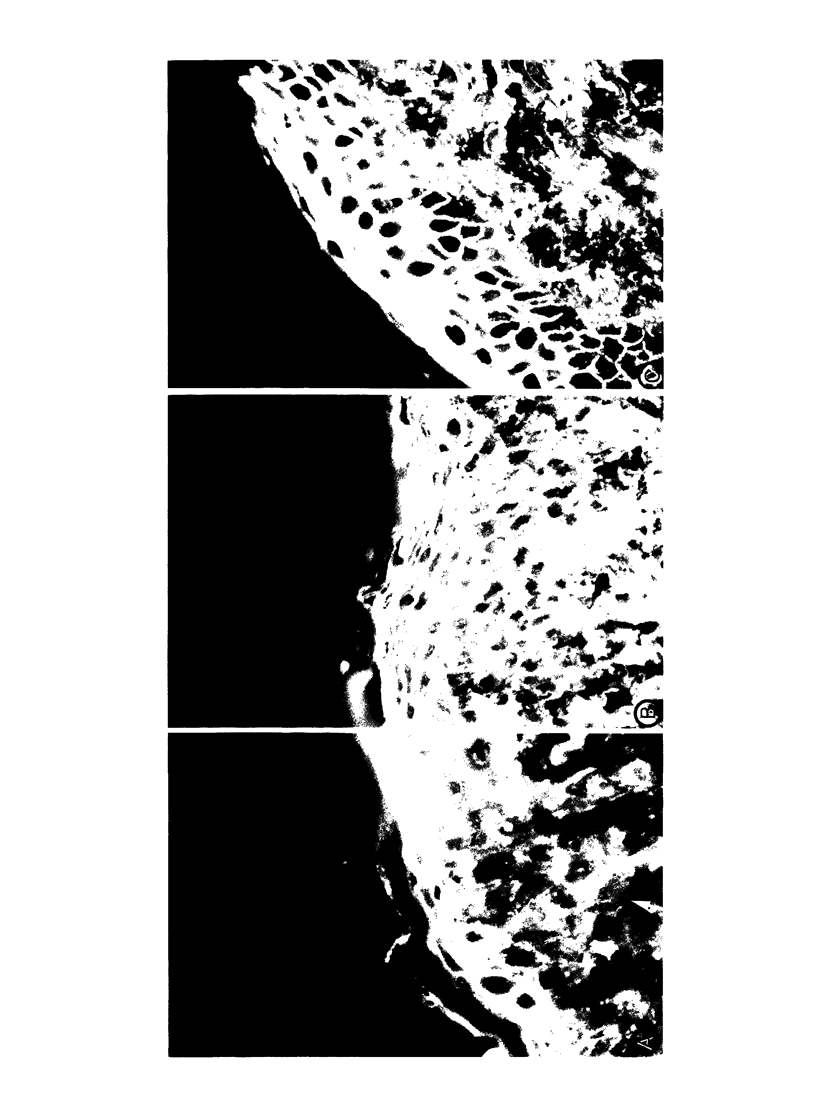
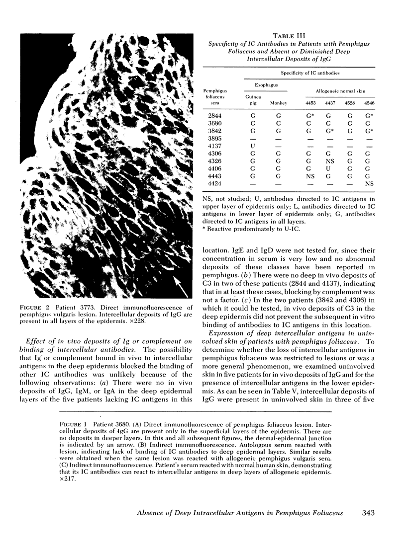
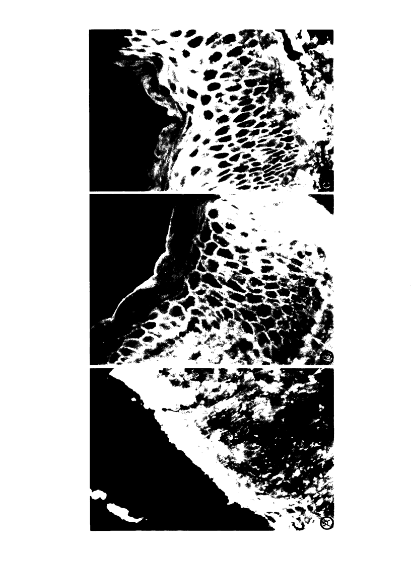
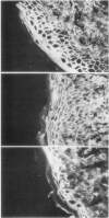
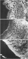
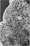
12 patients with pemphigus foliaceus, a form of pemphigus with lesions that arise in the intercellular substance in the superficial layers of the epidermis, and 7 patients with pemphigus vulgaris, where lesions are in the deep layers, were studied by immunofluorescence. Circulating antibodies to intercellular antigens (IC antibodies) were found in 11 pemphigus foliaceus and 5 pemphigus vulgaris patients. On direct immunofluorescence of skin lesions 75% (9 of 12), pemphigus foliaceus patients had intercellular deposits of IgG localized solely or predominantly in the superficial epidermal layers, whereas this was not the case in any of the patients with pemphigus vulgaris. Over 70% of the pemphigus foliaceus patients with predominantly superficial IgG deposits lacked in their lesions normal intercellular antigens usually expressed in the deep layers of the epidermis. This was shown by the inability of IC antibodies in autologous or allogeneic sera to bind to intercellular antigens in the lower epidermis of patient's skin, even though the same sera could bind to intercellular antigens in all layers of normal allogeneic skin. Lack of normal intercellular antigens deep in the epidermis may result in circulating IC antibodies binding to the superficial layers, a site which corresponds to, and thus in some patients may account for, the anatomical location of lesions in pemphigus foliaceus.
Full text
PDF
Images in this article
Selected References
These references are in PubMed. This may not be the complete list of references from this article.
- BEUTNER E. H., JORDON R. E. DEMONSTRATION OF SKIN ANTIBODIES IN SERA OF PEMPHIGUS VULGARIS PATIENTS BY INDIRECT IMMUNOFLUORESCENT STAINING. Proc Soc Exp Biol Med. 1964 Nov;117:505–510. doi: 10.3181/00379727-117-29622. [DOI] [PubMed] [Google Scholar]
- BEUTNER E. H., LEVER W. F., WITEBSKY E., JORDON R., CHERTOCK B. AUTOANTIBODIES IN PEMPHIGUS VULGARIS: RESPONSE TO AN INTERCELLULAR SUBSTANCE OF EPIDERMIS. JAMA. 1965 May 24;192:682–688. doi: 10.1001/jama.1965.03080210026006. [DOI] [PubMed] [Google Scholar]
- Beutner E. H., Jordon R. E., Chorzelski T. P. The immunopathology of pemphigus and bullous pemphigoid. J Invest Dermatol. 1968 Aug;51(2):63–80. [PubMed] [Google Scholar]
- Beutner E. H., Prigenzi L. S., Hale W., Leme C. de A., Bier O. G. Immunofluorescent studies of autoantibodies to intercellular areas of epithelia in Brazilian pemphigus foliaceus. Proc Soc Exp Biol Med. 1968 Jan;127(1):81–86. doi: 10.3181/00379727-127-32626. [DOI] [PubMed] [Google Scholar]
- Burnham T. K., Fine G. Indirect cutaneous immunofluorescence. I. Morphologic observations in bullous diseases, malignancies, and connective tissue diseases. Arch Dermatol. 1972 Jan;105(1):52–58. doi: 10.1001/archderm.105.1.52. [DOI] [PubMed] [Google Scholar]
- Bystryn J. C., Abel E., DeFeo C. Pemphigus foliaceus. Subcorneal intercellular antibodies of unique specificity. Arch Dermatol. 1974 Dec;110(6):857–861. doi: 10.1001/archderm.110.6.857. [DOI] [PubMed] [Google Scholar]
- Bystryn J. C., Abel E., Weidman A. Antibodies against the cytoplasm of human epidermal cells. Arch Dermatol. 1973 Aug;108(2):241–244. [PubMed] [Google Scholar]
- Chorzelski T. P., Beutner E. H. Factors contributing to occasional failures in demonstration of pemphigus antibodies by the immunofluorescence test. J Invest Dermatol. 1969 Sep;53(3):188–191. doi: 10.1038/jid.1969.132. [DOI] [PubMed] [Google Scholar]
- Chorzelski T. P., Von Weiss J. F., Lever W. F. Clinical significance of autoantibodies in pemphigus. Arch Dermatol. 1966 May;93(5):570–576. [PubMed] [Google Scholar]
- Hashimoto K., Lever W. F. An electron microscopic study on pemphigus vulgaris of the mouth and the skin with special reference to the intercellular cement. J Invest Dermatol. 1967 Jun;48(6):540–552. doi: 10.1038/jid.1967.86. [DOI] [PubMed] [Google Scholar]
- Inderbitzin T. M., Grob P. J. Destruction of epithelial cells in vivo by antiepithelial autoantibodies. J Invest Dermatol. 1967 Dec;49(6):642–645. doi: 10.1038/jid.1967.192. [DOI] [PubMed] [Google Scholar]
- Jordan R. E., Day N. K., Luckasen J. R., Good R. A. Complement activation in pemphigus vulgaris blister fluid. Clin Exp Immunol. 1973 Sep;15(1):53–63. [PMC free article] [PubMed] [Google Scholar]
- Jordon R. E., Sams W. M., Jr, Beutner E. H. Complement immunofluorescent staining in bullous pemphigoid. J Lab Clin Med. 1969 Oct;74(4):548–556. [PubMed] [Google Scholar]
- Jordon R. E., Schroeter A. L., Rogers R. S., 3rd, Perry H. O. Classical and alternate pathway activation of complement in pemphigus vulgaris lesions. J Invest Dermatol. 1974 Sep;63(3):256–259. doi: 10.1111/1523-1747.ep12680098. [DOI] [PubMed] [Google Scholar]
- Jordon R. E., Triftshauser C. T., Schroeter A. L. Direct immunofluorescent studies of pemphigus and bullous pemphigoid. Arch Dermatol. 1971 May;103(5):486–491. doi: 10.1001/archderm.103.5.486. [DOI] [PubMed] [Google Scholar]
- Peck S. M., Osserman K. E., Weiner L. B., Lefkovits A., Osserman R. S. Studies in bullous diseases. Immunofluorescent serologic tests. N Engl J Med. 1968 Oct 31;279(18):951–958. doi: 10.1056/NEJM196810312791801. [DOI] [PubMed] [Google Scholar]
- Sams W. M., Jr, Jordon R. E. Correlation of pemphigoid and pemphigus antibody titres with activity of disease. Br J Dermatol. 1971 Jan;84(1):7–13. doi: 10.1111/j.1365-2133.1971.tb14190.x. [DOI] [PubMed] [Google Scholar]
- Schiltz J. R., Michel B. Production of epidermal acantholysis in normal human skin in vitro by the IgG fraction from pemphigus serum. J Invest Dermatol. 1976 Aug;67(2):254–260. doi: 10.1111/1523-1747.ep12513454. [DOI] [PubMed] [Google Scholar]
- Thivolet J., Beyvin A. J. Anticorps anti-épiderme dans les dermatoses bulleuses. Documents cliniques et expérimentaux. Bull Soc Fr Dermatol Syphiligr. 1967;74(3):300–304. [PubMed] [Google Scholar]
- WILGRAM G. F., CAULFIELD J. B., MADGIC E. B. AN ELECTRON MICROSCOPIC STUDY OF ACANTHOLYSIS AND DYSKERATOSIS IN PEMPHIGUS FOLIACEUS: WITH A SPECIAL NOTE ON PECULIAR INTRACYTOPLASMIC BODIES. J Invest Dermatol. 1964 Nov;43:287–299. doi: 10.1038/jid.1964.159. [DOI] [PubMed] [Google Scholar]
- Wood G. W., Beutner E. H., Chorzelski T. P. Studies in immunodermatology. II. Production of pemphigus-like lesions by intradermal injection of monkeys with Brazilian pemphigus foliaceus sera. Int Arch Allergy Appl Immunol. 1972;42(4):556–564. [PubMed] [Google Scholar]
- van Joost T., Cormane R. H., Pondman K. W. Direct immunofluorescent study of the skin on occurrence of complement in pemphigus. Br J Dermatol. 1972 Nov;87(5):466–474. doi: 10.1111/j.1365-2133.1972.tb01595.x. [DOI] [PubMed] [Google Scholar]