Abstract
Background
Complications resulting from venipuncture include vein and nerve damage, hematoma, and neuropathic pain. Although the basic procedures are understood, few analyses of actual data exist. It is important to improve the safety standards of this technique during venipuncture. This study aimed to obtain data on actual needle movement during vacuum venipuncture in order to develop appropriate educational procedures.
Methods
Six experienced nurses were recruited to collect blood samples from 64 subjects. These procedures were recorded using a digital camera. Software was then used to track and analyze motion without the use of a marker in order to maintain the sterility of the needle. Movement along the X- and Y-axes during blood sampling was examined.
Results
Approximately 2.5 cm of the needle was inserted into the body, of which 6 mm resulted from advancing or moving the needle following puncture. The mean calculated puncture angle was 15.2°. Given the hazards posed by attaching and removing the blood collection tube, as well as by manipulating the needle to fix its position, the needle became unstable whether it was fixed or not fixed.
Conclusion
This study examined venipuncture procedures and showed that the method was influenced by increased needle movement. Focusing on skills for puncturing the skin, inserting the needle into the vein, and changing hands while being conscious of needle-tip stability may be essential for improving the safety of venipuncture.
Keywords: blood collection, nerve damage, motion analysis, patient safety, puncture angle, clinical education
Introduction
Venipuncture is a common procedure for reaching the venous bloodstream and plays an instrumental role as a special technique of phlebotomy in diagnosis and parenteral therapy.
All invasive procedures carry some degree of risk of damage to the normal structures in the proximity of the region at which the procedure is performed.1 Venipuncture is also an invasive procedure involving needle puncture. The complications resulting from venipuncture include nerve damage and neuropathic pain, local and systematic infections, vein damage, and hematoma, which may vary in seriousness. The epidemiology of needlestick and sharps injuries was investigated in a complete cross-section of 1,162 nurses in a large hospital in southern Japan. The prevalence of the devices used was 20.6% for syringe needles and 3.8% for butterfly needles.2 The cost of such accidents also needs to be calculated.
The immediate onset of symptoms with needle movement suggests direct needle-induced nerve trauma.3 Newman and Waxman,4 Newman et al,5 and Newman6 reported that the incidence of nerve damage caused by venipuncture during blood donation varies. Venipuncture-induced nerve injuries are rare; factors other than direct nerve contact appear necessary for the chronic pain syndrome to occur.7 Such injuries are typically mild or transient, and resolve spontaneously. Rarely, venipuncture-induced nerve injuries can be more severe, resulting in long-term disabling consequences, including Complex Regional Pain Syndrome type II.8 Nerve compression injury may result from large infiltrations and extravasations that can cause compartment syndrome.9 The importance of an accurate understanding of anatomy to prevent nerve damage and appropriate training from individuals experienced in venipuncture has previously been emphasized.10
Venipuncture is a serious medical procedure that should be carried out only by appropriately trained and competent medical staff.10 However, the procedure may often be performed not only by specialized technicians but also by novice practitioners.
The culture in which nurses and doctors have traditionally worked has often made it difficult for nurses to become competent at such skills. Nevertheless, the boundaries of medical and nursing roles continue to evolve, and a culture of shared roles that may confer substantial benefits for patients, medical staff, and nurses is emerging.11
Iatrogenic nerve injuries are not rare and occur in almost all branches of medicine, with malpositioning under general anesthesia and venipuncture as leading causes. Some of these injuries may be unavoidable, but greater awareness of which nerves are at risk and in what context should facilitate the development and/or wider implementation of preventive strategies.12 As for intramuscular injection, sciatic nerve injury in the upper outer quadrant of the buttock is an avoidable but persistent global problem. The consequences of this injury are potentially devastating, and should be promoted more widely by medical and nursing organizations.13
In addition, there are reports14 of infections being introduced into the blood by venipuncture. It is therefore important to improve safety standards for this technique in order to decrease the number of complications. To date, only a few studies15 have evaluated venipuncture techniques in detail. Accordingly, this study aimed to obtain data on actual needle movement during a venipuncture procedure using a vacuum collection system. This data could be used to develop appropriate educational practice programs.
Materials and methods
The study was conducted on three health check-up days scheduled during December 2011. If a subject was unwilling to cooperate on a particular day or the time was inconvenient, the check-up was scheduled for another day.
This study was approved by the ethics committee of the Graduate School of Health Management, Keio University, Kanagawa, Japan (June 23, 2010). The study objectives were explained to all participants. In addition, the participants were informed that they would not be penalized if they withdrew from the study and that only the researchers would view the video images. All participants signed a written consent form, which further explained that personal identification and individual evaluations would not be used and that questionnaire responses would be stored separately from the written consent forms.
Subjects
Sixty-four staff members, including health care workers and administrative staff, were subjected to venipuncture after providing consent for the examination to be recorded.
Six nurses who had worked in the hospital for longer than 10 years collected the blood samples. Only two nurses performed the venipunctures each day. The nurses performing the venipuncture were asked if the vacuum venipuncture technique was their first choice. They were also asked to select the best device for each individual.
The age and sex of the subjects undergoing venipuncture and other details of the procedure were recorded by the nurses. The circumference of the puncture site in the arm was measured after each venipuncture.
Venipuncture equipment
Four nurses used a tube holder (XX-VP010HD01) manufactured by Terumo Corporation, (Tokyo, Japan) for the vacuum tube procedures, with straight 21-gauge needles (MN-HD2138MS; Terumo Corporation). Two nurses used holders with straight 21-gauge needles (35209002; Nipro Corporation, Osaka, Japan). Blood collection tubes with conventional cap covers (NP-SP0725 or NP-EK0205; Nipro Corporation) were also used.
Video recording
A Digital HD Video camera recorder (HDR-HC9; Sony, Tokyo, Japan) mounted on a tripod and Hi-vision MiniDV tapes were used for recording the procedures. The maximum telephoto zoom (10 times) allowed a distance of 50 cm from the camera lens to the puncture point. This allowed optical image stabilization revision to avoid vibration. The puncture point was set directly under a pipe fluorescent lamp.
The blood collection procedure was captured using the video editing software EDIUS Pro 5 (Grass Valley KK, Hyogo, Japan), and the generated AVI files were transferred to a PC. The frame rate of the AVI file was 59.94 frames/second. To facilitate the acquisition process, we then converted these files into 24-bit true color with a video capture of 720 × 480 pixels whilst matching them on a monitor. A solid-state drive (SSD)-correlation tracking algorithm was activated during acquisition to adapt the quality of the image sequence and account for differences in color.
Video analysis
WINanalyze (v 2.1; Mikromak GmbH, Berlin, Germany) is a software application that assists in analyzing movements during video recording. This program uses calculations to generate analysis windows. The software is suitable for Windows AVI.
The needle movement can be visualized as a trajectory formed by using objects from all frames, in which the object sequences of the current image sequence are displayed in diagrammatic form with respect to time domain or converted to the tabulating data. It can be analyzed with other statistical programs. Data obtained from the X and Y coordinates were saved in an Excel spreadsheet (Microsoft Corporation, Redmond, WA, USA) and the data used for graphic making. The still images that emphasized the movement were shown using PowerPoint (Microsoft Corporation).
Venipuncture steps and measurements
The video assessed whether the nurses used their dominant hand to grasp the blood collection tube holder while puncturing the skin and then switched the tube holder to the nondominant hand, so that they were able to use their dominant hand to remove the tourniquet.
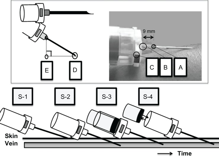
The welded part of the needle at the needle base was cream-colored and easy to distinguish from the surrounding materials (Figure 1). This was designated as point A and was used to track needle movements. Despite being covered by transparent green plastic, the needle base attached to the holder was recognizable and therefore designated as point B, which was used to measure distance. A seal 2 mm wide and 4 mm long was placed on the holder and was designated as point C. It was connected so that the needle hole was adjusted to the upper part of the seal and turned to the top.
Figure 1.
Location of the marker used and itemization of vacuum blood collection tube movement in the venipuncture procedure.
Notes: (A) The welded part of the needle at the needle base; (B) the needle base attached to the collection tube holder; (C) a point indicating where the blood collection tube holder was placed; (D) the point where the needle punctured the skin; (E) temporary point, interior angle of AED at 90°. The venipuncture procedure was divided into the following four steps based on the sequence of events: S-1: puncture; S-2: needle enters the vein; S-3: first collection tube; S-4: removal and insertion of collection tube.
Using WINanalyze, point D was assigned to the point where the needle punctured the skin, and point E was set as a temporary point at an angle of 90° to find the provisional value of the interior angle of ADE.
The vacuum venipuncture technique was assessed using video images and characterized by the point A trajectory. The procedure was subdivided into four steps (S-1–4), based on the following events:
S-1 – Puncture: first contact of the needle with the skin to entry of the needle into the vein.
S-2 – Needle enters the vein: entry of the needle into the vein to insertion of the first blood collection tube into the holder.
S-3 – First collection tube: insertion of the first blood collection tube into the holder and collection of blood to removal of the blood collection tube.
S-4 – Removal and insertion of collection tube: removal of the first blood collection tube to insertion of the second blood collection tube into the holder.
Measurement of movement during the venipuncture procedure
Given the size differences among the images obtained the study identified the coordinate representing the least difference between points A and B. The absolute value of the trajectory spanning from A to B was 0.9 cm. The distance in each picture was not always the same and therefore calculation bias was corrected using the distance between A and B. The X- and Y-axes represented backward-forward and upward-downward movements, respectively and the amount of motion occurring from one frame to the next was calculated and expressed as an absolute value.
Adjustments were made on the basis of the trajectories formed by points A and B calculated for each subject to derive the mean values for each step. The distance was calculated by the following formula:
| [1] |
Techniques with maximum movement values
For each step, the absolute values of the differences in measurements between frames were summed using [1] and expressed as movement distances. The maximum values for both the X and Y coordinates of each motion occurring during each step, as well as their characteristics, were extracted from the video images.
Observations of searching techniques
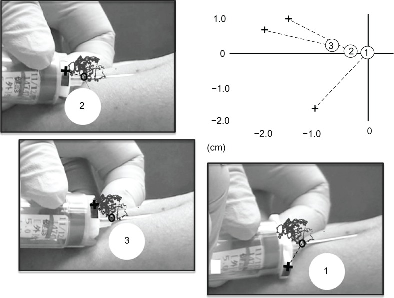
In one case, where the blood collection tube was inserted into the holder three times, point A and point C coordinates were extracted. The distance was measured, and the positional relationship of the coordinates was recorded.
Statistical analysis
Forty-six participants evaluated by video imaging were included in the analysis. If video imaging was blocked by placement of a finger over the needle during venipuncture, data from that section of the video recording were excluded from analysis. The recordings were reliable to a resolution of 1/10 mm. Descriptive statistics were summarized with SPSS for Windows (version 19.0; IBM Corporation, Armonk, NY, USA).
Results
Contributions
The mean age of the subjects was 42.3 years (standard deviation [SD] 12.2 years), and the mean circumference of their arm at the puncture site was 23.4 cm (SD 2.5 cm).
Six nurses performed the venipuncture, two of whom were aged in their 30s and four in their 40s. Four nurses always changed hands, whereas one nurse did not change hands at all. During the venipuncture procedures in 46 subjects, the nurses changed hands 82.6% of the time after puncture and 69.6% of the time when removing the tourniquet.
The needle travelled from the puncture site to the vein through a mean distance of 1.9 cm (SD 1.0 cm) in 29 subjects. This decrease in subject number was due to the video images being blocked by placement of a finger. The needle moved a mean distance of 0.6 cm (SD 0.3 cm) while entering the vein in 31 subjects. The mean puncture angle was 15.2° (SD 3.1°), with maximum and minimum angles of 21.8° and 10.7°, respectively (Table 1).
Table 1.
Subjects undergoing venipuncture, distance of needle movement, and puncture angle
| Nurse A | Nurse B | Nurse C | Nurse D | Nurse E | Nurse F | Total | ||||||||
|---|---|---|---|---|---|---|---|---|---|---|---|---|---|---|
| Number of subjects | n = 13 | n = 11 | n = 9 | n = 7 | n = 4 | n = 2 | n = 46 | |||||||
| Sex | ||||||||||||||
| Male | 4 | (30.8%) | 4 | (36.4%) | 4 | (44.4%) | 3 | (42.9%) | 1 | (25.0%) | 0 | (0.0%) | 16 | (34.8%) |
| Female | 9 | (69.2%) | 5 | (45.5%) | 4 | (44.4%) | 4 | (57.1%) | 3 | (75.0%) | 2 | (100%) | 27 | (58.7%) |
| No mention | 0 | (0.0%) | 2 | (18.2%) | 1 | (11.1%) | 0 | (0.0%) | 0 | (0.0%) | (50.0%) | 4 | (8.7%) | |
| Age (years, mean ± standard deviation) | 41.7 ± 12.5 | 42.3 ± 13.7 | 43.3 ± 10.3 | 43.4 ± 14.7 | 35.8 ± 7.6 | 52.0 ± 17.C | 42.3 ± 12.2 | |||||||
| Circumference puncture site (cm, mean ± standard deviation) | 23.2 ± 1.6 | 23.9 ± 1.8 | 23.5 ± 1.4 | 22.8 ± 1.7 | 25.0 ± 7.2 | 21.3± 1.1 | 23.4 ± 2.5 | |||||||
| Changed hands | ||||||||||||||
| After puncture | 13 | (100%) | 11 | (100%) | 9 | (100%) | 0 | (0.0%) | 3 | (75.0%) | 2 | (100%) | 38 | (82.6%) |
| Tourniquet removal | 11 | (84.6%) | 11 | (100%) | 5 | (55.6%) | 0 | (0.0%) | 3 | (75.0%) | 2 | (100%) | 32 | (69.6%) |
| Distance of the needle (cm, mean ± standard deviation) | ||||||||||||||
| From puncture to entry into vein | n = 3 | n = 8 | n = 8 | n = 6 | n = 4 | n = 29 | ||||||||
| 2.4 ± 0.3 | 1.3 ± 0.4 | 2.8 ± 1.1 | 1.9 ± 0.6 | 0.9 ± 0.3 | 1.9 ± 1.0 | |||||||||
| From entry into vein to entry into first tube | n = 3 | n = 10 | n = 8 | n = 6 | n = 4 | n = 31 | ||||||||
| 0.8 ± 0.2 | 0.6 ± 0.2 | 0.8 ± 0.4 | 0.3 ± 0.2 | 0.3 ± 0.1 | 0.6 ± 0.3 | |||||||||
| Angle of puncture (mean ± standard deviation) | n = 11 | n = 10 | n = 9 | n = 7 | n = 4 | n = 2 | n = 43 | |||||||
| Mean (°) | 14.4 ± 3.0 | 16.6 ± 2.8 | 16.2 ± 2.8 | 12.3 ± 1.1 | 15.3 ± 3.7 | 18.4 ± 4.9 | 15.2 ± 3.1 | |||||||
| Maximum (°) | 20.8 | 19.7 | 21.4 | 13.9 | 19.8 | 21.8 | 21.8 | |||||||
| Minimum (°) | 10.8 | 11.1 | 13.1 | 10.7 | 11.4 | 14.9 | 10.7 | |||||||
Note: *Estimated angle using temporary point.
Of the 33 subjects for whom all steps could be analyzed by video imaging, the needle moved a mean distance of 4.9 cm (SD 2.2 cm) along the X-axis and 3.2 cm (SD 1.4 cm) along the Y-axis. The needle moved a maximum of 12.4 cm and a minimum of 0.4 cm along the X-axis, whereas the Y-axis movement ranged from a minimum of 0.7 cm to a maximum of 10.3 cm.
In the case requiring insertion of the blood collection tube three times, the needle moved 6.3 cm along the X-axis and 3.7 cm along the Y-axis (Table 2).
Table 2.
Summation of total needle movement distance
| S-1 | S-2 | S-3 | S-4 | Total | ||||||
|---|---|---|---|---|---|---|---|---|---|---|
| Movement distances (cm) | n = 33 | n = 35 | n = 46 | n = 46 | n = 33 | |||||
| X-axis | ||||||||||
| Mean (Standard deviation) | 2.3 | (1.3) | 1.2 | (0.6) | 0.6 | (0.3) | 1.1 | (1.7) | 4.9 | (2.2) |
| Maximum | 5.3 | 2.8 | 1.5 | 11.8 | 12.4 | |||||
| Minimum | 0.8 | 0.3 | 0.1 | 0.2 | 0.4 | |||||
| Y-axis | ||||||||||
| Mean (Standard deviation) | 1.1 | (0.6) | 1.1 | (0.6) | 0.5 | (0.3) | 0.8 | (1.4) | 3.2 | (1.4) |
| Maximum | 3.2 | 2.4 | 1.4 | 9.6 | 10.3 | |||||
| Minimum | 0.4 | 0.4 | 0.2 | 0.2 | 0.6 | |||||
|
| ||||||||||
| Movement distances (a subject case) cm | ||||||||||
| X-axis | 3.0 | 1.4 | 0.8 | 1.1 | 6.3 | |||||
| Y-axis | 0.9 | 1.7 | 0.5 | 0.6 | 3.7 | |||||
Notes: S-1: puncture; S-2: needle enters the vein; S-3: first collection tube; S-4: removal and insertion; Total, subjects that were measured from S-1 to S-4.
Minimum and maximum needle movement
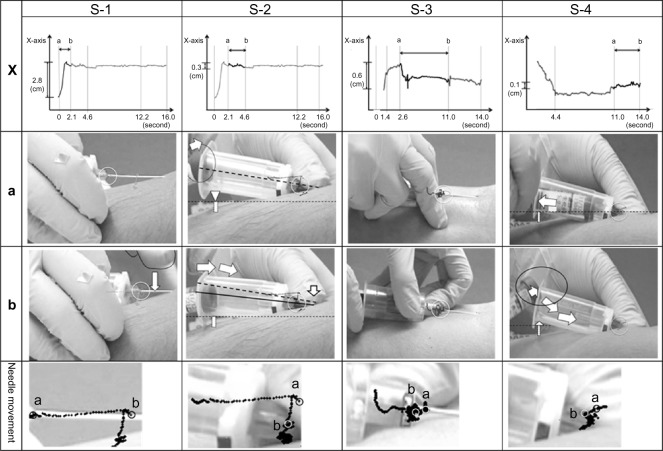
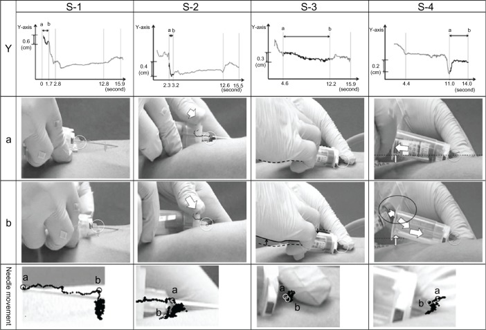
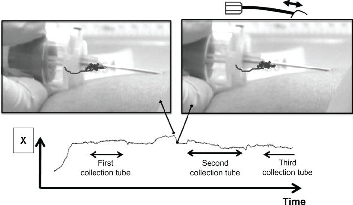
Figures 2 and 3 show the maximum accumulated movement that occurred at each step on the X- and Y-axes, respectively. Maximum movement was caused when the needle or holder was pressed from the top to the bottom, with the needle dropping less than 0.6 cm. In other cases, the blood collection tube contacted the opening of the holder. The needle-tip remained in the vein whilst the holder was raised to insert or remove the blood collection tube.
Figure 2.
X-axis: maximum of accumulated movement. (a) shows the start and (b) shows the end of the movement.
Notes: S-1: Needle was pressed from top to bottom by the first finger of the left hand. S-2: Holder traveled up and down during use of the first collection tube with the collection tube attached to the upper opening of the holder. S-3: Nurse changed hands after inserting the blood collection tube into the holder using the nondominant hand. S-4: The holder was pushed down during removal of the blood collection tube from the upper opening of the holder.
Figure 3.
Y-axis: maximum of accumulated movement. (a) shows the start and (b) shows the end of the movement.
Notes: S-1: The needle moved up and down. S-2: The holder was pressed from the top to the bottom using the first finger of the left hand. S-3: Needle was pushed by the left thumb with the collection tube attached to the upper opening of the holder. S-4: The holder was pushed down during removal of the blood collection tube from the upper opening of the holder.
Searching movements
A positional relationship was shown between points A and C in cases where blood flowed into the third blood collection tube that had been placed in the holder after puncture. The welded part of the needle moved 0.7 cm from the first to the second tube and 0.3 cm from the second to the third tube, as shown in Figure 4.
Figure 4.
Searching: location of the welded part of the needle at the point where the blood collection tube entered the tip of the holder, and the location of the sticker placed on the holder. (1) the first puncture is not successful; (2) the second puncture is not successful; (3) the third puncture is successful.
A case in which the hands were not changed
Notably, in one case, pulling of the skin or bending of the needle was observed during insertion and removal of the blood collection tubes. The needle area could be visualized clearly in cases in which the hands were not changed, as shown in Figure 5.
Figure 5.
A case in which the hands were not changed.
Notes: Pulling of the skin or bending of the needle was observed. The position of the holder is unstable.
Discussion
Successful venipuncture is characterized as reaching the venous bloodstream and not resulting in complications. This invasive procedure contains two critical steps: venipuncture site and vein selection, and venipuncture performance. In this study, we evaluated venipuncture performance from puncture to removal of the needle, which is especially related to venipuncture complications.
Unsuccessful venipuncture performance, which leads to complications, is related to improper movement of the needle. Therefore, the actual needle movement data for venipuncture performance are important to developing appropriate educational practice programs. The needle movement at the time of puncture is described as a puncture made at an approximate angle of 10°–30°.16 The needle is laid flat such that 1–2 mm of its tip can be inserted into the vein.17 These movements occur in a similar manner during venipuncture worldwide.
The length of the 21-gauge needle used in this study was 3.8 cm. When needle puncture occurred, the distance from the two coordinate points as the needle moved forward at an angle was calculated. As a result, approximately 1.9 cm of the needle was inserted after puncture, and it moved around 0.6 cm after insertion. Therefore, approximately 2.5 cm of the needle was inserted into the body. As the mean circumference of the puncture site was 0.6 cm, the distance for advancing the needle after the backflow of blood observed in this study was relatively longer than a distance of 1–2 mm.
This study calculated the puncture angle, ie, the interior angle of the needle and the puncture site. Although the puncture angles represented provisional values (Figure 1) due to differences in the position and orientation of the hand performing venipuncture, the mean, minimum, and maximum angles were 15.2°, 10.7°, and 21.8°, respectively. It is not feasible to check whether the needle has entered the vein during vacuum venipuncture until the blood collection tube is inserted. To the author’s knowledge, no other experimental studies are available; therefore, no comparisons with this study could be made. However, these findings indicate a puncture angle of approximately 15°. This estimate falls within the same range of standard teaching.
Moving the needle-tip to locate the vein under the skin following an unsuccessful puncture is termed “searching.” This problem occurs in frequently accessed venipuncture sites and also with errors when selecting the vein, but rarely as a result of venipuncture performance.
When the needle-tip did not enter the blood vessel, the nurse decided whether it was better to remove the needle or move the needle-tip to locate the blood vessel rather than repeat the puncture numerous times. When the needle-tip is moved, the holder moves at the same time. Observing the welded part of the needle, the study noted that a 0.7 cm movement occurred between the first and second tubes, whereas a 0.3 cm movement transpired between the second and third tubes, equaling a total of 1 cm. However, when the point marked on the holder was observed, the holder seemed to rotate. The needle bore may not have rotated to the top. Rotation changes the needle orientation, and blood does not flow back even if the needle has entered the vein. This leads to the notion that the needle has not entered the vein, irrespective of whether the vein was entered or not. At this stage, vein and nerve damage can occur if the needle is moved back and forth.
Vein and nerve damage during the actual puncture of the vein is an important concern. However, it should be emphasized that attaching and removing the blood collection tube as well as manipulating the needle to fix its position also bear risks.
The maximum summation of forward–backward movement in this study was 12.4 cm, whereas the maximum up–down movement was 10.3 cm, including movement during needle puncture. This summation value may be influenced of vibrations for the shaking of camera, tracking of data or back flow of blood. Movements were large when the needle was pressed down from top to bottom after its tip had entered the vein and when the blood collection tube was being prepared for insertion into the holder. There were also potential hazards for vein and nerve damage through attaching and removing the blood collection tube as well as by manipulating the needle to fix its position.
The problem of using the dominant versus nondominant hand remains. Collecting blood involves puncturing the skin, changing hands, grasping the holder, and inserting and removing blood collection tubes with the dominant hand, followed by removing the tourniquet when the hands are changed again. Alternatively, collection may involve puncture and insertion or removal of the blood collection tube with the nondominant hand without changing hands. In these cases, the needle-tip moved backward, and the skin looked visibly stretched while the blood collection tube was inserted and removed.
There are also individuals who hold the needle and needle base, which is a common practice in venipuncture. Holding the needle stops it from shaking and produces less movement which, in turn, presumably reduces the amount of stress on the patient. However, care must be taken to prevent infection when holding the needle.
Inserting or removing a blood collection tube caused the holder to move upward and downward when the tube contacted the area around the opening of the holder. If the blood collection tube contacted the holder, the point where the needle was held became a pivot point for raising the entire holder. Pushing the needle or needle base may cause the holder to appear raised. Therefore, inserting or removing the blood collection tube increases the risk of moving the needle-tip. In this regard, improving the quality of blood collection techniques is clearly needed, particularly in hospital wards.18 Training that focuses on needle stability during manipulation may be particularly beneficial.
Venipuncture is also used for diagnosis during an emergency. It is a skill that is required of nurses on duty at any time, and it may be expected even for dehydrated patients and patients in whom a vein cannot be located. Therefore, the high frequency of blood testing alone means that vein and nerve damage caused by venipuncture can potentially occur in anyone.
The participant hoped to undergo a health check-up at an early time in the day. Therefore, the number of participants that each nurse is involved with is only partially important. This study evaluated only a blood collection system. The number of nurses was not sufficient, and there were differences in venipuncture equipment. Variation between subjects, including where to place the fingers during venipuncture and movement of the hand, decreased with an increase in the number of videos analyzed.
Conclusion
This study examined the characteristics of techniques by reviewing the cases producing movements; individual differences and variations in techniques may have existed among the six nurses included in this study. Although an identical method was used, participants undergoing venipuncture may have moved at the time of puncture but otherwise may not have moved significantly. This study could therefore not determine the optimal technique. Further studies on how to assess venipuncture techniques are needed.
Footnotes
Disclosure
The author reports no conflicts of interest in this work.
References
- 1.Zubairy AI. How safe is blood sampling? Anterior interosseus nerve injury by venipuncture. Postgrad Med J. 2002;78:625. doi: 10.1136/pmj.78.924.625. [DOI] [PMC free article] [PubMed] [Google Scholar]
- 2.Smith DR, Mihashi M, Adachi Y, Nakashima Y, Ishitake T. Epidemiology of needlestick and sharps injuries among nurses in a Japanese teaching hospital. J Hosp Infect. 2006;64:44–49. doi: 10.1016/j.jhin.2006.03.021. [DOI] [PubMed] [Google Scholar]
- 3.Horowitz SH. What happens when cutaneous nerves are injured during venipuncture? Muscle Nerve. 2005;31:415–417. doi: 10.1002/mus.20287. [DOI] [PubMed] [Google Scholar]
- 4.Newman BH, Waxman DA. Blood donation-related neurologic needle injury: evaluation of 2 years’ worth of data from a large blood center. Transfusion. 1996;36:213–215. doi: 10.1046/j.1537-2995.1996.36396182137.x. [DOI] [PubMed] [Google Scholar]
- 5.Newman BH, Pichette S, Pichette D, Dzaka E. Adverse effects in blood donors after whole-blood donation: a study of 1000 blood donors interviewed 3 weeks after whole-blood donation. Transfusion. 2003;43:598–603. doi: 10.1046/j.1537-2995.2003.00368.x. [DOI] [PubMed] [Google Scholar]
- 6.Newman BH. Blood donor complications after whole-blood donation. Curr Opin Hematol. 2004;11:339–345. doi: 10.1097/01.moh.0000142105.21058.96. [DOI] [PubMed] [Google Scholar]
- 7.Horowitz SH. Venipuncture-induced causalgia: anatomic relations of upper extremity superficial veins and nerves, and clinical considerations. Transfusion. 2000;40:1036–1040. doi: 10.1046/j.1537-2995.2000.40091036.x. [DOI] [PubMed] [Google Scholar]
- 8.Horowitz SH. Venipuncture-induced nerve injury: a review. J Neuropathic Pain Symptom Palliation. 2005;1:109–114. doi: 10.1300/J426v01n01_05. [DOI] [PMC free article] [PubMed] [Google Scholar]
- 9.Masoorli S. Nerve injuries related to vascular access insertion and assessment. J Infus Nurs. 2007;30:346–350. doi: 10.1097/01.NAN.0000300310.18648.b2. [DOI] [PubMed] [Google Scholar]
- 10.McConnell AA, Mackay GM. Venipuncture: the medicolegal hazards. Postgrad Med J. 1996;72:23–24. doi: 10.1136/pgmj.72.843.23. [DOI] [PMC free article] [PubMed] [Google Scholar]
- 11.Campbell H, Carrington M, Limber C. A practical guide to venipuncture and management of complications. Br J Nurs. 1999;8:426–431. doi: 10.12968/bjon.1999.8.7.6645. [DOI] [PubMed] [Google Scholar]
- 12.Moore AE, Zhang J, Stringer MD. Iatrogenic nerve injury in a national no-fault compensation scheme: an observational cohort study. Int J Clin Pract. 2012;66:409–416. doi: 10.1111/j.1742-1241.2011.02869.x. [DOI] [PubMed] [Google Scholar]
- 13.Mishra P, Stringer MD. Sciatic nerve injury from intramuscular injection: a persistent and global problem. Int J Clin Pract. 2010;64:1573–1579. doi: 10.1111/j.1742-1241.2009.02177.x. [DOI] [PubMed] [Google Scholar]
- 14.WHO Guidelines Approved by the Guidelines Review Committee . WHO Best Practices for Injections and Related Procedures Toolkit. World Health Organization; 2010. [PubMed] [Google Scholar]
- 15.Knapp MB, Grytdal SP, Chiarello LA, Sinkowitz-Cochran RL, Zombeck A, Klein C, Warden B, Lyden J, Pearson ML. Evaluation of institutional practices for prevention of phlebotomy-associated percutaneous injuries in hospital setting. Am J Infect Control. 2009;37:490–494. doi: 10.1016/j.ajic.2008.06.012. [DOI] [PubMed] [Google Scholar]
- 16.Scales K. A practical guide to venipuncture and blood sampling. Nurs Stand. 2008;22:29–36. doi: 10.7748/ns2008.03.22.29.29.c6436. [DOI] [PubMed] [Google Scholar]
- 17.Lavery I, Ingram P. Venipuncture: best practice. Nurs Stand. 2005;19:55–65. doi: 10.7748/ns2005.08.19.49.55.c3936. [DOI] [PubMed] [Google Scholar]
- 18.Wallin O, Söderberg J, Van Guelpen B, Stenlund H, Grankvist K, Brulin C. Blood sample collection and patient identification demand improvement: a questionnaire study of preanalytical practices in hospital wards and laboratories. Scand J Caring Sci. 2010;24:581–591. doi: 10.1111/j.1471-6712.2009.00753.x. [DOI] [PubMed] [Google Scholar]


