Abstract
Variations in the branching pattern of the common, external, and internal carotid arteries can present as arteriovenous malformations, and their basis can be explained embryologically. Our case was a rare variation presenting as a congenital, very gradually increasing bluish painless swelling at the region of the left lobule of the ear arising from an abnormal vessel (from the postauricular artery) which was explored under general anesthesia through a postauricular curved incision. The abnormal vessel and other feeding vessels were ligated and a sclerosing agent injected. Anomalies of pharyngeal arch arteries like our case can be found resulting from the persistence of channels that normally disappear, and prior knowledge of these anomalies is essential before surgeries like mastoidectomy to prevent alarming hemorrhage.
Keywords: Congenital arteriovenous malformation, Auricular, Mastoidectomy
Introduction
In many studies, variations have been reported in the branching pattern of the external, internal, and common carotid arteries. By understanding the anatomic location and radiologic appearance, the basis of variation can be predicted embryologically. Variations can present as large facial arteriovenous malformations which are problematic for patients because of disfigurement, risk of rapid enlargement, and life-threatening rupture. Also, these variations are of importance for surgical approaches in the head and neck region, so the present case was studied and compared with those reported before.
Case Report
A 21-year-old male presented with a very gradually increasing bluish painless swelling around the lobule of the left ear since birth without any previous history of trauma or surgical intervention. There were no otological or neurological complaints associated with it. General physical examination did not show any significant findings. Local examination revealed a bluish pulsatile rounded swelling about 2 cm in diameter both on the medial and lateral aspects of the lobule of the left ear. The pulsation was synchronous with the carotid pulse. The swelling was compressible with rapid filling. The pulsations became feeble and the filling slower by applying pressure on the postauricular artery. An abnormal pulsatile vessel was also felt from the inferior border of the mastoid process to the post margin of the swelling. A weak bruit was heard, and the swelling was not transilluminant.
Systemic examination did not reveal any abnormality. Routine hematological parameters were within normal limits. Color Doppler showed significant arterial filling in the swelling.
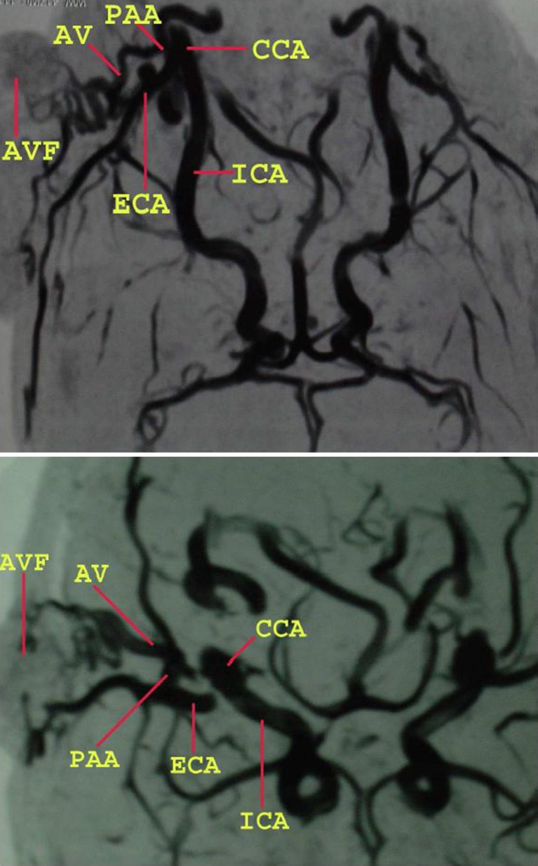
Magnetic resonance angiography of the region was performed using 3D time-of-flight technique with MOTSA and viewed on a 1.5-T scanner. It revealed an abnormal tortuous vessel in the region of the left lobule of the ear arising from the postauricular artery and showing early venous filling which is suggestive of an arteriovenous malformation (AVM) (Fig. 1). Bilateral intracranial carotid arteries, their branches, and the vertebrobasilar system were unremarkable, without any aneurysm, stenosis, and flow limitation.
Fig. 1.
MRA scan of the carotid system showing arteriovenous malformation on the left side. CCA common carotid artery, ICA internal carotid artery, ECA external carotid artery, PAA posterior auricular artery, AV abnormal vessel arising from posterior auricular artery, AVF arteriovenous malformation
The exploration was done under general anesthesia through a postauricular curved incision. The posterior auricular artery was very prominent, and an abnormal vessel was arising from it which was feeding the pulsatile swelling. As soon as this artery was ligated, the pulsation disappeared and the size of the swelling markedly reduced. The few smaller feeding vessels present were also ligated, a sclerosing agent was injected into the swelling after emptying it completely, and continuous manual pressure was applied at the site of surgery. The postoperative period was uneventful. Although the treatment of choice for an extracranial, congenital, and traumatic arteriovenous malformation is ideally total excision, embolization can also be done for extensive ones to prevent hemorrhage.
Discussion
Few people, like Matsushige et al., Shimoda et al., Ohno et al., and Takahashi et al. [1–4], have reported AVMs of the scalp, the precise natural course of which is not known. The posterior auricular artery is a branch of the external carotid artery which, according to Padget, arises embryologically as new branches from the ventral aspect of the third pharyngeal arch arteries which probably link up with endothelial channels left by the regression of the first and second arch arteries [5]. Frequent anomalies are found resulting from persistence of channels that normally disappear. Incomplete obliteration of vestigial organs or buried cell rests may result in abnormal presentations. A satisfactory embryological explanation, based on regressions and reversal of flux, may be proposed after correlation with basic anatomical and embryological facts. Prior knowledge of these anomalies is essential before surgeries like mastoidectomy to prevent alarming hemorrhage.
References
- 1.Matsushige T, Kiva K, Satoh H, Mizoue T, Kagawa K, Araki H. Arteriovenous malformation of the scalp: case report and review of the literature. Surg Neurol. 2004;62(3):253–259. doi: 10.1016/j.surneu.2003.09.033. [DOI] [PubMed] [Google Scholar]
- 2.Shimoda M, Matumae M, Shibuya N, Yamamoto I, Sato O. Three cases of arteriovenous malformations. No Shinkei Geka. 1987;15(2):173–178. [PubMed] [Google Scholar]
- 3.Ohno K, Tone O, Inaba Y, Terasaki T. Coexistent congenital arteriovenous malformation and aneurysms of the scalp (author's transl) No Shinkei Geka. 1981;9(10):1187–1191. [PubMed] [Google Scholar]
- 4.Takahashi S, Furuhashi N, Koshu K. Congenital arteriovenous malformations of the extracranial region—report of a case with review of literatures (author's transl) No Shinkei Geka. 1977;5(8):889–893. [PubMed] [Google Scholar]
- 5.Padget DH. The development of the cranial arteries in the human embryo. Contrib Embryo. 1948;32:205–262. [Google Scholar]


