

Abstract
Cutis laxa is a rare disease that may be either inherited or acquired. The acquired form is rarer than the inherited form. Pathogenesis of this disease is largely unknown. Two cases of acquired cutis laxa are reported here and neither of them had any systemic involvement or any history of drug intake. One of them had localized disease with history of preceding cutaneous inflammation. The other patient with generalized lesion lacked any history of preceding illness. The patient with localized lesion was treated satisfactorily by reconstructive surgery. The other patient had generalized involvement, for which no satisfactory treatment could be offered.
Keywords: Acquired, cutis laxa, elastase
Introduction
What was known?
Acquired cutis laxa is less common than congenital form. No treatment exists to prevent disease progression. Treatment of generalized form is usually unsatisfactory.
Cutis laxa is a rare connective tissue disorder, which may be inherited or acquired, and is characterized by progressive looseness of skin, associated with abnormalities of other organs and structures such as lung, vasculature or gastrointestinal tract[1] as they contain elastic tissues. The hereditary form of cutis laxa may be inherited as either autosomal dominant, X-linked recessive or autosomal recessive trait.[2] The acquired form is even rarer than the inherited form[3] and is often associated with some form of preceding or accompanying cutaneous eruptions like eczema, urticaria, erythema multiforme, Sweet's syndrome or multiple myeloma.[4,5,6] Sometimes, it may be drug induced.[4,6] The clinical appearance is characterized by inelastic, loose and pendulous skin that produces a wrinkled, prematurely aged appearance.[1] The primary change is in the elastic fiber, which is decreased or nearly absent in the dermis.[7] Here, we describe 2 cases of acquired cutis laxa.
Case Reports
Case 1
A 14-year-old Hindu female presented to us with complaint of ‘loose skin’ on the neck. An itchy eruption had developed on the anterior neck 5 years ago, and it lasted for about a month. Several months later, she noticed that the skin of that area was becoming wrinkled and loose. The wrinkling and looseness progressed for 4 to 5 months after which it became stable. She was otherwise well and had no history of dyspnoea, chest pain, palpitation or visual problems. She was not taking any medication. No similar changes were noted in any other part of her body. Family history of similar skin lesion was absent. Physical examination revealed a circumscribed palm sized area of lax, extensible, wrinkled skin localized to the anterior part of the neck [Figure 1a]. Stretching of the skin produced delayed recoil [Figure 1b]. No induration was noted. There was no hyper-extensibility of the joints and systemic examination was normal. Slit-lamp examination of the eyes revealed no angioid streaks.
Figure 1a.
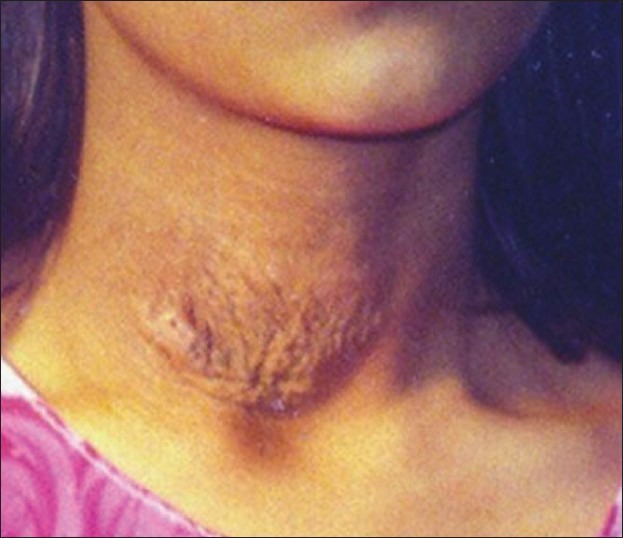
Lesion on the neck of a 14-year-old female
Figure 1b.
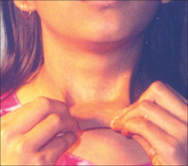
Lesion on the neck showing stretching of the lax skin
Case 2
A 40-year-old Muslim male presented to us with a complaint of loose skin all over his body for last 10 years. He was well till the age of 30, when he noticed that his skin was gradually becoming wrinkled and loose. It started from forehead and progressed slowly downwards over a couple of years after that the process stabilized. At the time of presentation, he was suffering from hypertension and cervical and lumbar spondylosis. He was having beta-blockers for hypertension. He had no episodes of dyspnoea, chest pain or palpitations. His family history was non-contributory. On examination, the patient appeared much older than his age. The skin of his forehead [Figure 2a], neck, abdomen and back [Figure 2b] was diffusely wrinkled and it hung in folds. There was blepharochalasis but no hyper-extensibility of joints. Eye examination did not reveal any angioid streaks.
Figure 2a.

Lesion on the scalp of a 40-year-old male
Figure 2b.

Lesion on the back of a 40-year-old male
In both the cases investigations revealed normal values for complete blood count, erythrocyte sedimentation rate, routine urine analysis, blood biochemistry, hepatic enzymes and serum electrophoresis. Chest X-ray, electrocardiography and gastroscopy were normal. Biopsy specimens taken from the involved area were stained with Hematoxylin-eosin (H and E), Von Kossa and Verhoeff-van Gieson stain. Histopathological findings were similar in both cases. H and E staining revealed normal epidermis with diminished elastic fibres and mild non-specific chronic lymphohistiocytic infiltrate in upper dermis [Figure 3a]. The elastic fibres present were fragmented, stained uneven and showed granular appearance, as evidenced in Verhoeff-van Gieson stain [Figure 3b]. Von-Kossa stain did not show any calcification.
Figure 3a.

Histopathology of skin lesion of case 1 showing normal epidermis with diminished elastic fibres and mild non-specific chronic lymphohistiocytic infiltrate in upper dermis (H and E, original magnification ×10)
Figure 3b.

Histopathology of skin lesion of case 2 showing fragmented and unevenly stained elastic fibres with granular appearance, (Verhoeff-van Gieson stain, original magnification ×10)
Discussion
Acquired cutis laxa usually begins in adulthood with insidious development of loose skin often starting at face and neck. The first case with localized lesion described above could be differentiated from pseudo xanthoma elasticum (PXE) by the absence of the characteristic yellow color of PXE and lack of calcification with Von Kossa stain. The second case could be differentiated from Elher Danlos syndrome, as the skin was lax and recoiled slowly, and there was no joint involvement. The girl (case 1) was referred to a reconstructive surgeon for treatment, and after successful excision of the loose skin only a fine linear scar was visible. But not much could be done for the older patient (case 2). He was advised to take one vitamin E capsule (400 mg) daily for 2 months. Emollients were given for topical application. There was not much improvement after 2 months and the patient was lost to follow up. One treatment option is injection of botulinum toxin. This option may be a less invasive way to improve facial defects in these patients.
The cause of acquired cutis laxa is largely unknown, though various hypotheses such as altered copper metabolism,[8] excess elastase,[9] and immune-mediated mechanisms[10] have been proposed. A unique hypothesis postulated by Reed et al.[4] is that elastolysis may result from release of granulocytic elastase because neutrophils contain large amounts of elastase, and is also found in macrophages and platelets. Up to 100-fold increase in the serum elastase level has been demonstrated in a patient with autosomal recessive cutis laxa. A suggested mechanism of cutis laxa is that, unknown causes stimulate mast cells to release mediators of inflammation, such as histamine, leucotrienes, neutrophil and eosinophil chemotactic factors. Consequently, there is a progressive infiltration of neutrophils and eosinophils around the blood vessels in the papillary dermis and between the collagen fibers in the mid- and reticular dermis. These neutrophils release elastase, and elastolysis first begins in areas with neutrophilic infiltration around the blood vessels and between collagen fibers, and then it gradually spreads to the entire dermis. These changes could be localized, as in the first case or they could be generalized leading to universal involvement, as in the second case.
What is new?
Localized cutis laxa responds well to plastic reconstruction.
Footnotes
Source of Support: Nil
Conflict of Interest: Nil.
References
- 1.Fitzsimmons JS, Fitzsimmons EM, Guibert PR, Zaldua V, Dodd KL. Variable clinical presentations of cutis laxa. Clin Genet. 1985;28:284–95. doi: 10.1111/j.1399-0004.1985.tb00402.x. [DOI] [PubMed] [Google Scholar]
- 2.Winter RM, Baraitser M. London: Chapman and Hall Medical; 1991. Multiple congenital anomalies: A diagnostic compendium; pp. 145–6. [Google Scholar]
- 3.Agha A, Sakati NO, Higginbottom MC, Jones KL, Jr, Bay C, Nyhan WL. Two forms of cutis laxa presenting in the newborn period. Acta Paediatr Scand. 1978;67:775–80. doi: 10.1111/j.1651-2227.1978.tb16260.x. [DOI] [PubMed] [Google Scholar]
- 4.Reed WB, Horowitz RE, Beighton P. Acquired cutis laxa. Primary generalized elastolysis. Arch Dermatol. 1971;103:661–9. [PubMed] [Google Scholar]
- 5.Turner RB, Haynes HA, Granter SR, Miller DM. Acquired cutis laxa following urticarial vasculitis associated with IgA myeloma. J Am Acad Dermatol. 2009;60:1052–7. doi: 10.1016/j.jaad.2008.09.059. [DOI] [PubMed] [Google Scholar]
- 6.Kumar Anil Gaikwad, MY Khedker. Acquired Cutis Laxa. Indian J Dermatol Venereol Leprol. 1989;55:185–6. [PubMed] [Google Scholar]
- 7.Nanko H, Jepsen LV, Zachariae H, Søgaard H. Acquired cutis Laxa (generalized elastolysis): Light and electron microscopic studies. Acta derma Venerol. 1979;59:315–24. [PubMed] [Google Scholar]
- 8.Goltz RW, Hult AM, Goldfarb M, Gorlin RJ. Cutis Laxa. A manifestation of generalized elastolysis. Arch Dermatol. 1965;92:373–87. doi: 10.1001/archderm.92.4.373. [DOI] [PubMed] [Google Scholar]
- 9.Lewis PG, Hood AF, Barnett NK, Holbrook KA. Postinflammatory elastolysis and cutis laxa: A case report. J Am Acad Dermatol. 1990;22:40–8. doi: 10.1016/0190-9622(90)70005-3. [DOI] [PubMed] [Google Scholar]
- 10.Newton JA, McKee PH, Black MM. Cutis laxa associated with amyloidosis. Clin Exp Dermatol. 1986;11:87–91. doi: 10.1111/j.1365-2230.1986.tb00430.x. [DOI] [PubMed] [Google Scholar]