Abstract
Although lead exposure has declined in recent years as a result of change to lead-free gasoline, several epidemiological have pointed out that it represents a medical and public health emergency, especially in young children consuming high amounts of lead-contaminated flake paints. A previous study in our laboratory indicated that lead exposure induces cytotoxicity in human liver carcinoma cells. In the present study, we evaluated the role of oxidative stress in lead-induced toxicity, and the protective effect of the anti-oxidant n-acetyl-l-cysteine (NAC). We hypothesized that oxidative stress plays a role in lead-induced cytotoxicity, and that NAC affords protection against this adverse effect. To test this hypothesis, we performed the MTT [3-(4, 5-dimethylthiazol-2-yl)-2, 5-diphenyltetrazolium bromide] assay and the trypan blue exclusion test for cell viability. We also performed the thiobarbituric acid test for lipid peroxidation. Data obtained from the MTT assay indicated that NAC significantly increased the viability of HepG2 cells in a dose-dependent manner upon 48 hours of exposure. Similar trend was obtained with the trypan blue exclusion test. Data generated from the thiobarbituric acid test showed a significant (p ≤ 0.05) increase of MDA levels in lead nitrate-treated HepG2 cells compared to control cells. Interestingly, the addition of NAC to lead nitrate-treated HepG2 cells significantly decreased cellular content of reactive oxygen species (ROS), as evidenced by the decrease in lipid peroxidation byproducts. Overall, findings from this study suggest that NAC inhibits lead nitrate-induced cytotoxicity and oxidative stress in HepG2 cells. Hence, NAC may be used as a salvage therapy for lead-induced toxicity in exposed persons.
Keywords: Lead nitrate, cytotoxicity, MDA, NAC, oxidative stress, HepG2 cells
Introduction
Lead is a naturally occurring heavy metal with toxic effects to organisms at very low levels. It can be found within the dust in homes, or maybe could be found in the parks or playgrounds where young children play, and might even be in tap water [1]. The EPA has decided that there is no safe level of lead intake which is very disturbing when we think about some states in America that exceed the legal limits of 15 parts per billion (ppm) of lead in drinking water [2]. Exposure to low levels of lead has been associated with behavioral abnormalities, learning impairment, decreased hearing, and impaired cognitive functions in humans and in experimental animals [3]. Other studies indicated that low-level of lead exposure could result in increased blood pressure, diminished male fertility, and nephrotoxicity [4].
Many investigators have shown that lead intoxication induced cellular damage mediated by the formation of reactive oxygen species (ROS) [5, 6]. Jiun and Hseien demonstrated that the levels of malondialdehyde (MDA) in blood were strongly correlated with lead concentration in the blood of exposed workers [7]. In erythrocytes from workers exposed to lead, the activities of the antioxidant enzymes, superoxide dismutase (SOD) and glutathione peroxidase were remarkably higher than that in non-exposed workers [8]. Gurer and his collaborators demonstrated that lead increased the pro-oxidant/antioxidant ratio in a concentration-dependent manner in lead-treated CHO cells and in rats [9–10]. Antioxidants such as NAC have been known to be cytoprotective after exposure to cellular damaging agents such as reactive oxygen species [11–13]. These findings indicate that antioxidants might play an important role in the treatment of lead poisoning.
NAC, a potent antioxidant has used clinically for decades for the treatment of many diseases. It has been also used as a chelator of heavy metal to protect against oxidative stress and prevent damage to cells. It derives from the amino acid L-Cysteine [14]. It plays an important role in the production of glutathione, which provides intracellular defense against oxidative stress [15], and it participates in the detoxification of many molecules [16]. During the last decade, numerous in vitro and in vivo studies had suggested that NAC had beneficial medicinal properties including inhibition of carcinogenesis, tumorigenesis, and mutagenesis, as well as the inhibition of tumor growth and metastasis [17, 18]. In light of the long history of therapeutic application of NAC, we suggest that use of this compound may be of interest in conditions where certain heavy metal-mediated forms of cell death and/or apoptosis via oxidative stress contribute significantly to disease.
Although NAC is an excellent scavenger of free radicals and chelator of heavy metal [19, 20], it remains unclear whether this compound affords cellular protection to HepG2 cells after lead nitrate treatment. Hence, the present study was designed to elucidate whether exposure to NAC could modulates oxidative stress associated with lead nitrate toxicity in human hepatocellular carcinoma (HepG2) cells.
Materials and Methods
Chemicals and Test Media
Reference solution (1000 ± 10 ppm) of lead nitrate (CAS No. 10099-74-8, Lot No. 981735-24) with a purity of 100% was purchased from Fisher Scientific in Fair Lawn, New Jersey. Dulbecco’s modified eagle’s medium (DMEM) was purchased from Life Technologies in Grand Island, New York. Ninety-six well plates were purchased from Costar (Cambridge, MA). Fetal bovine serum (FBS), n-aceltyl-l-cysteine, phosphate buffered saline (PBS), and MTT assay kit were obtained from Sigma Chemical Company (St. Louis, MO).
Tissue Culture
The human hepatocellular carcinoma (HepG2) cell line was purchased from the American Type Culture Collection -ATCC (Manassas, VA). This is a perpetual adherent cell line which was derived from the liver tissue of a 15 year old Caucasian male. It has been used for evaluations of the mechanism of toxicity [21, 22]. In the laboratory, HepG2 cells stored in liquid nitrogen. They were thawed by gentle agitation of their containers (vials) for 2 minutes in a water bath at 37°C. After thawing, the content of each vial was transferred to a 75cm2 tissue culture flask, diluted with DMEM supplemented with 10% fetal bovine serum (FBS) and 1% streptomycin and penicillin, and incubated for 24 hours at 37°C in a 5% CO2 incubator to allow the cells to grow, and form a monolayer in the flask. The growth medium was changed twice weekly. Cells grown to 75–85% confluence were washed with phosphate buffer saline (PBS), trypsinized with 3 mL of 0.25% (v) trypsin-0.0.3% /v) EDTA, diluted with fresh medium, and counted for experimental purposes.
Measurements of Cell Viability
The cell viability was assessed both by the trypan blue exclusion test (Life Technologies) using a hemocytometer to manually count the cells, and the ability of viable cells to reduce 3-[4, (5-dimethylthiasol-2-yl)-2, 4,-diphenyltetrazolium bromide] (MTT).
Trypan Blue Exclusion Test
Briefly, ten μl of a 0.5% solution of the dye were added to 100 μl of treated cells (1.0 × 10⁵/ml). The suspension was then applied to a hemocytometer. Both viable and nonviable cells were counted. A minimum of 200 cells were counted for each data point in a total of eight microscopic fields.
MTT Assay
In the experiment, 1 × 10⁴ cells were plated in each well of 96-well plates, and were placed in the humidified 5% CO2 incubator at 37°C to allow them to attach to the substrate for 24-h period. Cells were exposed to different concentrations of NAC and placed in the humidified 5% CO2 incubator for 48 hr. Cells incubated in culture medium alone served as a control for cell viability (untreated wells). Cell viability was determined using the MTT assay [23, 24]. In brief, 50 μL aliquots of MTT solution (5 mg/mL in PBS) were added to each well and re-incubated for 30 min at 37°C. Then, the supernatant culture medium were carefully aspirated and 200 μL aliquots of dimethylsulfoxide (DMSO) were added to each well to dissolve the formazan crystals, followed by incubation for 10 minutes to dissolve air bubbles. The culture plate was placed on a Biotex Model micro-plate reader and the absorbance was measured at 550 nm. The amount of color produced is directly proportional to the number of viable cell. All assays were performed in six replicates for each concentration and means ± SD values were used to estimate the cell viability. Cell viability rate was calculated as the percentage of MTT absorption as follows:
From a recently published experiment, we reported that lead nitrate is cytotoxic to HepG2 cells, showing a 48 hr LD50 of 37.5 ± 9.2 μg/mL [25]. Hence, to examine the effect of NAC on lead nitrate-induced cytotoxicity, cells were co-exposed to NAC at 0.125, 0.25, and 0.5 mM plus 30 μg/mL lead nitrate following by incubation in a humidified 5% CO2 incubator at 37°C for 48 hr. The cell viability was tested according to the MTT assay protocol described above.
Assay of Lipid Peroxidation
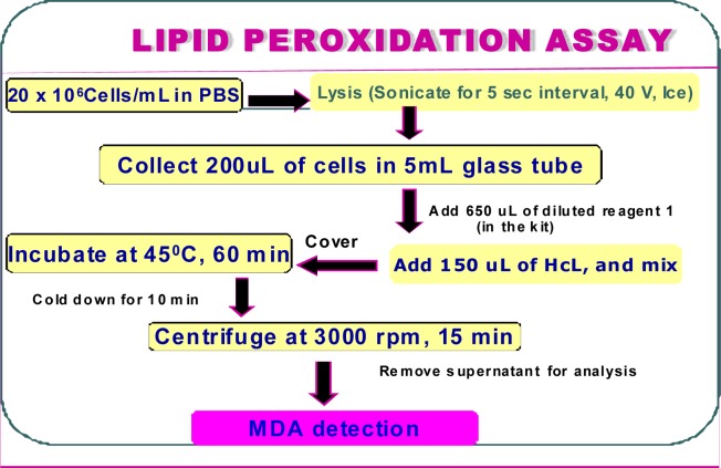
Malondialdehyde (MDA) is formed during lipid peroxidation. The concentration of MDA was measured by using a lipid peroxidation assay kit (Calbiochem-Novabiochem, San Diego, CA). Briefly, 2 × 10⁶ HepG2 cells/mL untreated as a control and co-treated with NAC at 0.125, 0.25, and 0.5 mM and 30 μg/mL lead nitrate were cultured in a total volume of 10 ml growth medium for 48 hours. After the incubation period, cells were collected in 15 mL tube, followed by low-speed centrifugation. The cell pellets were re-suspended in 0.5 ml of Tris-HCl, pH 7.4, and lysed using a sonicator (W-220; Ultrasonic, Farmingdale, NY) under the conditions of duty cycle 25% and output control 40% for 5 sec on ice. The protein concentration of the cell suspension was determined using a protein assay kit (BioRad, Hercules, C.A.). A 200μl aliquot of the culture medium or 2 mg of cell lysate protein was assayed for MDA according to the lipid peroxide assay kit protocol (Calbiochem-Novabiochem, San Diego, CA). The absorbance of the sample was monitored at 586 nm, and the concentration of MDA was determined from a standard curve. Figure 1 illustrates the steps involved in the lipid peroxidation test protocol [26].
Figure 1:
Schematic representation of the steps in lipid peroxidation assay
Statistical Analysis
Data were presented as means ± SDs. Statistical analysis was done using one way analysis of variance (ANOVA Dunnett’s test) for multiple samples and Student’s t-test for comparing paired sample sets. P-values less than 0.05 were considered statistically significant. The percentages of cell viability and MDA levels were presented graphically in the form of histograms, using Microsoft Excel computer program.
Results
Measurements of Cell Viability
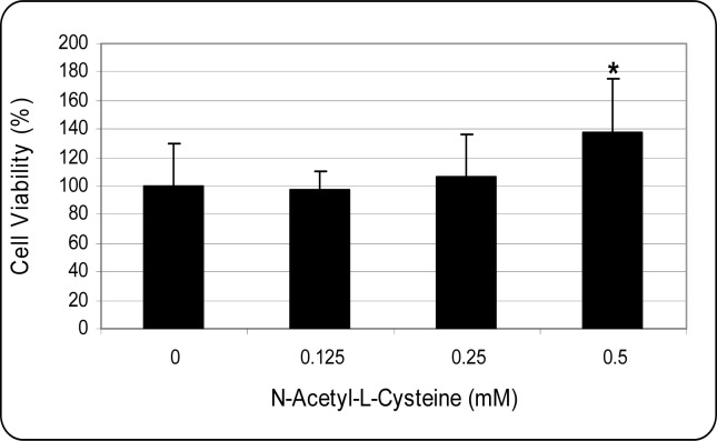
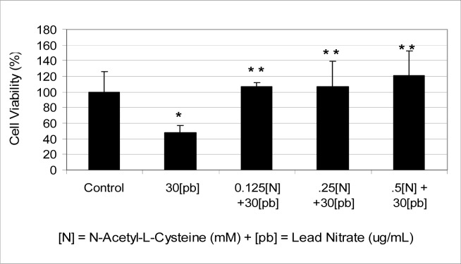
Using the MTT assay, we found that treatment of HepG2 cells by NAC increased cell viability in a dose-dependent manner (Figure 2) whereas treatment of cells with 30 μg/mL of lead nitrate decreased cell viability to about 52% (Figure 3). The viability of HepG2 cells co-exposed to NAC (0.125, 0.25, and 0.5 mM) plus 30 μg/mL lead nitrate resulted in cell growth and proliferation compared to cells treated with lead nitrate alone, indicating the stimulatory effect of this antioxidant (Figure 3). For instance, in cells treated with 0.125 mM NAC and 30μg/mL lead nitrate, the viability was 102% compared to 42% for cells treated with lead nitrate alone. Similar results were also obtained using the trypan blue exclusion test (data not shown).
Figure 2:
Effect of n-acetyl-l-cysteine (NAC) to human hepatocellular liver carcinoma (HepG2) cells. HepG2 cells were cultured with different doses of NAC for 48 hours as indicated in the Materials and Methods. Cell viability was determined based on the MTT assay. Each point represents a mean value and standard deviation of 3 experiments with 6 replicates per dose.
*Significantly different from the control by ANOVA Dunnett’s test; p<0.05.
Figure 3:
Potential effect of co-administration of n-acetyl-l-cysteine (NAC) and lead nitrate to human hepatocellular liver carcinoma HepG2 cells. HepG2 cells were cultured in the absence or presence of NAC and lead nitrate or in combination of NAC and lead nitrate for 48 hr as indicated in the Materials and Methods. Cell viability was determined based on the MTT assay. Each point represents a mean value and standard deviation of 3 experiments with 6 replicates per dose. *Significantly different from the control by ANOVA Dunnett’s test; p < 0.05. **Significantly different from lead nitrate by ANOVA Dunnett’s test; p < 0.05.
Lipid Peroxidation Assay
Experiment done to develop a standard curve for MDA determination resulted in a correlation coefficient (r2 value) of 0.96, indicative of a strong positive correlation between MDA concentration levels and the optimal density readings set at 586 nm. This result (data not shown) showed a strong concentration-dependent response with regard to MDA amounts in the samples.
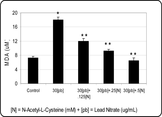
The treatment of HepG2 cells with lead nitrate resulted in a marked increase of MDA production, an indication of oxidative stress and cell injury. In contrary, incubation of HepG2 cells with increasing concentrations of NAC decreased the amount of MDA production in dose-dependent manner (Figure 4). The MDA production level was greatly reduced when cells were treated with 0.5 mM NAC and 30μg/mL of lead nitrate compared to lead nitrate alone. Findings from these studies suggest that the antioxidant property of NAC in vitro inhibits the generation of reactive oxygen species (ROSs) that contributes to direct mechanism of lead nitrate toxicity.
Figure 4:
Protective effect of NAC on lead nitrate-induced oxidative stress in HepG2 cells. Cells were incubated for 48 hr with 30 μg/mL lead nitrate and various concentrations of NAC (0.125, 0.25, and 0.5 mM). Malondialdehyde formation was determined as described in Materials and Methods. *Significantly different from the control by ANOVA Dunnett’s test; p < 0.05. **Significantly different from ATO alone by ANOVA Dunnett’s test; p < 0.05. Data are representative of 3 independent experiments.
Discussion
Measurements of Cell Viability
Data obtained from the present study indicate that lead nitrate at 30μg/mL of exposure is highly cytotoxic to human hepatocellular carcinoma (HepG2) cells. These results are in agreement with other cytotoxicity studies on HepG2 cells and other cell lines in our laboratory that reported a concentration and time-dependent decrease in cell viability based on the MTT assay [27– 30]. In the present study, we also investigated the protective effect of NAC on lead nitrate-treated HepG2 cells. We found that cells co-exposed to both compounds resulted in a significant (p<0.05) increase of cell growth and proliferation. Similarly, NAC increased survival in cultured human bronchial cells and counteracted the toxicity of cigarette smoke and their non-volatile and semi-volatile fraction in rat hepatocytes and lung cells [31]. These findings clearly showed evidence that NAC acts as a potential chelator of heavy metal that attenuates lead nitrate toxicity in HepG2 cells (Fig 3). Recent studies have suggested that NAC may have potential as an anti-tumorigenic agent with efficacy in preventing initial tumor take and metastasis along with a repression of VEGF expression in an experimental Karposi sarcoma model [32–33].
Lipid Peroxidation Assay
In the present study, the induction of lipid peroxidation in lead nitrate-treated HepG2 cells in the absence or presence of NAC was determined by estimating the levels of malondialdehyde (MDA) [34]. We found that NAC decreased malondialdehyde (MDA) level, a by-product of lipid peroxidation in HepG2 cells treated with lead nitrate in a concentration-dependent manner. This finding suggests that NAC protects cells against death and oxidative stress associated with exposure to lead nitrate in vitro. Using this assay, we observed a maximum cell protection from oxidative stress at 0.5 mM NAC. In contrary, the treatment of cells with lead nitrate alone resulted in a marked increase of MDA production in HepG2 cells. Our results are consistent with recent studies reporting that lead-induced oxidative stress is primarily due to increased hydroxyl radical generation in both intact animals and cultured endothelial cells [35]. There is a growing amount of evidence indicating that cellular damage mediated by oxidative stress may be involved in some of the pathologies associated with lead toxicity [36–37]. Numerous studies also indicate that lead induces the generation of reactive oxygen species (ROSs) that contribute significantly to cell killing. Recent studies in our laboratory demonstrated that the toxic effects of heavy metals including arsenic trioxide and lead nitrate are particularly mediated through oxidative stress and the release of lipid peroxidation of membrane lipids [26, 36]. This process results in the production of lipid radicals and in the formation of a complex mixture of lipid degradation products such as MDA and other aldehydes, which are extremely toxic for the cells [34]. If not neutralised, these oxidative radicals cause cellular oxidative stress, increase lipid peroxidation and cause instability of cellular membranes.
Antioxidants such as NAC have been known to be cytoprotective after exposure to cellular damaging agents such as reactive oxygen species [11–13]. NAC has been known to act as an antioxidant/free radical scavenger or reducing agent [38] that protects cells against cell death [39]. Moreover, NAC increased intracellular GSH and protected cultured pulmonary endothelial cells from injury produced by hyperoxia [40], and exerted a dose-dependent inhibition in primary cultures of aortic endothelial cells incubated in the presence of the hypoxanthine-xanthine oxidase system [41]. The antioxidant activities of NAC have been examined by various methods in vitro and in vivo. Using the lipid peroxidation assay protocol, we found that NAC attenuates oxidative stress associated with lead nitrate toxicity in HepG2 cells. This suggests that antioxidants (NAC) may play an important role in the treatment of lead poisoning as a kind of excellent scavenger of free radicals and chelator of heavy metal.
Conclusion
In this study, lead nitrate treatment decreased cell viabilities and increased lipid peroxidation levels, which indicate that free radicals play an important role in lead nitrate-induced damage to HepG2 cells. In contrary, NAC increased cellular proliferation in cells co-treated with lead nitrate and reversed the effects of lead nitrate on oxidative stress parameters in a concentration-dependent manner. Therefore, it can be deduced that the increased cell viability in NAC-treated cells, along with improved lipid peroxidation levels, reflects the antioxidant action of NAC in lead nitrate-treated cells. Overall, findings from these studies suggest that NAC attenuates lead nitrate-induced cytotoxicity and oxidative stress in HepG2 cells by inhibiting the generation of reactive oxygen species that contribute to cell killing. To our knowledge, NAC showed inhibitory activity to lead nitrate in the in vitro studies, we believe that its protective effect on oxidative damage in HepG2 cells exposed to lead nitrate might be related to both its ability to scavenge free radicals and to chelate metal ions.
Acknowledgments
This research was financially supported in part by a grant from the National Institutes of Health (Grant No. 1G12RR13459), through the RCMI-Center for Environmental Health, and in part by a grant from the U.S. Department of the Army (Cooperative Agreement No. W912H2-04-2-0002) through the CMCM Program at Jackson State University. The authors thank Dr. Abdul Mohamed, Dean Emeritus of College of Science, Engineering, and Technology for his technical support in this research.
References
- 1.ATSDR . The nature and extent of lead poisoning in the United States. A report to Congress. Atlanta, GA: Agency for Toxic Substances and Disease Registry; 1988. [Google Scholar]
- 2.U.S. Environmental Protection Agency (EPA) Evaluation of the Potential Carcinogenicity of Lead and Lead Compounds. Office of Health and Environmental Assessment; 1989. EPA/600/8-89/045A. [Google Scholar]
- 3.Cory-Slechta DA, Pound JG. Lead neurotoxicity. In: Chang LW, Dyer RS, editors. Handbook of Neurotoxicology. Dekker; New York: 1995. pp. 61–89. [Google Scholar]
- 4.Goyer RA. Lead toxicity: current concerns. Environ Health Perspect. 1993;100:177–187. doi: 10.1289/ehp.93100177. [DOI] [PMC free article] [PubMed] [Google Scholar]
- 5.Bechara EJH, Medeiros MHG, Monteiro HP, Her-mes-Lim M, Pereira B, Demasi M, Costa CA, Ab-dalla DSP, Onuk J, Wendel CMA, Di Mascio P. A free-radical hypothesis of lead poisoning and inborn porphyrias associated with 5-aminolevulinic acid overload. Quim. Nova. 1993;16:385–92. [Google Scholar]
- 6.Hermes-Lima M, Pereira B, Bechara EJH. Are free radicals involved in lead poisoning? Xenobiotica. 1991;21:1085–1090. doi: 10.3109/00498259109039548. [DOI] [PubMed] [Google Scholar]
- 7.Jiun YS, Hsien LT. Lipid peroxidation in workers exposed to lead. Archiv. Environ. Health. 1994;49:256–259. doi: 10.1080/00039896.1994.9937476. [DOI] [PubMed] [Google Scholar]
- 8.Monteiro HP, Abdalla DSP, Arcuri AS, Bechara EJH. Oxygen toxicity related to exposure to lead. Clin. Chem. 1985;31:1673–1676. [PubMed] [Google Scholar]
- 9.Gurer H, Ozgunes H, Oztezcan S, Ercal N. Antioxidant role of α-lipoic cid in lead toxicity. Free Radic. Biol. Med. 1999;27:75–81. doi: 10.1016/s0891-5849(99)00036-2. [DOI] [PubMed] [Google Scholar]
- 10.Gurer H, Ercal N. Can antioxidants be beneficial in the treatment of lead poisoning? Free Radic. Biol. Med. 2000;29:927–945. doi: 10.1016/s0891-5849(00)00413-5. [DOI] [PubMed] [Google Scholar]
- 11.Baas P, Oppelaar H, van der Valk MA, van Zandwijk N, Stewart FA. Partial protection of photodynamic-induced skin reactions in mice by N-acetylcysteine: a preclinical study. Photochem Photobiol. 1994;59:448–454. doi: 10.1111/j.1751-1097.1994.tb05063.x. [DOI] [PubMed] [Google Scholar]
- 12.Emonet-Piccardi N, Richard MJ, Ravanat JL, Signorini N, Cadet J, Beani JC. Protective effects of antioxidants against UVA-induced DNA damage in human skin fibroblasts in culture. Free Radic. Res. 1998;29:307–313. doi: 10.1080/10715769800300341. [DOI] [PubMed] [Google Scholar]
- 13.De Flora S, Izzotti A, D’Agostini F, Balansky RM. Mechanisms of N-acetylcysteine in the prevention of DNA damage and cancer, with special reference to smoking-related end-points. Carcinogenesis. 2001;22:999–1013. doi: 10.1093/carcin/22.7.999. [DOI] [PubMed] [Google Scholar]
- 14.De Vries N, De Flora S. N-acetyl-L-cysteine. J. Cell. Biochem. 1993;17:270–277. doi: 10.1002/jcb.240531040. [DOI] [PubMed] [Google Scholar]
- 15.Shan XQ, Aw TY, Jones DP. Glutathione-dependent protection against oxidative injury. Pharmacol Ther. 1990;47:61–71. doi: 10.1016/0163-7258(90)90045-4. [DOI] [PubMed] [Google Scholar]
- 16.Thomas SH. Paracetamol (acetaminophen) poisoning. Pharmacol Ther. 1993;60:91–120. doi: 10.1016/0163-7258(93)90023-7. [DOI] [PubMed] [Google Scholar]
- 17.De Flora S, Cesarone CF, Balansky RM, Albini A, D’Agostini F, Bennicelli C. Chemopreventive properties and mechanisms of N-acetylcysteine: the experimental background. J. Cell. Biochem. 1995;22:33–41. doi: 10.1002/jcb.240590806. [DOI] [PubMed] [Google Scholar]
- 18.Pendyala L, Creaven PJ. Pharmacokinetic and pharmacodynamic studies of N-acetylcysteine, a potential chemopreventive agent during phase 1 trial. Cancer Epidemiol. Biomarkers Prev. 1995;4:245–251. [PubMed] [Google Scholar]
- 19.Guo BY, Cheng QK. Chelating capability of tea components with metal ion. J. Tea Science. 1991;11:139–144. [Google Scholar]
- 20.Kumamoto M, Sonda T, Nagayama K, Tabata M. Effects of pH and metal ions on antioxidative activities of catechins. Biosci. Biotechnol. Biochem. 2001;65:126–132. doi: 10.1271/bbb.65.126. [DOI] [PubMed] [Google Scholar]
- 21.Borenfreund E, Babich H, Martin-Alguacil N. Rapid chemosensitivity assay with human normal and tumor cells in vitro. In Vitro Cell Dev. Biology. 1990;26:1030–1034. doi: 10.1007/BF02624436. [DOI] [PubMed] [Google Scholar]
- 22.Marinovitch M, Lorenzo J, Flaminio L, Ganata A, Galli C. The HepG2 cell line as a possible alternative to isolated hepatocytes in cytotoxicity studies. ATLA. 1988;16:16–22. [Google Scholar]
- 23.Mosmann T. Rapid colorimetric assay for cellular growth and survival: application to proliferation and cytotoxicity assays. J. Immunol. Methods. 1983;65:55–63. doi: 10.1016/0022-1759(83)90303-4. [DOI] [PubMed] [Google Scholar]
- 24.Tchounwou PB, Wilson B, Schneider J, Ishaque A. Cytogenic assessment of arsenic trioxide toxicity in the Mutatox, Ames II and CAT-Tox assays. Metal Ions Biol. Med. 2000;6:89–91. [Google Scholar]
- 25.Tchounwou PB, Yedjou CG, Foxx D, Ishaque A, Shen E. Lead induced cytotoxicity and transcriptional activation of stress genes in human liver carcinoma cells. Mol. Cell. Biochem. 2004;255:161–170. doi: 10.1023/b:mcbi.0000007272.46923.12. [DOI] [PubMed] [Google Scholar]
- 26.Yedjou C, Steverson M, Tchounwou P. Lead nitrate-induced oxidative stress in human liver carcinoma (HepG2) cells. Metal Ions Biol. Med. 2006;9:293–297. [PMC free article] [PubMed] [Google Scholar]
- 27.Tchounwou PB, Wilson BA, Abdelgnani AA, Ishaque AB, Patlolla AK. Differential cytotoxicity and gene expression in human liver Carcinoma (HepG2) cells exposed to arsenic trioxide and monosodium acid methanearsonate (MSMA) Int J Mol. Sci. 2002;3:1117–1132. [Google Scholar]
- 28.Tchounwou PB, Yedjou CG, Dorsey WC. Arsenic Trioxide-Induced Transcriptional Activation and Expression of Stress Genes in Human Liver Carcinoma Cells (HepG2) Cellular and Molecular Biology™. 2003;49(7):1071–1079. [PubMed] [Google Scholar]
- 29.Yedjou CG, Moore P, Tchounwou PB. Dose and Time-Dependent Response of Human Acute Promyelocytic Leukemia HL-60 Cells to Arsenic Trioxide Treatment. Int. J. Environ. Res. Public Health. 2006;3(2):136–140. doi: 10.3390/ijerph2006030017. [DOI] [PMC free article] [PubMed] [Google Scholar]
- 30.Cai T, Fassina G, Morini M, Aluigi MG, Masiello L, Fontanini G, D’Agostini F, De Flora S, Noonan DM, Albini A. N-acetylcysteine inhibits endothelial cell invasion and angiogenesis. Lab Invest. 1999;79:1151–1159. [PubMed] [Google Scholar]
- 31.Moldeus P, Berggren M, Grafstrom R. N-acetylcysteine protection against the toxicity of cigarette smoke and cigarette smoke condensates in various tissues and cells in vitro. Eur. J. Resp. Dis. 1985;39(suppl):123–129. [PubMed] [Google Scholar]
- 32.Aluigi MG, De Flora S, D’Agostini F, Albini A, Fassina G. Antiapoptotic and antigenotoxic effects of N-acetylcysteine in human cells of endothelial origin. Anticancer Res. 2000;20:3183–3187. [PubMed] [Google Scholar]
- 33.Halliwell B, Gutteridge JMC. Oxygen toxicity, oxygen radicals, transition metals and disease. Biochem. J. 1984;219:1–14. doi: 10.1042/bj2190001. [DOI] [PMC free article] [PubMed] [Google Scholar]
- 34.Ding Y, Gonick HC, Vaziri ND. Lead promotes hydroxyl radical generation and lipid peroxidation in cultured aortic endothelial cells. Am J Hypertens. 2000;13:552–555. doi: 10.1016/s0895-7061(99)00226-5. [DOI] [PubMed] [Google Scholar]
- 35.Adonaylo VN, Oteiza PI. Lead intoxication: Antioxidant defenses and oxidative damage in rat brain. Toxicology. 1999;135:77–85. doi: 10.1016/s0300-483x(99)00051-7. [DOI] [PubMed] [Google Scholar]
- 36.Sandhir R, Gill KD. Effect of lead on lipid peroxidation in liver of rats. Biol. Trace Elem. Res. 1995;48:91–97. doi: 10.1007/BF02789081. [DOI] [PubMed] [Google Scholar]
- 37.Yedjou CG, Tchounwou PB. Oxidative stress in human leukemia (HL-60), human liver carcinoma (HepG2), and human jurkat T-cells exposed to arsenic trioxide. Metal Ions Biol. Med. 2006;9:298–303. [PMC free article] [PubMed] [Google Scholar]
- 38.Cotgreave IA. N-acetylcysteine: Pharmacological considerations and experimental and clinical applications. Adv. Pharmacol. 1997;38:205–22. [PubMed] [Google Scholar]
- 39.Mayer M, Noble M. N-Acetyl-L-cysteine is a pluripotent protector against cell death and enhancer of trophic factor-mediated cell survival in vitro. Proc. Natl. Acad. Sci. U.S.A. 1994;91:7496–7500. doi: 10.1073/pnas.91.16.7496. [DOI] [PMC free article] [PubMed] [Google Scholar]
- 40.Suttorp N, Kastle S, Neuhof H. Glutathione redox cycle is important defense system of endothelial cells against chronic hyperoxia. Lung. 1991;169:203–214. doi: 10.1007/BF02714155. [DOI] [PubMed] [Google Scholar]
- 41.Junod AF, Jurnot L, Grichting G. Comparative study on selenium-and-n-aceltylcysteine-related effects on the toxic action of hyperoxia, paraquat and enzyme reaction hypoxanthine-xanthine oxidative in culture endothelial cells. Agents Actions. 1987;22:176–183. doi: 10.1007/BF01968835. [DOI] [PubMed] [Google Scholar]


