Abstract
The underlying mechanism for the biological activity of inorganic mercury is believed to be the high affinity binding of divalent mercuric cations to thiols of sulfhydryl groups of proteins. A comprehensive analysis of published data indicates that inorganic mercury is one of the most environmentally abundant toxic metals, is a potent and selective nephrotoxicant that preferentially accumulates in the kidneys, and is known to produce cellular injury in the kidneys. Binding sites are present in the proximal tubules, and it is in the epithelial cells of these tubules that toxicants such as inorganic mercury are reabsorbed. This can affect the enzymatic activity and the structure of various proteins. Mercury may alter protein and membrane structure and function in the epithelial cells and this alteration may result in long term residual effects. This research was therefore designed to evaluate the dose-response relationship in human renal proximal tubule (HK-2) cells following exposure to inorganic mercury. Cytotoxicity was evaluated using the MTT assay for cell viability. The Annexin-V assay was performed by flow cytometry to determine the extent of phosphatidylserine externalization. Cells were exposed to mercury for 24 hours at doses of 0, 1, 2, 3, 4, 5, and 6 μg/mL. Cytotoxicity experiments yielded a LD50 value of 4.65 ± 0.6 μg/mL indicating that mercury is highly toxic. The percentages of cells undergoing early apoptosis were 0.70 ± 0.03%, 10.0 ± 0.02%, 11.70 ± 0.03%, 15.20 ± 0.02%, 16.70 ± 0.03%, 24.20 ±0.02%, and 25.60 ± 0.04% at treatments of 0, 1, 2, 3, 4, 5, and 6 μg/mL of mercury respectively. This indicates a dose-response relationship with regard to mercury-induced cytotoxicity and the externalization of phosphatidylserine in HK-2 cells.
Keywords: Mercury, cytotoxicity, MTT, HK-2 cells, apoptosis, flow cytometry, Annexin V
Introduction
Inorganic mercury is toxic and exerts its effects in a number of organs, tissues, and cell systems, but the kidneys constitute the primary target organ for inorganic mercury toxicity and mercuric ion accumulation in humans and mammals [1 – 4]. In vivo, inorganic mercury has been shown to be a potent and specific nephrotoxicant that accumulates predominantly in the kidneys and selectively in the proximal tubule cells [5, 6]. The intrarenal content of mercury has been shown to correlate with the severity of mercury-induced nephropathy and cellular injury, but only up to the point where injury occurs [7 – 9]. A variety of renal systems have shown an extremely steep dose response relationship for inorganic mercury [10 – 14]. In some of these systems, a threshold effect has been generally observed in that no cellular death is observed up to a certain dose. However, above the dose where an effect is observed, cell death progresses rapidly, and in some but not all of these systems, an all or none response is observed [15 – 19]. This suggests that subtoxic doses of mercury have biochemical and physiological effects, but not to an extent where they can be measured. It is known that endogenous ligands such as glutathione bind to and may compete for mercury to prevent functional changes from occurring [20]. Accordingly, above a certain dose or concentration of mercury, the ligands are depleted, and the mercuric ions can bind readily to critical nucleophilic groups in the cell causing functional impairment. It is likely that intracellular sulfhydryl-containing proteins such as metallothionein or low-molecular weight thiols such as glutathione function in the capacity of ligands that bind to and compete for mercuric ions.
Alterations in cellular function in the kidney may have many diverse roles that modify the susceptibility to mercury-induced renal injury. Understanding biochemical and molecular actions of mercury in the kidney and the ability of intracellular ligands to compete for mercury is vital in diagnosing and combating renal failures. Knowledge of the molecular and cellular mechanisms of mercury induced toxicity in the target organ (kidney) and the target cells (the epithelial cells lining the proximal tubule) is essential for early diagnosis and treatment regimens.
Materials and Methods
Chemicals and Growth Medium
Reference solution of mercury (10,000 μg/mL) was purchased from EM Science (Gibbstown, New Jersey). Gibco keratinocyte-SFM medium, supplements for keratinocyte-SFM medium (recombinant epidermal growth factor 5 ng/mL and bovine pituitary extract 0.05 mg/mL), Dulbecco’s phosphate buffered saline solution without calcium chloride and without magnesium chloride were purchased from Invitrogen Corporation (Carlsbad. California). Hanks balanced salt solution, trypsin – EDTA without calcium and magnesium, and the commercially available MTT cell viability assay kit were purchased from American Type Culture Collection (Manassas, Virginia).
Cell Culture
The HK-2 cell line used is an immortalized proximal tubule epithelial cell line from normal adult human kidney. It has been established by transduction with HPV 16 E6/E7 genes. It is well differentiated on the basis of its histochemical, immune, cytochemical, and functional characteristics. But most importantly, it has been shown to be able to reproduce experimental results obtained with freshly isolated proximal tubule cells. The HK-2 cells are a powerful tool for the study of the physiology, pathophysiology, and mechanisms of renal cell injury and repair [21].
Human renal proximal tubule (HK-2) cells were purchased from American Type Culture Collection, Manassas, VA. The cells were stored in liquid nitrogen until use. The content of each vial was transferred to a 75 cm2 tissue culture flask diluted with Keratinocyte-serum free medium with 10% fetal bovine serum (FBS), 1% streptomycin and penicillin, 5 ng/mL recombinant epidermal growth factor, 0.05 mg/ml bovine pituitary extract, and incubated at 37°C under an atmosphere of 5% CO2 with humidified air to allow the cells to grow and form a monolayer in the flask. Subsequently, cells grown to 85–95% confluency were washed with Hanks balanced salt solution, trypsinized with 5 mL of 0.25% (w/v) EDTA, diluted, counted, and seeded in 96-well microtiter tissue culture plates (5 × 10⁵ cells/well) for cell viability experiments.
Cell Viability Experiments
The MTT cell viability assay was carried out to establish cell viability in human renal proximal tubule HK-2 cells treated with concomitant doses of mercury. The administered doses ranged from 0 to 6.0 μg/mL for an exposure period of 24 hours. Prior to exposure, cells (5 × 10⁵) were maintained in Keratinocyte-serum free medium with 10% fetal bovine serum (FBS), 1% streptomycin and penicillin, 5 ng/mL recombinant epidermal growth factor, 0.05 mg/ml bovine pituitary extract, and incubated at 37°C under an atmosphere of 5% CO2 with humidified air. On the day of exposure, the medium with supplements were removed and replaced with Keratinocyte-serum free medium only. In the concomitant experiments, the cells were dosed with 0, 1, 2, 3, 4, 5, and 6 μg/mL of mercury for a 24 hour period. Cell viability was evaluated using a colorimetric assay in which the reduction of a tetrazolium salt [3-(4,5-dimethylthiazol-2-yl)-2,5-diphenyltetrazolium bromide] (MTT) by the mitochondrial dehydrogenase of living cells was detected. In this assay, metabolically active cells were able to convert MTT to water-insoluble dark-blue formazan crystals. Viable cells were quantified by dissolution in 100% dimethyl sulfoxide and measured by absorbance with the wavelength set at 550 nm; using an Labsystems Multiskan Ascent Plate Reader (Thermo Labsystems, Helsinki, Finland). The toxic effect of mercury at different doses was expressed as the percentage of the absorbance determined for control cells incubated with the corresponding vehicle.
Annexin V Experiment
Annexin V identifies cells at an earlier stage of apoptosis than assays based on DNA fragmentation because externalization of phosphatidylserine occurs earlier than the nuclear changes associated with apoptosis. Since translocation of phosphatidylserine to the external surface also occurs during necrosis, the Annexin V-FITC is typically used in conjunction with a vital dye such as propidium iodide (PI) to distinguish apoptotic cells from necrotic cells. Since apoptosis leads to cell death, apoptotic cells in culture eventually lose membrane integrity. Once cells externalize phosphatidylserine, it remains on the cell surface and cells that have died are Annexin V and PI positive. Therefore, the assay does not distinguish between cells that have already undergone an apoptotic death and those that have died a necrotic death because they both stain similarly with Annexin V-FITC. Gating, quadrant marker, and histogram markers are used to separate apoptotic from necrotic cells. The Annexin V assay relies on the property of cells to lose membrane asymmetry in the early phase of apoptosis. Annexin V is a calcium dependent phospholipids-binding protein that has a high affinity for phosphatidylserine, and is very useful for identifying apoptotic cells with exposed phosphatidylserine. Propidium (PI) is a standard flow cytometric viability probe, and is used to distinguish viable from non-viable cells. Viable cells with intact membranes exclude propidium iodide, whereas the membranes of dead and damaged cells are permeable to propidium iodide.
The Annexin V assay was carried out using a commercially available staining kit (Annexin V-FITC Apoptosis Detection Kit – BD Biosciences) San Diego, CA. After exposure, mercury stimulated cells were washed twice with Hanks balanced salt solution and collected by trypsinization. To analyze for Annexin V cells were washed twice with cold PBS and resuspended in 1× binding buffer at a concentration of 1 × 10⁶ cells/ml. One hundred μl of the resuspended solution was transferred to a 5 ml culture tube. Five μl Annexin V-FITC and propidium iodide was added. The cells were gently vortexed and incubated in the dark at room temperature for 15 minutes. Four hundred μl of the binding buffer was added to each tube (one per concentration). The cells were analyzed within 1 hour using histogram analysis and the BD FACS Calibur flow cytometer.
Statistical Analysis
Absorbance readings at 550 nm from cell viability experiments were transformed into percentages to compare the viability of treated cells to that of untreated (control) cells. Graphs were made to illustrate the dose-response relationship with respect to cytotoxicity or cell viability. Standard deviations were determined, and the Student’s t-test values were computed to determine if there were significant differences in cell viability in mercury treated cells compared to control cells. A value of p<0.05 was considered significant. All flow cytometric results were analyzed statistically analysis done using the Cell Quest Pro software BD Biosciences (Becton Dickinson, and Company) San Jose, CA.
Discussion
Cytotoxicity
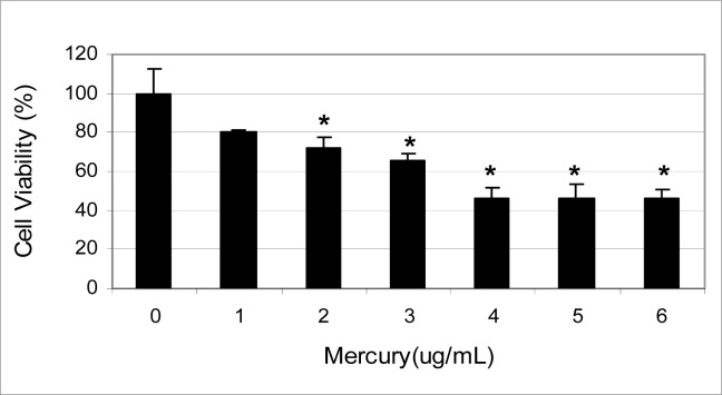
The effects of inorganic mercury on the viability of human renal proximal tubule (HK-2) cells are shown in figure 1.
Figure 1:
Cytotoxicity of mercury to human renal proximal tubule (HK-2) cells
Data presented in this figure indicate a strong dose-response relationship with respect to the cytotoxicity of mercury. Upon 24 hours of exposure, the average percentages of cell viability were 100 ± 0%, 80.0 ± 0.07%, 72 ± 0.04%, 66 ± 0.02%, 46 ± 0.04%, 46 ±0.06%, and 46 ± 0.03% in 0, 1, 2, 3, 4, 5, and 6 μg/mL mercury respectively. The LD50 value for inorganic mercury was computed to be 4.65 ± 0.6 μg/mL upon 24 hours of exposure, indicating the mercury is highly toxic to the cells.
The cytotoxicity of mercury compounds begins with the binding to proteins on the surface of cell membranes which disrupts the transport of materials in and out of the cells. Once inside the cell, mercury dissolves in the blood plasma and from there has access to diffuse into any cell in the body. Mercury compounds have a profound effect on the cytoskeleton of cells because the cytoskeleton is made up, in part, of hollow tube like structures of fibrous materials composed of distinct proteinaceous globules called tubules [22]. Their building blocks are peptide units called tubulin, and each tubulin pair contains 14 sulfhydryl groups which act as points of attachment for mercury compounds. These attachments facilitate the formation of complexes with one or more of the sulfhydryl groups, which in turn results in a disassociation of the tubulin building blocks. This disruption drastically affects the overall structure and activities of the cell [23 – 25].
The kidneys are a primary target organ for mercuric ions where they accumulate due the biotransformation of ionic ions to mercuric ions [26]. Mercuric ions are highly reactive and rapidly combine with intracellular sulfhydryl ligands, potentially disrupting enzymes and proteins essential to normal kidney function [27]. Mercury is an extremely active agent for the destruction of the microtubules responsible for mitotic spindle formation. Since this prevents cell division and blocks cell cycle progression, it markedly reduces cell viability. Inactivation of microtubules within the cytoplasm also affects the structure, migration, and phagocytic activity of cells. These actions, which are not overly observable, explain the effects of mercury on the embryo, and the reproductive, hematopoietic, immune, and neurological systems of the adults [28, 29]
Inorganic mercury has been known to induce membrane damage, to be mutagenic, generate DNA strands breaks and chromosomal aberrations, to affect mitochondrial activity by inducing the development of a membrane permeability transition, cause alterations in protein structure, calcium transport, inhibit glucose transport and enzyme function, interfere with essential nutrients by replacing essential minerals such as zinc at sites in enzymes, alter stress proteins in whole kidneys and induce regional and cell specific stress proteins in rat kidneys, as well as disrupt the calldherin genes that encode for transmembrane proteins that regulate calcium (Ca2+) dependent homotypic intracellular adhesions [30 – 42].
Urinary excretions indicating tubular cytotoxicity, such as increased tubular antigens and enzymes, altered levels of biochemical enzymes such as decreased urinary output of eicosanoids and glycoosaminoglycans; and a more acidic pH are all early renal changes seen in workers with chronic low level exposure to inorganic mercury [43]. Autoimmune glomeruler nephritis has been induced in genetically susceptible strains of rats and mice. When rodents were injected with mercuric chloride, they produced antibodies which attacked the kidneys and caused autoimmune glomeruler nephritis [44]. The concentration of inorganic mercury in the kidney is directly related to the amount take in [45].
Apoptosis
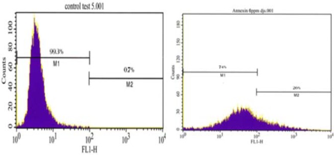
Figure 2 shows the histogram analysis in which the Annexin positive cells are shown in M2 and the Annexin negative cells are shown in M1 for the control and 6 μg/for HK-2 cells in response to mercury exposure. The percentage of Annexin positive cells were 0.7 ± 0.3%, 10.0 ± 0.02%, 11.70 ± 0.03%, 15.20 ± 0.02%, 16.70 ± 0.03%, 24.20 ± 0.02, and 25.60 ± 0.04% in 0, 1, 2, 3, 4, 5, and 6 μg/mL mercury respectively..
Figure 2:

Annexin V Histogram Analysis showing Annexin positive cells in M2.
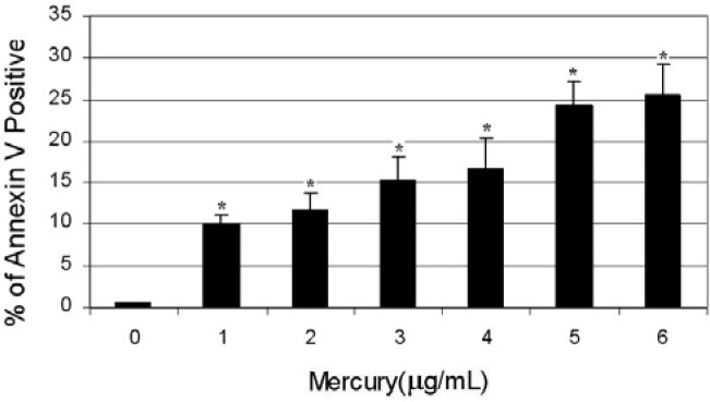
Figure 3 shows a dose dependent response of Annexin positive cells in response to mercury exposure. The percentage of Annexin positive cells increases as the concentration of mercury increases in a dose-dependent manner. These results clearly establish a dose-response relationship with respect to the externalization of phosphatidylserine human renal proximal tubule (HK-2) cells exposed to mercury.
Figure 3:
Percentage of Annexin Positive cells with respect to mercury concentration.
The maintenance of multi-cellular biological systems depends on the sophisticated system of apoptosis due to its role in development, differentiation, proliferation, homeostasis, regulation and function of the immune system, and in the removal of harmful cells [46]. Thus, dysfunction or dysregulation of the apoptotic program is implicated in a wide variety of pathological conditions such as cancer, autoimmune disease and spreading of viral infections, neurodegenerative disorders, AIDS, and ischemic diseases [47]. Dysregulation of apoptotic signaling can play primary or secondary roles in many diseases [48]. Malfunction of the apoptosis process results from the mutations of genes that code for factors that are directly or indirectly involved in the initiation, mediation, or execution of this process. Gaining insight into the cellular and molecular mechanisms and alterations by which the apoptosis process contributes to pathogenic processes and diseases should allow for the development of more effective, higher specific, and therefore better-tolerable therapeutic processes.
Changes in the plasma membrane of the cell surface are one of the earliest features of cells undergoing apoptosis [49]. In apoptotic cells, the membrane phospholipid phosphatidylserine is translocated from the inner to the outer leaflet of the plasma membrane, thereby exposing phosphatidylserine to the external cellular environment. [50]. Annexin V is a 35–36 kDA Ca2+ dependent phospholipid-binding protein that has a high affinity for phosphatidylserine, and binds to cells with exposed phosphatidylserine. Annexin V is a sensitive probe for identifying cells that are undergoing apoptosis because phosphatidylserine exposure occurs early in the apoptotic process. It is typically conjugated to a flurochrome such a fluorescein isothiocyanate (FITC), for easy identification of apoptotic cells by flow cytometry. These cells may also be identified with a vital dye alone such as propidium iodide if proper gating and quadrant marking is used in the flow cytometric analysis [51].
Inorganic mercury compounds have been shown to induce apoptosis in a dose dependent manner in HepG2 cells, HEK-293 cells, SHSY5 cells, rat kidney epithelial cells, cultured neurons and fibroblast, t-lymphocytes, and the porcine renal cell line LLC-PK1. These compounds have also been shown to induce acute renal tubular necrosis in the proximal tubules in the JCL-SD rat strain [52 – 61].
Conclusions
Findings from this study indicate that (a): acute exposure to inorganic mercury significantly (p < 0.05) reduces the viability of human renal proximal tubule (HK-2) cells; and (b): early stage apoptosis (the externalization of phosphatidylserine) is induced in human renal proximal tubule (HK-2) cells exposed to mercury in a dose dependent manner.
Acknowledgments
This research was financially supported in part by a grant from the National Institutes of Health (Grant No. 1G12RR13459) through the Center for Environmental Health and in part by a grant from the Department of Defense through the U. S. Army Engineer Research and Development Center (Vicksburg, MS); Contract #W912HZ-04-2-0002.
References
- 1.Zalups RK, Barfuss DW. Nephrotoxicity of inorganic mercury co–administered with L-cysteine. Toxicology. 1996;109:15–29. doi: 10.1016/0300-483x(95)03297-s. [DOI] [PubMed] [Google Scholar]
- 2.Cannon VT, Zalups RK, Barfuss DW. Amino acid transporters involved in luminal transport of mercuric conjugates of cysteine in rabbit proximal tubule. J Pharmacol Exp Ther. 2001;298:780–789. [PubMed] [Google Scholar]
- 3.Lash LH, Putt DA, Zalups RK. Role of extracellular thiols in accumulation and distribution of inorganic mercury in rat renal proximal and distal tubular cells. J Pharmacol Exp Ther. 1998;285:1039–1050. [PubMed] [Google Scholar]
- 4.Cannon VT, Barfuss DW, Zalups RK. Molecular homology and luminal transport of Hg2+ in the renal proximal tubule. J. of the American Society of Nephrology. 2000;11:394–402. doi: 10.1681/ASN.V113394. [DOI] [PubMed] [Google Scholar]
- 5.Zalups RK. Autometallographic localization of inorganic mercury in the kidney of rats: Effect of unilateral nephrectomy and compensatory renal growth. Exp. Mol. Pathol. 1991;54:10–21. doi: 10.1016/0014-4800(91)90039-z. [DOI] [PubMed] [Google Scholar]
- 6.Zalups RK. Method for studying the in vivo accumulation of inorganic mercury in segments of the nephron in the kidney of rats treated with mercuric chloride. J. Pharmacol Methods. 1991;26:89–104. doi: 10.1016/0160-5402(91)90058-d. [DOI] [PubMed] [Google Scholar]
- 7.Bohets HH, Van Thielen MN, Van Der Biest I, Van landeghem GF, D’Haese PC, Nouwen EJ, De Broe ME, Dierlickx PJ. Cytotoxicity of mercury compounds in LLC-PK1, MDCK and human proximal tubular cells. Kidney Int. 1995;47:395–403. doi: 10.1038/ki.1995.52. [DOI] [PubMed] [Google Scholar]
- 8.Burton CA, Hatlelid K, Divine K, Carter DE, Fernando Q, Brendel K, Gandolfi AJ. Glutathionine effects on toxicity and uptake of mercuric chloride and sodium arsenite in rabbit renal cortical slices. Environ Healt Perspec. 1995;103(Suppl. 1):81–84. doi: 10.1289/ehp.95103s181. [DOI] [PMC free article] [PubMed] [Google Scholar]
- 9.Girardi G, Elias MM. Effectiveness of N-acetylcystein in protecting against mercuric chloride-induced nephrotoxicity. Toxicology. 1991;67:155–164. doi: 10.1016/0300-483x(91)90139-r. [DOI] [PubMed] [Google Scholar]
- 10.Zalups RK, Diamond GL. Mercuric chloride induced nephrotoxicity in the rat after unilateral nephrectomy and compensatory renal growth. Virchows Arch B. 1987;53:336–346. doi: 10.1007/BF02890261. [DOI] [PubMed] [Google Scholar]
- 11.Zalups RK, Parks L, Cannon VT, Barfuss DW. Mechanisms of action of 2,3,-dimercaptopropane-1sulfonate and the transport, disposition, and toxicity of inorganic mercury in isolated perfused segments of rabbit proximal tubules. Mol Pharmacol. 1998;54:353–363. doi: 10.1124/mol.54.2.353. [DOI] [PubMed] [Google Scholar]
- 12.Zalups RK, Lash LH. Effects of uninephrectomy and mercuric chloride on renal glutathionine homeostasis. J Pharmacol Exp Ther. 1990;254:962–970. [PubMed] [Google Scholar]
- 13.Zalups RK. Renal accumulation and intrarenal distribution of inorganic mercury in the rabbit: Effects of unilateral nephrectomy and dose of mercuric chloride. J Toxicol Environ Health. 1991;33:213–228. doi: 10.1080/15287399109531519. [DOI] [PubMed] [Google Scholar]
- 14.Reugg CE, Gandolfi AJ, Nagle RB, Brendel K. Differential patterns of injury to the proximal tubule of renal cortical slices following in vitro exposure to mercuric chloride, potassium dichromate, or hypoxic conditions. Toxicol Appl Pharmacol. 1987;90:261–273. doi: 10.1016/0041-008x(87)90334-6. [DOI] [PubMed] [Google Scholar]
- 15.Barfus DW, Robinson MK, Zalups RK. Inorganic mercury transport in the proximal tubule of the rabbit. Journal of the American Society of Nephrology. 1990;1:910–917. doi: 10.1681/ASN.V16910. [DOI] [PubMed] [Google Scholar]
- 16.Zalups RK. Early aspects of the intrarenal distribution of mercury after the intravenous administration of mercuric chloride. Toxicology. 1993;79:215–228. doi: 10.1016/0300-483x(93)90213-c. [DOI] [PubMed] [Google Scholar]
- 17.Lash LH, Zalups RK. Mercuric chloride-induced cytotoxicity and compensatory hypertrophy in rat kidney proximal tubular cells. J Pharmacol Exp Ther. 1992;261:819–829. [PubMed] [Google Scholar]
- 18.Smith MA, Acosta D, Bruckner JV. Development of a primary culture system of rat kidney cortical cells to evaluate the nephrotoxicity of xenobiotics. Food Chem Toxicol. 1986;24:551–556. doi: 10.1016/0278-6915(86)90112-2. [DOI] [PubMed] [Google Scholar]
- 19.Lash LH, Putt DA, Zalups RK. Influence of exogenous thiols on inorganic mercury-induced injury in renal proximal and distal tubular cells from normal and uninephrectomized rats. J Pharmacol Exp Ther. 1999;29:492–502. [PubMed] [Google Scholar]
- 20.Zalups RK, Lash LJ. Depletion of glutathione in the kidney and the renal disposition of administered inorganic mercury. Drug Metab Dispos. 1997;25:516–523. [PubMed] [Google Scholar]
- 21.Ryan MJ, Johnson G, Kirk J, Fuerstenbert SM, Zager RA, Torok-Storb B. HK-2: An immortalized proximal tubule epithelial cell line from normal adult human kidney. Kidney International. 1994;45:48–57. doi: 10.1038/ki.1994.6. [DOI] [PubMed] [Google Scholar]
- 22.Margolis RL, Wilson L. Microbutule treadmills – possible molecular machinery. Nature. 1981;293:705–711. doi: 10.1038/293705a0. [DOI] [PubMed] [Google Scholar]
- 23.Graff RD, Falconer MM, Brown DL, Reuhl KR. Altered sensitivity of posttranslationally modified microtubules to methylmercury in differentiating embryonal carcinoma-derived neurons. Pharmacol Toxicol. 1997;77:41–47. doi: 10.1006/taap.1997.8138. [DOI] [PubMed] [Google Scholar]
- 24.Moszczynski P. Mercury compounds and the immune system: A review. Int J Occup Med Environ Health. 1997;10:24–58. [PubMed] [Google Scholar]
- 25.Miura K, Imura N. Mechanisms of cytotoxicity of methymercury. With special reference to microtubule disruption. Biol Trace Elem Res. 1989;18:161–188. doi: 10.1007/BF02917269. [DOI] [PubMed] [Google Scholar]
- 26.Zalups RK, Barfuss DW. Nephrotoxicity of inorganic mercury co–administered with L-cysteine. Toxicology. 1996;109:15–29. doi: 10.1016/0300-483x(95)03297-s. [DOI] [PubMed] [Google Scholar]
- 27.Zalups RK. Molecular interactions of mercury in the kidneys. Pharmacological Reviews. 2000;52:113–144. [PubMed] [Google Scholar]
- 28.Tchounwou PB, Ayensu WK, Ninashvili N, Sutton DJ. Environmental exposure to mercury and its toxicopathologic implications for public health. Env. Toxicol. 2003;18:149–175. doi: 10.1002/tox.10116. [DOI] [PubMed] [Google Scholar]
- 29.Sutton DJ, Tchounwou PB, Ninashvili N, Shen E. Mercury induces cytotoxicity and transcriptionally activates stress genes in human liver carcinoma (HepG2) cells. Int. J. Mol. Sci. 2002;3:965–984. [Google Scholar]
- 30.Ferrat L, Romeo M, Gnassia-Barelli M, Pergent-Martini C. Effects of mercury on antioxidant mechanisms in the marine phanerogam Posidonia ocenica. Dis. Aquat. Organ. 2002;50:157–160. doi: 10.3354/dao050157. [DOI] [PubMed] [Google Scholar]
- 31.Ben-Ozer E, Rosenpire A, MaCabe M, Worth R, Kindzelski A, Warra N, Petty H. Mercuric chloride damages cellular DNA by a non-apoptotic mechanism. Muta. Res. 2000;470:19–27. doi: 10.1016/s1383-5718(00)00083-8. [DOI] [PubMed] [Google Scholar]
- 32.Schurz F, Sabater-Vilar M, Fink-Gremmels J. Mutagenecity of mercury chloride and mechanisms of cellular defense: The role of metal binding proteins. Mutagenesis. 2000;15:525–530. doi: 10.1093/mutage/15.6.525. [DOI] [PubMed] [Google Scholar]
- 33.Finney LA, O’Halloran TV. Transition metal speciation in the cell: Insights from the chemistry of metal ion receptors. Science. 2003;300:931–936. doi: 10.1126/science.1085049. [DOI] [PubMed] [Google Scholar]
- 34.Goyer RA. Nutrition and metal toxicity. Am J. Clin Nutr. 1995;61:646S–650S. doi: 10.1093/ajcn/61.3.646S. [DOI] [PubMed] [Google Scholar]
- 35.Sutton DJ, Tchounwou PB, Ninashvili N, Shen E. Mercury induces cytotoxicity and transcriptionally activates stress genes in human liver carcinoma (HepG2) cells. Int. J. Mol. Sci. 2002;3:965–984. [Google Scholar]
- 36.Olivieri G, Brack C, Muller-Sphan F, Stahelin HB, Hermann M, Renard P, Brockhaus M, Hock C. Mercury induces cell cytotoxicity and oxidative stress and increases ß-amyloid secretion and tau phosphorylation in SHSY5Y neuroblastoma cells. J. Neurochemistry. 2000;74:231–236. doi: 10.1046/j.1471-4159.2000.0740231.x. [DOI] [PubMed] [Google Scholar]
- 37.Baskin DS, Ngo H, Didenko V. Thimerosal induces DNA breaks, Caspase-3 activation, membrane damage, and cell death in cultured human neurons and fibroblasts. Toxicological Sciences. 2003;74:361–368. doi: 10.1093/toxsci/kfg126. [DOI] [PMC free article] [PubMed] [Google Scholar]
- 38.Ziemba SE, Mattingly RR, McCabe MJ, Rosenspire AJ. Inorganic mercury inhibits the activation of LAT in T-cell receptor-mediated signal transduction. Toxicological Sciences. 2006;89(1):145–153. doi: 10.1093/toxsci/kfj029. [DOI] [PubMed] [Google Scholar]
- 39.Matsouka M, Wispriyono B, Iryo Y, Igisu H. Mercury chloride activates c-Jun N-terminal kinase and induces c-jun expression in LLC-PK1 cells. Toxicological Sciences. 2000;53:361–368. doi: 10.1093/toxsci/53.2.361. [DOI] [PubMed] [Google Scholar]
- 40.Hajela RK, Peng SQ, Atchinson WD. Comparative effects of methylmercury and Hg2+ on neuronal N-and R-type high voltage activated calcium channels transiently expressed in human embryonic kidney 293 cells. The J. Pharmac. And Exp. Therap. 2003;306:1129–1136. doi: 10.1124/jpet.103.049429. [DOI] [PubMed] [Google Scholar]
- 41.Hare MF, Atchinson WD. Comparative action of methylmercury and divalent inorganic mercury on nerve terminal and intraterminal mitochondrial membrane potentials. The J. Pharmac. And Exp. Therap. 1992;261:166–172. [PubMed] [Google Scholar]
- 42.Bridges CC, Zalups RK. Homocystein, system b0,+ and the renal epithelial transport and toxicity of inorganic mercury. Am J. Pathology. 2004;165:1385–1394. doi: 10.1016/S0002-9440(10)63396-2. [DOI] [PMC free article] [PubMed] [Google Scholar]
- 43.Cardenas A, Roells H, Bernard AM, Barbon R, Buchet JP, Lauwerys RR. Markers of early renal changes induced by industrial pollutants. Application to workers exposed to mercury vapor. Journal of Industrial Medicine. 1993;50:17–27. doi: 10.1136/oem.50.1.17. [DOI] [PMC free article] [PubMed] [Google Scholar]
- 44.Zalups RK, Diamond GL. Mercuric chloride induced nephrotoxicity in the rat after unilateral nephrectomy and compensatory renal growth. Virchows Arch B. 1987;53:336–346. doi: 10.1007/BF02890261. [DOI] [PubMed] [Google Scholar]
- 45.Zalups RK. Molecular interactions of mercury in the kidneys. Pharmacological Reviews. 2000;52:113–144. [PubMed] [Google Scholar]
- 46.Meier P, Finch A, Evan G. Apoptosis in development. Nature. 2000;407:796–801. doi: 10.1038/35037734. [DOI] [PubMed] [Google Scholar]
- 47.Mullauer L, Gruber P, Sebinger D, Buch J, Wohlfart S, Chott A. Mutations in apoptosis genes: a pathogenic factor for human disease. Mut Res, 2001;488:211–231. doi: 10.1016/s1383-5742(01)00057-6. [DOI] [PubMed] [Google Scholar]
- 48.Reed JC. Apoptosis-based therapies. Nat Rev Drug Discov. 2002;1(2):111–121. doi: 10.1038/nrd726. [DOI] [PubMed] [Google Scholar]
- 49.Darzynkiewics S, Juan G, Li X, Gorczyca W, Murakami T, Traganos F. Cytometry in cell necrobiology. Analysis of apoptosis and accidental cell death (necrosis) Cytometry. 1997;27:1–20. [PubMed] [Google Scholar]
- 50.Sartprois U, Schmitz I, Krammer PH. Molecular mechanisms of death-receptor-mediated apoptosis. Chembiochem. 2001;2(1):20–29. doi: 10.1002/1439-7633(20010105)2:1<20::AID-CBIC20>3.0.CO;2-X. [DOI] [PubMed] [Google Scholar]
- 51.Martin SJ, Reutelingsperger CM, Green DR, Annexin V. In: A specific probe for apoptotic cells, In: Techniques in apoptosis: A User Guide. Cotter TG, Martin SJ, editors. Portland Press; London: 1996. pp. 107–120. [Google Scholar]
- 52.Sutton DJ, Tchounwou PB, Ninashvili N, Shen E. Mercury induces cytotoxicity and transcriptionally activates stress genes in human liver carcinoma (HepG2) cells. Int. J. Mol. Sci. 2002;3:965–984. [Google Scholar]
- 53.Olivieri G, Brack C, Muller-Sphan F, Stahelin HB, Hermann M, Renard P, Brockhaus M, Hock C. Mercury induces cell cytotoxicity and oxidative stress and increases ß-amyloid secretion and tau phosphorylation in SHSY5Y neuroblastoma cells. J. Neurochemistry. 2000;74:231–236. doi: 10.1046/j.1471-4159.2000.0740231.x. [DOI] [PubMed] [Google Scholar]
- 54.Baskin DS, Ngo H, Didenko V. Thimerosal induces DNA breaks, Caspase-3 activation, membrane damage, and cell death in cultured human neurons and fibroblasts. Toxicological Sciences. 2003;74:361–368. doi: 10.1093/toxsci/kfg126. [DOI] [PMC free article] [PubMed] [Google Scholar]
- 55.Ziemba SE, Mattingly RR, McCabe MJ, Rosenspire AJ. Inorganic mercury inhibits the activation of LAT in T-cell receptor-mediated signal transduction. Toxicol. Sciences. 2006;89(1):145–153. doi: 10.1093/toxsci/kfj029. [DOI] [PubMed] [Google Scholar]
- 56.Matsouka M, Wispriyono B, Iryo Y, Igisu H. Mercury chloride activates c-Jun N-terminal kinase and induces c-jun expression in LLC-PK1cells. Toxicol. Sciences. 2000;53:361–368. doi: 10.1093/toxsci/53.2.361. [DOI] [PubMed] [Google Scholar]
- 57.Hajela RK, Peng SQ, Atchinson WD. Comparative effects of methylmercury and Hg2+ on neuronal N-and R-type high voltage activated calcium channels transiently expressed in human embryonic kidney 293 cells. The J. Pharmac. And Exp. Therap. 2003;306:1129–1136. doi: 10.1124/jpet.103.049429. [DOI] [PubMed] [Google Scholar]
- 58.Hare MF, Atchinson WD. Comparative action of methylmercury and divalent inorganic mercury on nerve terminal and intraterminal mitochondrial membrane potentials. The J. Pharmac. And Exp. Therap. 1992;261:166–172. [PubMed] [Google Scholar]
- 59.Bridges CC, Zalups RK. Homocystein, system b0, + and the renal epithelial transport and toxicity of inorganic mercury. Am J. Pathology. 2004;165:1385–1394. doi: 10.1016/S0002-9440(10)63396-2. [DOI] [PMC free article] [PubMed] [Google Scholar]
- 60.Goering PL, Fisher BR, Chaudhary PP, Dick CA. Relationship between stress protein induction in rat kidney by mercuric chloride and nephrotoxicity. Toxicol. Appl. Pharmacol. 1992;113:184–1921. doi: 10.1016/0041-008x(92)90113-7. [DOI] [PubMed] [Google Scholar]
- 61.Goering PL, Fisher BR, Noren BT, Papaconstantinou A, Rojko JL, Marler RJ. Mercury induces regional and cell-specific stress protein expression in rat kidney. Toxicol. Sciences. 2000;53:447–457. doi: 10.1093/toxsci/53.2.447. [DOI] [PubMed] [Google Scholar]


