

Abstract
Purpose
To report the outcomes of intraocular lens (IOL) dislocation management in 6 cases with Retinitis Pigmentosa (RP).
Setting
Private practice, Los Angeles, USA.
Design
Retrospective interventional case series.
Methods
The medical reports of six eyes of four RP patients with capsule bag fixated posterior chamber IOL dislocation were retrospectively reviewed. Pre-operative data included demographics, systemic or ocular disorders, history of trauma, previous intraocular surgery and pre-operative visual acuity. Outcome measures included the type of surgery, surgical complications, elevation of intraocular pressure (IOP), ocular inflammation, cystoid macular edema (CME) and IOL dislocation at 3 months or greater post-operatively.
Results
The medical records of six eyes of four patients operated on between December 2009 and May 2011 were evaluated. In four cases, dislocated PC IOL implants were sutured to the sclera. In two eyes of one patient anterior chamber IOLs (AC IOLs) were implanted after PC IOLs were explanted. One eye developed CME during the follow-up period. Despite modest tilt in one case and modest decentration in another, stability and centration of the IOLs was excellent during the follow-up period. No eyes had intraocular inflammation requiring long term medical treatment, new onset glaucoma or retinal detachment. Mean follow-up time was 6.9 months (range 3-20).
Conclusions
Cataract surgeons should be aware of the increased risk for decentration and malposition of PC IOLs in patients with RP. Satisfactory results can be achieved by fixation of the PC IOL or AC IOL implantation.
Keywords: Retinitis pigmentosa, IOL dislocation, IOL dislocation management
Introduction
RP is a clinically and genetically diverse group of retinal dystrophies initially affecting rod photoreceptors with subsequent degeneration of the cones. The condition is characterized by peripheral and night vision loss and has the classical triad of arteriolar attenuation, bone spicule pigmentation, and waxy disc pallor.1 Cataract, most commonly the posterior subcapsular form, develops at a relatively early age in patients with RP and apart from the risks of a conventional cataract surgery these patients carry a higher risk of having poor visual outcomes.2
One of the most common complications of cataract surgery in RP patients is the risk of spontaneous IOL dislocation. The rate of late “in-the-bag” IOL dislocation has increased since continuous curvilinear capsulorrhexis (CCC) became popular among surgeons.3 This risk is even higher in RP patients as a result of capsule contraction and subsequent late zonular dehiscence.4,5
Herein we report case series of six eyes in four RP patients who were operated on for dislocated PC IOLs.
Patient 1 (Case 1)
A 64-year-old male was referred for evaluation in November 2009 by a prior surgeon. Retinitis pigmentosa was diagnosed at age 19 and he had bilateral cataract extraction with “in-the-bag” or capsule fixated single piece acrylic PC IOLs in 2003, at age 58. Systemic medications include atorvastatin (Lipitor), ezetimibe (zetia), pantoprazole (protonix), ranitidine (zantac), and baby asprin.
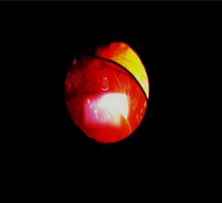
The patient noted worsening of vision and monocular diplopia in the right eye resulting from a malpositioned PC IOL (Fig. 1). Diamox was prescribed for high intraocular pressure in the eye with the dislocated IOL.
Figure 1.

Case 1. Irregular and ovoid pupil secondary to partial anteriorly malpositioned single piece acrylic lens. Note that the haptic is encased within the capsule bag.
On initial evaluation, best-corrected visual acuity (BCVA) was hand movements (HM) in both eyes. There was no relative afferent pupillary defect (RAPD) and external examination and extraocular motility were normal. However, confrontational visual fields were grossly constricted in both eyes.
In the right eye (RE), the pupil was irregular and ovoid secondary to a malpositioned single piece acrylic lens and capsule bag encased temporal loop prolapsed into the anterior chamber (Fig. 1). In the left eye (LE), a well-positioned single piece acrylic lens was noted to be in-the-bag, and the posterior capsule was intact. Irido-pseudophacodonesis was noticed in both eyes.
Fundus examination revealed waxy appearance of the optic disc with narrowed, attenuated vessels and extensive bone spicules in both eyes.
After dilation it was observed the lens was significantly loose, with minimal sphincter defect and an intact posterior capsule. However, there was 5 clock hours of zonular absence noted temporally.
In December 2008 repositioning of the dislocated PC IOL was performed using scleral suture fixation with 10-0 polyester suture (PC-7 [Alcon Laboratories, Inc., Fort Worth, TX]) employing the Hoffman technique.6 The Hoffman technique involves creation of reverse scleral pocket or corneal–scleral pockets without the need for conjunctival dissection. Additionally, an anterior capsulectomy was performed via anterior vitrectomy. During the operation it became evident that the nasal loop was also significantly loose. Therefore, both loops were sutured to the sclera.
Twenty months post-operatively, the monocular diplopia was resolved, BCVA was unchanged as a result of the extensive retinal disease, and the sutured PC IOL was stable.
Patient 2 (Case 2)
A 60-year-old male was referred for our care in April 2011. He was known to have Usher Syndrome, systemic hypertension and hypercholesterolemia. Systemic medications include benazepril (lotensin).
He had bilateral cataract surgery with “in-the-bag” capsule fixated single piece polymethyl methacrylate (PMMA) PC IOLs in the 1980s. Shortly after surgery, the left eye required a repositioning of the lens to the ciliary sulcus.
The patient recently noticed progressive diminution in vision in the right eye for 1 month and was referred for evaluation of a dislocated PC IOL in the right eye.
On initial exam BCVA was 20/50 RE and 20/25 LE. Intraocular pressures measured 14 mmHg OU. Slit lamp examination of the RE revealed a single piece, “in-the-bag” capsule fixated, PMMA lens that was inferiorly subluxated with significant pseudophacodonesis. Iris transillumination defects in the inferotemporal quadrant and disinsertion of the superior zonules were noted. (Fig. 2)
Figure 2.
Case 2. Inferotemporal malpositioned PMMA PC-IOL and disinsertion of the superior zonules.
For the LE, there was a well-centered three-piece PMMA lens in the sulcus, and the posterior capsule was open.
Repositioning of the right dislocated PC IOL was performed. Surgery consisted of scleral suture fixation of the existing lens by means of Hoffman pockets employing 8-0 Gore-Tex suture (off-label use) (W.L. Gore & Associates, Inc., Flagstaff, AZ).
On post-operative week 1, the BCVA in the right eye was 20/40. The IOL was visible in pupillary space with modest tilt. In addition, the patient developed cystoid macular edema (CME) that was treated successfully with topical steroidal and non-steroidal agents.
By 3 months post-operatively, the CME was markedly improved and BCVA was 20/30 with normal IOP and a well-centered and stable PC-IOL.
Patient 3 (Cases 3 and 4)
A 62-year-old man was referred for our management in January 2011.
RP was diagnosed at age 30. He had had bilateral cataract surgery in 1995. The referring ophthalmologist noted gradual and progressive zonulopathy with bilateral pseudophacodonesis and IOL subluxation. The patient complained of decreased vision in the RE. His medical history is significant for a history of lung cancer and emphysema and systemic medications included zolpidem (ambien), omeprazole (prilosec) and naproxen (aleve).
At the initial exam, visual acuities were HM RE and 20/30 LE and intraocular pressures measured 13 mmHg OU. In the RE, there was an “in-the-bag” capsule fixated plate haptic silicone PC IOL with marked inferior subluxation owing to extensive zonular loss; the superior pole of the PC IOL and Soemmering’s ring could be visualized in the pupil. The posterior capsule was open (Fig. 3).
Figure 3.

Case 3. Pre-operative photo of the “in-the-bag” capsule fixated plate haptic silicone PC IOL with marked subluxation noted owing to extensive zonular loss.
In the LE, a temporally displaced plate haptic silicone lens was observed with significant zonular weakness and extensive Soemmering’s ring formation.
Fundus examination revealed a waxy appearance of the optic discs with attenuated vessels, equatorial bone spicules and macular sparing in both eyes.
There was a poor view of the fundus due to the displacement of the IOL/capsule bag complex in the RE.
Surgery for the RE was performed in February 2011. The existing silicone plate haptic IOL lens was brought into the anterior chamber via a pars plana approach and removed through a 5.5 mm sclera–corneal tunnel incision (Fig. 4). Pars plana anterior vitrectomy was performed and following vitrectomy an anterior chamber lens (MTA4UO, Alcon Laboratories, Inc., Fort Worth, TX]) was implanted (Fig. 5).
Figure 4.

Case 3. Intraoperative photo of removal of dislocated plate haptic silicone “in-the-bag” capsule fixated PC IOL. The plate haptic is grasped by the MST forceps while the optic is elevated with a spatula.
Figure 5.

Case 3. Post-operative photo of well-positioned AC IOL. Note mid-peripheral location of the iridectomy.
The left eye was operated in May 2011 in similar fashion.
At 3 months post-operatively, the BCVA was 20/40 RE and 20/50 LE. The AC IOLs have remained centered through the follow-up period.
Patient 4 (Cases 5 and 6)
A 74-year-old male with RP was referred as a result of significant decentration of three-piece PMMA PC IOLs with marked pseudophacodonesis OU. His medical history includes benign prostate hyperplasia. Systemic medications include tamsulosin (flomax) and valproic acid.
On initial examination, uncorrected visual acuities were HM RE and 20/200 LE. However, BCVA was 20/50 OU with aphakic correction for the RE and pseudophakic myopic correction for the LE. The RE revealed a “tab-optic” three-piece PMMA lens with infero-nasal dislocation. The LE revealed a similar “tab-optic” PMMA PC IOL that was subluxated nasally; the posterior capsules were open in both eyes. The LE had a significant Soemmering’s ring. Both PC IOLs appeared to be in the sulcus rather than “in-the-bag” capsule fixated.
Dilated fundus examination revealed macular sparing of classic advanced RP changes in both eyes.
Surgery was performed for the RE in March 2011. The existing IOL loops were scleral sutured fixated with 8-0 Gore-Tex suture (off-label use) (W.L. Gore & Associates, Inc.; Flagstaff, Ariz) using the Hoffman technique; an anterior vitrectomy was required. The LE was operated 1 month later using the same technique. However, the capsule and lens remnants were removed by a pars plana approach with 23-gauge vitrectomy.
Final BCVAs at 4 months post-operatively were 20/60 RE and 20/40 LE. Intraocular pressures were normal and the PC IOLs remained well-centered throughout the follow-up period.
Discussion
Spontaneous late PC IOL dislocation rate has increased since the development and popularization of CCC.3 In an earlier study, possible predisposing factors for “in-the-bag” dislocation were found to be pseudoexfoliation (44.7%), retinitis pigmentosa (10.5%), post-vitrectomy (5.3%), trauma (5.3%), and a long axial length (5.3%), whereas those for “out-of-the-bag” dislocation were secondary IOL implantation (45.8%), surgical complications (12.5%), mature cataract (12.5%), and pseudoexfoliation (8.3%).7
PC IOL dislocation in RP patients has been presented in prior case reports and series.8–10 The proposed mechanisms for IOL dislocation include surgical trauma, capsule contraction, zonular weakness and Nd:YAG laser capsulotomy.10
Some authors recommend creating larger capsulorrhexis during cataract surgery in RP patients because of their susceptibility to anterior capsule contraction and avoid using silicone IOLs.
Some others advice the use of capsular tension rings,9 although there are papers reporting IOL dislocation despite CTRs in RP patients, and the benefit of a standard CTR to prevent anterior capsule contraction has not been established.8
For all case types with a predilection for capsule contraction, RP in particular, we recommend meticulous cleaning of the anterior subcapsular lens epithelial cells (LECs) at the time of cataract surgery, early and frequent post-operative visits to discern capsule contraction, and radial Nd:YAG laser relaxing incisions to the capsule when contraction is observed. CTR placement may be useful as a later adjunct to facilitate “lasso” scleral suturing for stability of the bag/IOL complex.
In our current series we opted to employ the Hoffman reverse or corneal–scleral pocket technique for scleral suture fixation in four of six eyes. These cases all had previously implanted looped PC IOLs, facilitating the procedure. However, for eyes (Cases 3 and 4) with plate haptic silicone PC IOLs, removal was the best option. Based upon anatomy and surgical considerations, we opted to place AC IOLs in these eyes. All patients in this small current series benefitted from surgical management of their dislocated PCIOLs (see Table 1).
Table 1.
Case descriptions and surgical method.
| Cases | Malpositioned PC IOL | Gender | Eye | Surgical method | Stability of IOL at 3 months or greater post-operatively? |
|---|---|---|---|---|---|
| Case 1 | “In-the-bag” capsule fixated single piece acrylic PC IOL | Male | OD | Hoffman technique, scleral suture fixation 10-0, polyester (PC-7 Alcon) | Yes |
| Case 2 | “In-the-bag” capsule fixated single piece PMMA PC IOL | Male | OD | Hoffman technique, Scleral suture fixation 8-0, Gore-Tex (off-label) | Yes |
| Case 3 | “In-the-bag” capsule fixated plate haptic silicone PC IOL | Male | OD | IOL exchange, AC IOL MTA4UO | Yes |
| Case 4 | “In-the-bag” capsule fixated plate haptic silicone PC IOL | Male | OS | IOL exchange, AC IOL MTA4UO | Yes |
| Case 5 | “Sulcus fixated” three-piece PMMA PC IOL | Male | OD | Hoffman technique, scleral suture Fixation 8-0, Gore-Tex (off-label) | Yes |
| Case 6 | “Sulcus fixated” three-piece PMMA PC IOL | Male | OS | Hoffman technique, scleral suture fixation 8-0, Gore-Tex (off-label) | Yes |
Conclusion
RP patients are susceptible to capsule contraction have increased risk for PC IOL dislocation. Since these patients already carry a poor visual acuity potential because of their retinal problems, these patients have to be warned about the possible late complications after the surgery. Care should be taken during the operation to decrease trauma to the zonules, subcapsular LECs should be removed and patients should be monitored closely post-operatively.
Financial disclosure
Dr. Masket is a consultant to Alcon Laboratories. Drs. Bostanci Ceran and Fram do not have any financial interest.
References
- 1.Kanski Jack J., Bowling B. 7th ed. Elsevier Saunders; 2011. Clinical ophthalmology, a systemic approach. p. 651 (Chapter 15) [Google Scholar]
- 2.Bastek J.V. Cataract surgery in retinitis pigmentosa patients. Ophthalmology. 1982;89(8):880–884. doi: 10.1016/s0161-6420(82)34700-4. [DOI] [PubMed] [Google Scholar]
- 3.Masket S. Postoperative complications of capsulorrhexis. J Cataract Refract Surg. 1993;19:721–724. doi: 10.1016/s0886-3350(13)80340-9. [DOI] [PubMed] [Google Scholar]
- 4.Jackson H. Outcome of cataract surgery in patients with retinitis pigmentosa. Br J Ophthalmol. 2001;85:936–938. doi: 10.1136/bjo.85.8.936. [DOI] [PMC free article] [PubMed] [Google Scholar]
- 5.Lee D.H. Intraocular lens implantation in retinitis pigmentosa patients. J Korean Ophthalmol Soc. 1990;31:1520–1522. [Google Scholar]
- 6.Hoffman R.S. Scleral fixation without conjunctival dissection. J Cataract Refract Surg. 2006;32(11):1907–1912. doi: 10.1016/j.jcrs.2006.05.029. [DOI] [PubMed] [Google Scholar]
- 7.Hayashi K. Possible predisposing factors for in-the-bag and out-of-the-bag intraocular lens dislocation and outcomes of intraocular lens exchange surgery. Ophthalmology. 2007;114(5):967–969. doi: 10.1016/j.ophtha.2006.09.017. [DOI] [PubMed] [Google Scholar]
- 8.Rachipalli R. Capsulorrhexis phimosis in retinitis pigmentosa despite capsular tension ring implantation. J Cataract Refract Surg. 2001;27:1691–1694. doi: 10.1016/s0886-3350(01)00869-0. [DOI] [PubMed] [Google Scholar]
- 9.Najjar D.M. Late capsular bag contraction and intraocular lens subluxation in retinitis pigmentosa: a case report. J Med Case Rep. 2011;14(5):65. doi: 10.1186/1752-1947-5-65. [DOI] [PMC free article] [PubMed] [Google Scholar]
- 10.Gimbel H.V. Late in-the-bag intraocular lens dislocation: incidence, prevention, and management. J Cataract Refract Surg. 2005;31(11):2193–2204. doi: 10.1016/j.jcrs.2005.06.053. [DOI] [PubMed] [Google Scholar]