



Abstract
Shrinkage and whitening of the anterior capsule opening – capsular contraction syndrome – is a well-known complication after continuous curvilinear capsulorrhexis.
A 72-year-old women underwent continuous curvilinear capsulorrhexis, phacoemulsification, and implantation of posterior chamber intraocular lens with polymethylmethacrylate haptics. Four months postoperatively, the patient reported deterioration in visual acuity that was resulted due to complete occlusion of anterior capsular opening by fibrotic tissue. The fibrous membrane was excised surgically in capsulorrhexis fashion.
Keywords: Capsular contraction syndrome, Capsulorrhexis, Intraocular lens, Capsular tension ring and YAG laser
1. Introduction
Cataract surgery is the most common surgical procedure performed among ophthalmologist. Continuous tear capsulotomy was the preferred method of anterior capsulotomy among 80% of members of American Society of Cataract and Refractive Surgery (Leaming, 1993). Distinct complication like capsular bag hyperdistention, shrinkage of the anterior capsule opening with visual loss and/or intraocular lens (IOL) decentration, and lens epithelial cell hyperproliferation on the posterior lens capsule have been reported following anterior capsulotomy (Masket, 1993).
Shrinkage and whitening of the anterior capsule opening – capsular contraction syndrome (CCS) – is a well-distinct known complication after continuous curvilinear capsulorrhexis. It has been described as an exaggerated fibrotic response that can lead to reduction in the size of the anterior capsulotomy and capsular bag diameter following capsulorrhexis. Such changes can lead to impaired visual function secondary to the opacity that involving the capsular bag in the pupillary area or the decentration of IOL within the capsular bag (Davison, 1993).
CCS can be provoked by many factors like zonular weakness (Hayashi et al., 1998), small capsulorrhexis (Sugimoto et al., 1998), IOL design, and biomaterial (Ursell et al., 1997).
CCS was initially reported in eyes with pseudoexfoliation (Davison, 1993; Hayashi et al., 1998), uveitis (Davison, 1993; Spang et al., 1999), myotonic dystrophy (Davison, 1993; Newman, 1998). Then, subsequently reported in eyes with retinitis pigmentosa (Hayashi et al., 1998) and diabetic retinopathy (Hayashi et al., 1997).
CCS to the extent of complete occlusion of the capsulorrhexis opening had been hypothesized to be secondary to proliferated actin-positive lens epithelial cells (Spang et al., 1999). Anterior capsule polishing (Bolz et al., 2006), insertion of capsular tension ring (CTR) or capsule-bending ring (CBR) to maintain the integrity and the contour of the capsular bag, might prevent the anterior capsule opening contraction (Sun and Gimbel, 1998; Nishi et al., 1998).
Documented progressive shrinkage of anterior capsule opening in CCS can be managed with early Nd:YAG radial anterior capsulotomy to interrupt the contraction forces (Davison, 1993) or surgically by vitrector (Yeh et al., 2002) or anterior repeat capsulorrhexis (Spang et al., 1999).
Complete occlusion of anterior capsular opening had been rarely reported before (Spang et al., 1999; Behrendt and Wetzel, 1994; Martínez Toldos et al., 1996; Faschinger and Eckhardt, 1999; Höhn and Spraul, 2004; Patton et al., 2004; Edrich et al., 2005). We are reporting a case of capsular phimosis with complete occlusion of the anterior capsular opening after intact capsulorrhexis despite the use of three-pieces SENSAR® IOL (polymethylmethacrylate monofilament haptics and acrylic optic).
2. Case report
A 65-year-old woman known to have immature cataract and age related macular degeneration with history of active classic subfoveal choroidal neovascular membrane in her right eye that was treated with photodynamic therapy. The patient underwent phacoemulsification with implantation of IOL in the bag. There were no intraoperative complication, capsulorrhexis opening size was around 5 mm and all cortical material was aspirated.
Postoperatively, topical prednisolone acetate 1% was given for 6 weeks with ofloxacin for 2 weeks. Visual acuity improved from counting finger one foot to 20/200 and remained so up to 6 weeks postoperatively (second visit). Four months later the patient reported a marked reduction of vision. Slitlamp examination revealed capsular contraction with fibrosis occluding the anterior capsule opening completely. The IOL was central and enclosed in the capsular bag. Numerous linear fibrous folds radiated from the thick central fibrosis (Fig. 1). As the fibrous membrane was very thick the patient was taken to operating room and the fibrous membrane was excised in repeat continuous curvilinear capsulorrhexis fashion with restoration of vision (Fig. 2). The capsular opening remained widely open till 6 months postoperatively (Fig. 3).
Figure 1.
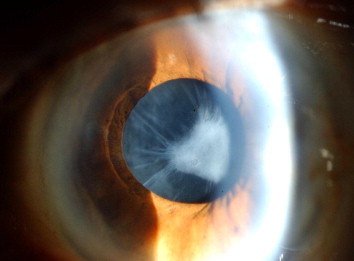
Showing complete occlusion of the anterior capsule opening with fibrous membrane that contains a radiating folds.
Figure 2.

Showing clear visual axis after removal of fibrous membrane with no recurrence after 6 months.
Figure 3.

Showing the repeat capsulorrhexis technique.
3. Discussion
Shrinkage and whitening of the anterior capsule opening is a well-known complication after continuous curvilinear capsulorrhexis, presumably as a result of imbalance between centrifugal forces of the zonules and the tensile force of the loop haptic of the IOL on the one hand and the centripetal forces of the proliferative and metaplastic residual lens epithelial cells (LECs) on the other hand. Marked capsule contraction can reduce the vision if the anterior capsule remnants obscure the visual axis or if the IOL is decentered by asymmetric bag contracture which occurs more often after implantation of polypropylene loop supports, which lose their loop shape memory with time (Masket, 1993; Zambarakji et al., 1997).
Anterior capsule seem to remain more stable in the presence of hydrophobic biomaterials than PMMA and silicon (Ursell et al., 1997). However, some reports suggest that PMMA haptics maintains the capsulorrhexis diameter more effectively than polypropylene haptics (Masket, 1993; Zambarakji et al., 1997). But it was not the case in one comparative study, where both type did not differ significantly in preventing capsular contraction (Gallagher and Pavilack, 1999).
To the best of our knowledge there are very few reports in the literature with capsular contraction that led to complete occlusion of the capsular opening.
We are reporting a case of CCS with complete occlusion of the capsulorrhexis opening with the use of three-pieces SENSAR® IOL in a patient without any known ocular risk factor for the development of capsular phimosis. This case might be explained by the age of patient in whom the zonules are weakening with aging.
Those patients with preoperative risk factor like pseudoexfoliation (Davison, 1993; Hayashi et al., 1998), uveitis (Davison, 1993; Spang et al., 1999), myotonic dystrophy, retinitis pigmentosa (Hayashi et al., 1998), diabetic retinopathy (Hayashi et al., 1997), high myopia, old age and Marfan’s syndrome or those who had intraoperative complication leading to the weakening of the zonules might benefit from insertion of capsular tension ring (CTR) or capsule-bending ring (CBR) to prevents the development of CCS (Sun and Gimbel, 1998; Nishi et al., 1998). However there are few reports of capsular contraction syndrome despite insertion of CTR (Faschinger and Eckhardt, 1999; Waheed et al., 2001; Sudhir and Rao, 2001; Moreno-Montanes et al., 2002).
Davison was the first one to describe the use of Nd:YAG radial anterior capsulotomy to interrupt the contraction forces without need of surgery to prevent IOL decentration and complete occlusion of the capsular opening (Davison, 1993). Then the same technique was used by many investigators with high success rate (Hayashi et al., 1998, 1997; Gallagher and Pavilack, 1999; Sudhir and Rao, 2001).
Yeh and coauthors have described the removal of fibrotic membrane in CCS by vitrector to minimize depries floating in the anterior chamber and the increase in intraocular pressure after Nd:YAG capsulotomy, but has the disadvantage of enlarging the anterior capsular opening to a scalloped-edge pattern (Yeh et al., 2002). Spang and coauthors preferred surgical excision of the fibrotic tissue. Our patient had removal of the fibrous membrane in a form of repeat capsulorrhexis without any sequel.
In conclusion, CCS is a well-known and recognized complication after intact capsulorrhexis. It is more common in a small size anterior capsule opening, in patients with week zonules and in patients with risk factors. Patient at risk should be recognized and managed accordingly intraoperatively or postoperatively. YAG radial anterior capsulotomy should be performed before both complete occlusion of the opening and/or ciliary body detachment occurred. In case of complete occlusion surgical excision as vitrectorrhexis or capsular peeling can be performed.
References
- Behrendt S., Wetzel W. Complete occlusion of the capsulorrhexis incision by anterior capsule shrinkage. Ophthalmology. 1994;91(4):526–528. [PubMed] [Google Scholar]
- Bolz M., Menapace R., Findl O., Sacu S., Buehl W., Wirtitsch M., Leydolt C., Kriechbaum K. Effect of anterior capsule polishing on the posterior capsule opacification-inhibiting properties of a sharp-edged, 3-piece, silicone intraocular lens: three- and 5-year results of a randomized trial. J. Cataract Refract. Surg. 2006;32(9):1513–1520. doi: 10.1016/j.jcrs.2006.04.020. [DOI] [PubMed] [Google Scholar]
- Davison J.A. Capsule contraction syndrome. J. Cataract Refract. Surg. 1993;19:582–589. doi: 10.1016/s0886-3350(13)80004-1. [DOI] [PubMed] [Google Scholar]
- Edrich C.L., Ghanchi F., Calvert R. Anterior capsular phimosis with complete occlusion of the capsulorrhexis opening. Eye. 2005;19(11):1229–1232. doi: 10.1038/sj.eye.6701738. [DOI] [PubMed] [Google Scholar]
- Faschinger C.W., Eckhardt M. Complete capsulorrhexis opening occlusion despite capsular tension ring implantation. J. Cataract Refract. Surg. 1999;25(7):1013–1015. doi: 10.1016/s0886-3350(99)00070-x. [DOI] [PubMed] [Google Scholar]
- Gallagher S.P., Pavilack M.A. Risk factors for anterior capsule contraction syndrome with polypropylene or poly(methyl methacrylate) haptics. J. Cataract Refract. Surg. 1999;25:1356–1361. doi: 10.1016/s0886-3350(99)00228-x. [DOI] [PubMed] [Google Scholar]
- Hayashi K., Hayashi H., Nakao F., Hayashi F. Reduction in the area of the anterior capsule opening after poly-methylmethacrylate, silicone, and soft acrylic intraocular lens implantation. Am. J. Ophthalmol. 1997;123:441–447. doi: 10.1016/s0002-9394(14)70169-2. [DOI] [PubMed] [Google Scholar]
- Hayashi K., Hayashi H., Matsuo K. Anterior capsular contraction and intraocular lens dislocation after implant surgery in eyes with retinitis pigmentosa. Ophthalmology. 1998;105:1239–1243. doi: 10.1016/S0161-6420(98)97028-2. [DOI] [PubMed] [Google Scholar]
- Hayashi H., Hayashi K., Nakao F., Hayashi F. Anterior capsular contraction and intraocular lens dislocation in eyes with pseudoexfoliation syndrome. Br. J. Ophthalmol. 1998;82:1429–1432. doi: 10.1136/bjo.82.12.1429. [DOI] [PMC free article] [PubMed] [Google Scholar]
- Höhn S., Spraul C.W. Complete occlusion of the frontal capsule after cataract-operation in a patient with pseudoexfoliation syndrome. Klin. Monatsbl. Augenheilkd. 2004;221(6):495–497. doi: 10.1055/s-2004-813293. [DOI] [PubMed] [Google Scholar]
- Leaming D.V. Practice styles and preferences of ASCRS members – 1992 survey. J. Cataract Refract. Surg. 1993;19:600–606. doi: 10.1016/s0886-3350(13)80007-7. [DOI] [PubMed] [Google Scholar]
- Martínez Toldos J.J., Artola Roig A., Chipont Benabent E. Total anterior capsule closure after silicone intraocular lens implantation. J. Cataract Refract. Surg. 1996;22(2):269–271. doi: 10.1016/s0886-3350(96)80230-6. [DOI] [PubMed] [Google Scholar]
- Masket S. Postoperative complications of capsulorrhexis. J. Cataract Refract. Surg. 1993;19:721–724. doi: 10.1016/s0886-3350(13)80340-9. [DOI] [PubMed] [Google Scholar]
- Moreno-Montanes J., Scanchez-Tocino H., Rodriguez-Conde R. Complete anterior capsule contraction after phacoemulsification with acrylic intraocular lens and endocapsular ring implantation. J. Cataract Refract. Surg. 2002;28(4):717–719. doi: 10.1016/s0886-3350(01)01231-7. [DOI] [PubMed] [Google Scholar]
- Newman D.K. Severe capsulorrhexis contracture after cataract surgery in myotonic dystrophy. J. Cataract Refract. Surg. 1998;24:1410–1412. doi: 10.1016/s0886-3350(98)80238-1. [DOI] [PubMed] [Google Scholar]
- Nishi O., Nishi K., Menapace R. Capsule-bending ring for the prevention of capsular opacification. Ophthalmic Surg. Lasers. 1998;29(9):749–753. [PubMed] [Google Scholar]
- Patton N., Ironside J.W., Aslam T.M., Bennett H.G., Singh J. Complete occlusion of the anterior capsular opening by fibrocellular membrane associated with retained silicone oil: a clinicopathologic correlation. Retina. 2004;24(3):483–487. doi: 10.1097/00006982-200406000-00031. [DOI] [PubMed] [Google Scholar]
- Spang K.M., Rohrbach J.M., Weidle E.G. Complete occlusion of the anterior capsular opening after intact capsulorrhexis: clinicopathologic correlation. Am. J. Ophthalmol. 1999;127:343–345. doi: 10.1016/s0002-9394(98)00323-7. [DOI] [PubMed] [Google Scholar]
- Sudhir R.R., Rao S.K. Capsulorrhexis phimosis in retinitis pigmentosa despite capsular tension ring implantation. J. Cataract Refract. Surg. 2001;27(10):1691–1694. doi: 10.1016/s0886-3350(01)00869-0. [DOI] [PubMed] [Google Scholar]
- Sugimoto Y., Takayanagi K., Tsuzuki S. Postoperative changes over time in size of anterior capsulorrhexis in phacoemulsification/aspiration. Jpn. J. Ophthalmol. 1998;42:495–498. doi: 10.1016/s0021-5155(98)00052-5. [DOI] [PubMed] [Google Scholar]
- Sun R., Gimbel H.V. In vitro evaluation of the efficacy of the capsular tension ring for managing zonular dialysis in cataract surgery. Ophthalmic Surg. Lasers. 1998;29:502–505. [PubMed] [Google Scholar]
- Ursell P.G., Spalton D.J., Pande M.V. Anterior capsule stability in eyes with intraocular lenses made of poly (methyl methacrylate), silicone, and AcrySof. J. Cataract Refract. Surg. 1997;23:1532–1538. doi: 10.1016/s0886-3350(97)80025-9. [DOI] [PubMed] [Google Scholar]
- Waheed K., Eleftheriadis H., Liu C. Anterior capsular phimosis in eyes with a capsular tension ring. J. Cataract Refract Surg. 2001;27(10):1688–1690. doi: 10.1016/s0886-3350(01)00766-0. [DOI] [PubMed] [Google Scholar]
- Yeh P.C., Goins K.M., Lai W.W. Managing anterior capsule contraction by mechanical widening with vitrector-cut capsulotomy. J. Cataract Refract. Surg. 2002;28:217–220. doi: 10.1016/s0886-3350(01)01031-8. [DOI] [PubMed] [Google Scholar]
- Zambarakji H., Rauz S., Reynolds A., Jhoshi N., Simcock P.R., Kinnear P.E. Capsulorrhexis phimosis following uncomplicated phacoemulsification surgery. Eye. 1997;11:635–638. doi: 10.1038/eye.1997.168. [DOI] [PubMed] [Google Scholar]