Abstract
Background:
We present the magnitude and determinants of age-related macular degeneration (ARMD) among the 50 year and older population that visited our hospital.
Materials and Methods:
This was a cohort of eye patients with ARMD, seen from 2006 to 2009. Optometrist noted the best-corrected vision. Ophthalmologists examined eyes using a slit-lamp bio-microscope. The ARMD was confirmed by fluoresceine angiography and optical coherent tomography. The age, sex, history of smoking, sun exposure, family history of ARMD, diet, body mass index (BMI), hypertension, and diabetes were associated with ARMD.
Result:
Of the 19,140 persons of ≥ 50 years of age-attending eye clinic in our hospital, 302 persons had ARMD in at least one eye. The proportion of overall ARMD was 1.38% (95% CI 1.21--1.55). The proportion of age-related maculopathy (ARM) and late ARMD was 1.14% (95% CI 0.99--1.29) and 0.24% (95% CI 0.21–0.24) respectively. ARM was unilateral and bilateral in 64 (29.2%) and 155 (70.8%) persons respectively. Dry ARMD was found in 47.8%. On regression analysis, old age (OR = 1.05), male (OR = 0.54), and history of smoking (OR = 2.32) were significant risk factors of ARMD. A total of 4.2% of persons with ARMD were blind (vision <3/60). Only 43% of persons with ARMD had J6 grade of the best-corrected near vision.
Conclusion:
ARMD does not seem to be of public health magnitude in the study area. Early stages of ARMD were common among patients. ge, being male, and history of smoking were significant risk factors for ARMD.
Keywords: Age-related macular degeneration, blindness, low vision, prevalence, retina
Age-related macular degeneration (ARMD) accounts for 8.7% of the total blindness globally, and is the third common cause of visual impairment. It is the primary cause of visual impairment in industrialized countries.[1,2] The persons with ARMD are likely to increase from three to six million by the year 2020. This could be due to a decline in avoidable blindness, due to anterior segment pathologies, and the increasing life expectancy of the global population. It has therefore been included in the action plan of the World Health Organization, to address avoidable blindness in VISION 2020 program.[3]
The literature exhibits a number of studies to determine the magnitude and risk factors of ARMD including that in Indian subcontinent.[4,5,6,7,8] However, to our knowledge, no such data have been generated in the state of Maharashtra - the second most populous in India (Census 2010). Apart from the metropolitan cities of Mumbai and Pune, the population of this state comprises mainly the Aryan race. They are part of mainly Hindu or Muslim communities. The study area experiences three main seasons in a year; harsh summer (up to 45°C), winter (lowest 5°C) and rainfall of 40-50 in for 3 months.
This study was conducted to determine the magnitude of ARMD among the native population aged 50 years and older in a part of Maharashtra who visited the ophthalmology department of our institute.
Materials and Methods
The ethical and research committee of our hospital approved this study. Informed verbal consent was obtained from each participant. This study was conducted between June 2006 and May 2009.
This was a cross-sectional type of descriptive study. The study population comprised all eye patients aged 50 years and older that visited our institution in last 4 years for their eye ailments. Those suspected to suffer from ARMD, either due to history or clinical examination, were recruited in our cohort [Fig. 1]. Patients with dense corneal and lens opacities, obstructing the view of central retina, were excluded from the study. Their baseline information was used for the present manuscript.
Figure 1.
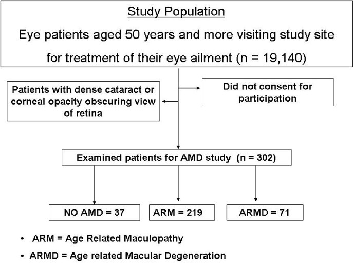
Flow chart showing study population and examined sample for age-related macular degeneration study
In a population of 19,140 persons of 50 years and older age who attended the eye clinic, we calculated the sample size needed for our study. We assumed that the proportion of ARMD would be 4.8%.[4] To achieve 95% confidence interval, acceptable error margin of 3% and design effect of two, we need to examine 387 persons of this age group.
Two ophthalmologists and two optometrists were our field staff. To assess distant vision we used Snellen's illiterate “E” chart, held at 6 m distance from the patient. If a person could not correctly identify “E” of the top line, the test was repeated at 3 m distance. The near vision was tested using a Jager near vision chart, held at 25-33 cm from the face. A person's vision was tested with his/her spectacles on. The anterior segment of the eye was evaluated using slit lamp bio-microscope (Appasamy, India). The lens status was defined as immature cataract, aphakia, pseudophakia, and clear lens. The retina and optic nerve head were examined by using +90 D Volk lens and slit lamp bio-microscope. A red free filter was applied to distinguish drusen from exudates and hemorrhage. A slit beam was used to determine macular edema. If central lenticular opacity was present, we used indirect ophthalmoscope and +30 D lens to view central retina.
ARMD was graded as per the international classification.[9] The digital image using fundus photographs were evaluated by the ophthalmologist without any prior information about risk factors. The mild ARMD was called ARM and it included the presence of soft drusen of more than 63 mc size, presence of hyper or hypo pigmentation of retinal pigment epithelium or both of these above mentioned conditions [Fig. 2]. Dry ARMD includes geographic atrophy. The eye with chorio-retinal atrophy secondary to other obvious cause like high myopia and chronic grenulomatous eye disease in the past were excluded while labeling eye with dry ARMD. Wet ARMD included neo-vascular vessels of chorio-capillary plexus in macular area (CNVM), exudative pigment epithelial defect (PED), macular scar or combinations of any of these three conditions [Fig. 3]. If a person had blood pressure more than 150/90 mm Hg and other persons are taking medication to control blood pressure, they were defined to suffer from hypertension. If a person's fasting capillary blood sugar level was >7 mmol/l, he/she was considered to suffer from diabetes. Those already taking medicine to control diabetes were also included in the pool of diabetics. Height and weight were measured. Body mass index was calculated using the formula: weight (in kilograms)/(height × height) (in meters). The person's diet was enquired about person consumed vegetarian, nonvegetarian or mixed diet in the last 2 weeks. Depending on the number of hours a person is working in sun exposure, we grouped participants into those exposed to sun (more than 8 h/day) and other. We inquired if any other elderly family member was told by the eye doctor about having a central retina problem that had caused visual impairment.
Figure 2.
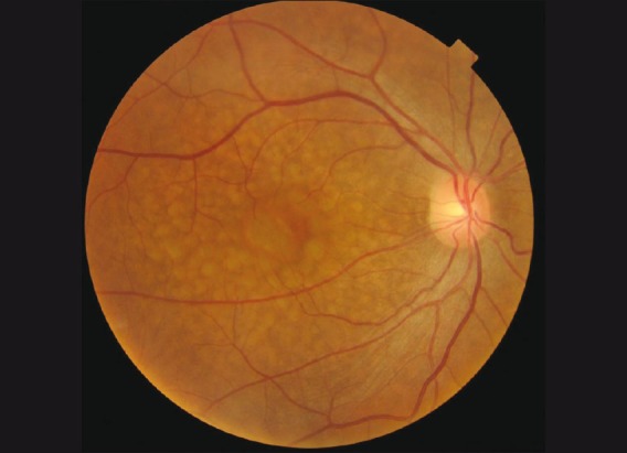
Multiple soft drusen in eye with age-related maculopathy (ARM)
Figure 3.
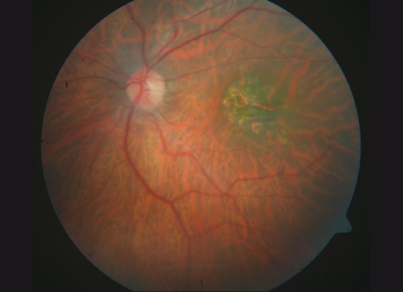
Geographic chorio-retinal atrophy in eye with age-related macular degeneration (ARMD)
Result
During the study period, 19,140 persons of 50 years and older age with some eye ailment attended our eye hospital. On comprehensive examination, 302 of them were found to have age-related macular degeneration and were included in our study for further detailed examination. The study population and examined sample by gender and age group are given in Table 1.
Table 1.
Comparison of study population and examined participants for the ARMD study
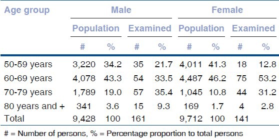
The examined sample, frequency of ARMD, late form of ARMD (AMD) and total ARMD, adjusted percentage proportion with 95% confidence interval were calculated [Table 2]. The proportion of age-related maculopathy (ARM) was 1.14% (95% CI 0.99-1.29). The proportion of ARMD was 1.38% (95% CI 1.21-1.55).
Table 2.
Prevalence of age-related maculopathy (ARM), age-related macular degeneration (ARMD) among 50 year and older eye patients

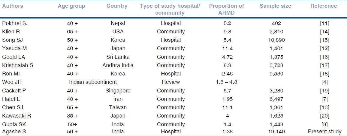
We compared the proportions of ARMD from different studies [Table 3]. Variation in methods and target population resulted in wide variation in the proportion of ARMD in different studies.
Table 3.
Proportion of age-related macular degeneration in different studies
ARM was unilateral in 64 (29.2%) persons and bilateral in 155 (70.8%) persons. In 132 persons with AMD only drusen were noted in the macular area and 87 persons had retinal pigment epithelium pathology. Among persons with late ARMD, dry type was noted in 27 persons (38%), while 40 (56.4%) persons had neovascularization (with/without pigment epithelial defect) and four (5.6%) persons had macular scar.
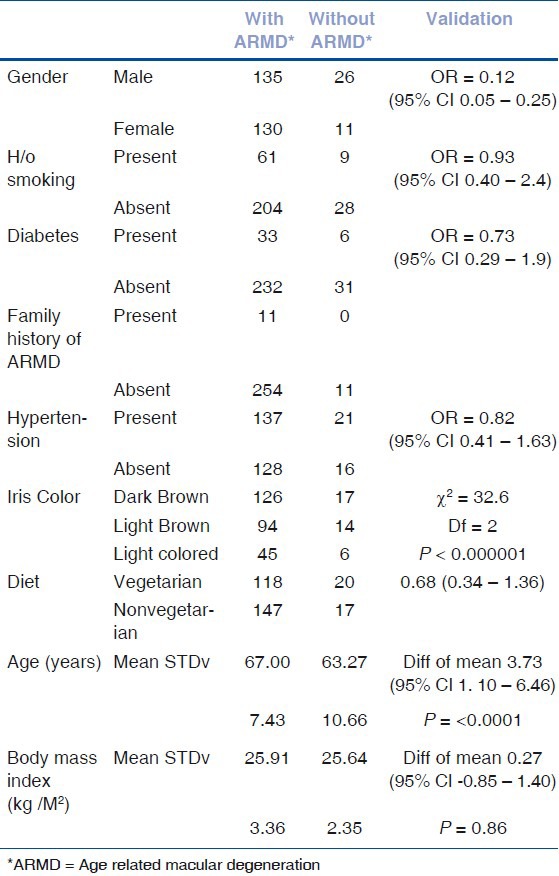
To associate different variables with the ARMD, we performed univariate analysis [Table 4]. ARMD was significantly associated with the female sex, old age, positive family history of ARMD, and dark brown color of iris.
Table 4.
Risk factors of age-related macular degeneration (ARMD)
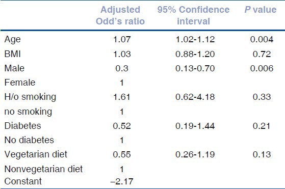
To understand the interaction of the different variables while influencing ARMD, we conducted binary logistic regression analysis [Table 5]. Old age and female sex were significant risk factors for ARMD.
Table 5.
Risk factor of age-related macular degeneration (regression analysis model)
Based on the best-corrected visual status for distance in the better eye, the persons with ARMD were grouped into bilateral blindness (<3/60), severe visual impairment (<6/60 to 3/60), moderate visual impairment (6/18 to 6/60), and normal vision (>6/18) [Fig. 4]. A total of 4.2% of persons with ARMD were bilateral blind.
Figure 4.
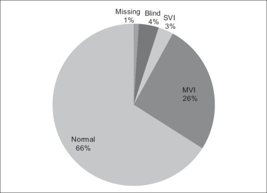
Proportion of visual disabilities among persons with age-related macular degeneration (ARMD)
The best corrected near visual acuity was J6 in better eye in 114 (43%) patients with ARMD. In 92 (34.7%) cases, it was J8-J10. Near visual acuity was J12-J18 in 24 (9.1%) cases with ARMD. In 35 (13.2%) cases it was J36 or less. Among 37 cases without clinically significant ARMD the near vision was J6 in 26 (70.3%) persons. In seven (18.9%) cases, near vision acuity was J8–J10. Thus most of the ARMD cases had significant poorer level of near vision compared to those without ARMD.
Of the 604 eyes of 302 persons, we grouped (1) those with lens (immature cataract or clear lens) and (2) those without lens (aphakia and pseudophakia). In our study, 280 phakic eyes had ARMD while 100 phakic eyes did not have ARMD. In 120 aphakic/peudophakic eyes, ARMD was noted while in 50 aphakic/peudophakic eyes ARMD was not present. We associated the presence of ARMD with cataract surgeries. Thus odds of aphakia/pesudophakia among ARMD was 1.08 (95% CI 0.73–1.62).
Discussion
India is the second most populous country in the world, with over 1.18 billion people (estimate for April, 2010), more than a sixth of the world's population. With 17.3% of the world's population already, India is projected to be the world's most populous country by 2025. By 2050 it is projected to surpass China, its population will exceed 1.6 billion people.[10] With rapidly increasing aging population in India (life expectancy of: 69.9 years) and with blindness due to cataract being addressed more vigorously (cataract surgery rate of 4,500 / million population in 2005), the age-related blinding causes of posterior segment will be more important in the coming years.[11] The demography of India is remarkably diverse. Epidemiological findings of aging population in western India could differ from other parts. Our study in Maharashtra therefore will add to the knowledge of epidemiology of ARMD in India and also enable the health planners to determine risk factors and propose preventive measures.
Our study had a large sample of eye patients of 50 years and older age. Therefore the proportion of ARMD is less likely to be influenced by systematic error. The epidemiologically sound method has minimized the effect of bias in our study. One should, however, note the limitation of a hospital-based study while interpreting the results for community-based action. The posterior segment examination using indirect ophthalmoscope and slit lamp bio-microscope, as used in our study differed from digital fundus photography to evaluate ARMD in many other studies. It is possible that we may have under estimated early stages of ARMD in our study. The objective method of noting early ARMD could be less accurate, with digital photography, the spotting of early drusen could be more reliable, and thus yield better internal validity. Therefore we suggest undertaking future study using digital images and internationally standard grading system of ARMD.
The overall proportion of ARMD was 1.38%. (ARMD -- 1.34% and late ARMD -- 0.37%). Gupta et al. had reported the proportion of early and late ARMD as 47% and 1.4% respectively.[6] The proportion of early and late ARMD was 5.1% and 0.34% in South Korea.[12] The magnitude of late ARMD of Korea matched with results of our study. It is postulated by Kawasaki et al. that early ARMD is less common in Asians.[13] This may explain why our population had a low proportion of ARMD. In view of the difference in age groups and target populations, varying ARMD proportion should be interpreted with caution.[7,12,13,14,15,16,17,18,19,20,21] The denominator used for calculating the proportion of ARMD was the number of eye patients of same age group attending our hospital during the study period. Patients from rural areas coming for cataract surgery would have dense lens opacities and could not be examined to evaluate posterior segment. This could result in a low proportion of ARMD. It may be also possible that the risk of ARMD is different for different geographic areas.
Less than 5% persons with ARMD had bilateral blindness. Pokharel et al. has noted that 15% of ARMD cases with less than 3/60 visual acuity in Nepal.[14] It seems that few persons with advanced ARMD could have resulted in fewer blind people in our study. Many persons with ARMD had severe visual impairment. They experienced difficulties in performing visual tasks in daily living and could be benefited by low vision care.
Older age was an independent risk factor for ARMD in our study. This was also observed in other studies.[7,12,13,14,15,16,17,18,19,20,21,22] The comparison of ARMD in the younger and the older age group should be done with caution as mortality in the older age group with ARMD (significantly associated with smoking) is higher.[22] Hence the older age group may artificially show lower proportions of ARMD.
Females had significantly higher risk for ARMD than males in our study. This was also noted Pokhrel et al. and Hazin et al.[11,23] But it was more in male in USA and in Japan.[15,16] The risk of ARMD did not differ in male and females in Sri Lanka.[17] Variation in smoking habits, better longevity among females, and exposure to sunlight among females might be the reason for this variation in ARMD by sex.[24]
Smoking was surprisingly not associated with ARMD in our study. This was in contrast to other studies with larger cohorts.[15,16,21,22] Getting information only about current smoking and not knowing about type and amount of tobacco consumption could have affected the association of smoking and ARMD in our study. Further studies to investigate this association are recommended.
In our study, we did not find an association between diet and ARMD. A diet high in unsaturated fatty acids and olive oil had lower risk of ARMD.[25,26] Risk of ARMD also differed by the type of meat consumed.[27] The differential method of collecting information on diet in our study, compared to these studies, could be the reason for this variation. Further detailed information on diet and studies to identify nutrients as risk for ARMD are needed.
Hypertension was associated with ARMD in Korea and India.[12,6] Both hypertension and body mass index (BMI) were not associated with ARMD in USA.[15] In our study also, we could not find a significant association of hypertension with ARMD.
We did not find a significant association between ARMD and cataract surgery. This positive association was noted by Chew et al. and Ho et al.[28,29] Longitudinal studies are suggested to understand the relationship of ARMD following cataract surgery.
The proportion of ARMD in our population in Maharashtra seems to be low. Visual disability of blinding due to ARMD is of low magnitude. Older age, family history of ARMD, and the color of iris were factors associated with ARMD. The preventable risk factors like smoking and alteration of diet need further assessment in order to propose preventive measures.
Acknowledgments
We thank the Ethical and Research Committee of HV Desai eye hospital for their approval and support in this survey. Dr. Andurkar's encouragement and help in logistics was enormous. Dr. Radha Shenoy of Armed Force Hospital, Muscat, Oman had provided guidance to improve the manuscript. We thank all of them.
Footnotes
Source of Support: Nil,
Conflict of Interest: None declared.
References
- 1.World Health Organization. Age Related Macular Degeneration: Priority eye diseases. [Last accessed on 2011 Feb 6]. Available from: http://www.who.int/blindness/causes/priority/en/index8.html .
- 2.Białek-Szymańska A, Misiuk-Hojło M, Witkowska K. Risk factors evaluation in age-related macular degeneration. Klin Oczna. 2007;109:127–30. [PubMed] [Google Scholar]
- 3.World Health Organization. Age Related Macular Degeneration: Disease control and prevention of visual impairment in global initiative for the elimination of Avoidable Blindness Action Plan 2006–2011. Printed in France. 2007. [Last accessed on 2010 Nov 26]. pp. 32–3. Accessed from: http://www.who.int/blindness/Vision2020_report.pdf .
- 4.Woo JH, Sanjay S, Au Eong KG. The epidemiology of age-related macular degeneration in the Indian subcontinent. Acta Ophthalmol. 2009;87:262–9. doi: 10.1111/j.1755-3768.2008.01376.x. [DOI] [PubMed] [Google Scholar]
- 5.Tan JS, Mitchell P, Kifley A, Flood V, Smith W, Wang JJ. Smoking and the long-term incidence of age-related macular degeneration: The Blue Mountains Eye Study. Arch Ophthalmol. 2007;125:1089–95. doi: 10.1001/archopht.125.8.1089. [DOI] [PubMed] [Google Scholar]
- 6.Krishnaiah S, Das TP, Kovai V, Rao GN. Associated factors for age-related maculopathy in the adult population in southern India: The Andhra Pradesh Eye Disease Study. Br J Ophthalmol. 2009;93:1146–50. doi: 10.1136/bjo.2009.159723. [DOI] [PubMed] [Google Scholar]
- 7.Hatef E, Fotouhi A, Hashemi H, Mohammad K, Jalali KH. Prevalence of retinal diseases and their pattern in Tehran: The Tehran eye study. Retina. 2008;28:755–62. doi: 10.1097/IAE.0b013e3181613463. [DOI] [PubMed] [Google Scholar]
- 8.Gupta SK, Murthy GV, Morrison N, Price GM, Dherani M, John N, et al. Prevalence of early and late age-related macular degeneration in a rural population in northern India: The INDEYE feasibility study. Invest Ophthalmol Vis Sci. 2007;48:1007–11. doi: 10.1167/iovs.06-0712. [DOI] [PubMed] [Google Scholar]
- 9.American Academy of Ophthalmology. Age Related Macular Degeneration. Preferred Practice Patterns. [Last accessed on 2010 Nov 29]. Accessed from: http://one.aao.org/ce/practiceguidelines/ppp_content.aspx?cid=f413917a-8623-4746-b441-f817265eafb4#section3 .
- 10. [Last accessed on 2010 Dec 17]. India. Available from: http://en.wikipedia.org/wiki/India .
- 11.Shrivastava RK, Jose R, Rammamoorthy K, Bhushan B, Tiwari AK, Malik KP, et al. NPCB: Achievements and targets. NPCB India. 2006;1:5–6. [Google Scholar]
- 12.Pokharel S, Malla OK, Pradhananga CL, Joshi SN. A pattern of age-related macular degeneration. JNMA J Nepal Med Assoc. 2009;48:217–20. [PubMed] [Google Scholar]
- 13.Klein R, Cruickshanks KJ, Nash SD, Krantz EM, Javier Nieto F, Huang GH, et al. The prevalence of age-related macular degeneration and associated risk factors. Arch Ophthalmol. 2010;128:750–8. doi: 10.1001/archophthalmol.2010.92. [DOI] [PMC free article] [PubMed] [Google Scholar]
- 14.Song SJ, Youm DJ, Chang Y, Yu HG. Age-related macular degeneration in a screened South Korean population: Prevalence, risk factors, and subtypes. Ophthalmic Epidemiol. 2009;16:304–10. [PubMed] [Google Scholar]
- 15.Yasuda M, Kiyohara Y, Hata Y, Arakawa S, Yonemoto K, Doi Y, et al. Nine-year incidence and risk factors for age-related macular degeneration in a defined Japanese population the Hisayama study. Ophthalmology. 2009;116:2135–40. doi: 10.1016/j.ophtha.2009.04.017. [DOI] [PubMed] [Google Scholar]
- 16.Goold LA, Edussuriya K, Sennanayake S, Senaratne T, Selva D, Sullivan TR, et al. Prevalence and determinants of age-related macular degeneration in central Sri Lanka: The Kandy Eye Study. Br J Ophthalmol. 2010;94:150–3. doi: 10.1136/bjo.2009.163808. [DOI] [PubMed] [Google Scholar]
- 17.Roh MI, Kim JH, Byeon SH, Koh HJ, Lee SC, Kwon OW. Estimated prevalence and risk factor for age-related maculopathy. Yonsei Med J. 2008;49:931–41. doi: 10.3349/ymj.2008.49.6.931. [DOI] [PMC free article] [PubMed] [Google Scholar]
- 18.Cackett P, Tay WT, Aung T, Wang JJ, Shankar A, Saw SM, et al. Education, socio-economic status and age-related macular degeneration in Asians: The Singapore Malay Eye Study. Br J Ophthalmol. 2008;92:1312–5. doi: 10.1136/bjo.2007.136077. [DOI] [PubMed] [Google Scholar]
- 19.Chen SJ, Cheng CY, Peng KL, Li AF, Hsu WM, Liu JH, et al. Prevalence and associated risk factors of age-related macular degeneration in an elderly Chinese population in Taiwan: The Shihpai Eye Study. Invest Ophthalmol Vis Sci. 2008;49:3126–33. doi: 10.1167/iovs.08-1803. [DOI] [PubMed] [Google Scholar]
- 20.Kawasaki R, Wang JJ, Ji GJ, Taylor B, Oizumi T, Daimon M, et al. Prevalence and risk factors for age-related macular degeneration in an adult Japanese population: The Funagata study. Ophthalmology. 2008;115:1376–81. doi: 10.1016/j.ophtha.2007.11.015. 1381. [DOI] [PubMed] [Google Scholar]
- 21.Hazin R, Freeman PD, Kahook MY. Age-related macular degeneration: A guide for the primary care physician. J Natl Med Assoc. 2009;101:134–8. doi: 10.1016/s0027-9684(15)30825-7. [DOI] [PubMed] [Google Scholar]
- 22.Coleman HR, Chan CC, Ferris FL, 3rd, Chew EY. Age-related macular degeneration. Lancet. 2008;372:1835–45. doi: 10.1016/S0140-6736(08)61759-6. [DOI] [PMC free article] [PubMed] [Google Scholar]
- 23.Arnarsson A, Sverrisson T, Stefánsson E, Sigurdsson H, Sasaki H, Sasaki K, et al. Risk factors for five-year incident age-related macular degeneration: The Reykjavik Eye Study. Am J Ophthalmol. 2006;142:419–28. doi: 10.1016/j.ajo.2006.04.015. [DOI] [PubMed] [Google Scholar]
- 24.Plestina-Borjan I, Klinger-Lasić M. Long-term exposure to solar ultraviolet radiation as a risk factor for age-related macular degeneration. Coll Antropol. 2007;1:33–8. [PubMed] [Google Scholar]
- 25.Parekh N, Voland RP, Moeller SM, Blodi BA, Ritenbaugh C, Chappell RJ, et al. Association between dietary fat intake and age-related macular degeneration in the Carotenoids in Age-Related Eye Disease Study (CAREDS): An ancillary study of the Women's Health Initiative. Arch Ophthalmol. 2009;127:1483–93. doi: 10.1001/archophthalmol.2009.130. [DOI] [PMC free article] [PubMed] [Google Scholar]
- 26.Chong EW, Robman LD, Simpson JA, Hodge AM, Aung KZ, Dolphin TK, et al. Fat consumption and its association with age-related macular degeneration. Arch Ophthalmol. 2009;127:674–80. doi: 10.1001/archophthalmol.2009.60. [DOI] [PubMed] [Google Scholar]
- 27.Chong EW, Simpson JA, Robman LD, Hodge AM, Aung KZ, English DR, et al. Red meat and chicken consumption and its association with age-related macular degeneration. Am J Epidemiol. 2009;169:867–76. doi: 10.1093/aje/kwn393. [DOI] [PubMed] [Google Scholar]
- 28.Chew EY, Sperduto RD, Milton RC, Clemons TE, Gensler GR, Bressler SB, et al. Risk of advanced age-related macular degeneration after cataract surgery in the Age-Related Eye Disease Study: AREDS report 25. Ophthalmology. 2009;116:297–303. doi: 10.1016/j.ophtha.2008.09.019. [DOI] [PMC free article] [PubMed] [Google Scholar]
- 29.Ho L, Boekhoorn SS, Liana, van Duijn CM, Uitterlinden AG, Hofman A, et al. Cataract surgery and the risk of aging macula disorder: The rotterdam study. Invest Ophthalmol Vis Sci. 2008;49:4795–800. doi: 10.1167/iovs.08-2066. [DOI] [PubMed] [Google Scholar]


