Abstract
This study was carried out to evaluate the anti-obesity effect of Vernonia amygdalina Del. (VA) supplemented diet. VA leaf powder was fed at 5% and 15% to diet-induced obese rats for 4 weeks and its effect compared with orlistat (5.14 mg/kg p.o.), an anti-obesity drug. Food intake, body and organ weights, total body fat, some lipid components and amino transaminase activities in serum, hepatocytes and brain; as well as serum glucose, were measured during or at end of the study. Result showed respective decrease of 12.78% and 38.51% in body weight gain, of VA fed rats against 17.45% of orlistat at end of study (P < 0.05); but with no effect on food intake. Total body fat was lowered by 28.04% and 30.02% vs. obese control rats (CDC) (P < 0.05). Furthermore, serum triacylglycerol (TG), serum and brain total cholesterol (TCHOL), were down regulated at 15% VA supplementation (P < 0.05). Serum glucose which increased in obese rats by 46.26% (P < 0.05) vs. NC, indicating intolerance, was restored by VA (38.75% and 34.65%) and orlistat (31.80%) vs. CDC (P < 0.05). VA diet also exerted hepato-protection, via lowering serum alanine amino transaminase (ALT) (41.35% and 27.13%) and aspartate amino transaminase (AST) (17.09% and 43.21%) activities (P < 0.05). Orlistat had no effect on these enzymes. Histology of adipose tissue corroborated the changes on total body fat. We concluded that, diet supplemented with VA can attenuate dietary obesity as well as ameliorates the potential risks of hepato-toxicity and glucose intolerance associated with obesity.
Keywords: Vernonia amygdalina Del., Adipose tissue, Histology, Total body fat, Lipid profile, Glucose intolerance, Diet-induced obesity
1. Introduction
Obesity is fast becoming a medical condition of global concern, particularly due to its consistent association with increased prevalence of cardiovascular diseases, diabetes mellitus, hypertension and some forms of cancers (Xia et al., 2010). It is now certain that genetic predisposition and consumption of high energy foods are the commonest pathogenetic factors (Thompson et al., 2011). Weight reduction, via dietary modulation is therefore the target of choice of many therapeutic measures. Subramine and orlistat the approved conventional drugs, promote about 5–10% loss of weight, and best, only when used in conjunction with diet, exercise, and behavior change regimens (Kumar et al., 2011). This is grossly inadequate and unsatisfactory given the present size of the market and current status for development of these drugs (Shrestha et al., 2007). The reported minor and grave side effects of the drugs, as well as weight rebounds when discontinued (Ellrichmann et al., 2008), have necessitated a new dimensional approach into the search for anti-obesity medicines. Incidentally, numerous preclinical and clinical studies, with various herbal medicines have reported significant improvement in controlling body weight, without any noticeable adverse effects (Kumar et al., 2011).
Vernonia amygdalina Del. (VA) is a nutritionally and pharmacologically responsive vegetable, commonly called “African bitter leaf” used in West Africa particularly, Nigeria in the preparation of the popular “bitter leaf soap” served at homes and restaurants. Its nutritional and medicinal uses and scientific studies have respectively been articulated in two extensive reviews by Ijeh and Ejike (2010) and Farombi and Owoeye (2011). A number of earlier studies with extracts from the plant (Ekpo et al., 2007; Ezekwe and Obidia, 2001) and the leaves incorporated into diet by weight (Ugwu et al., 2009, 2011) have documented the lipid modulating effect in normal adult rats. In Pharmacologically induced hyperlipidemic models, Adaramoye et al. (2008) have shown the anti-hyperlipidemic activity of extracts from the plant extract.
However, these studies are neither in-depth nor modeled the practical societal problem of obesity, using diet induced obese models, combined with a dietary approach to intervention. The present study is therefore conducted to investigate the possible anti-obesity effect of VA leaves incorporated by weight in diet of diet-induced obese rat models.
2. Materials and methods
2.1. Experimental diets
Cafeteria diet (CD), the fattening diet, was formulated according to the method of Kumar et al. (2011) with some modifications. The CD is comprised of three sets of diets A, B, and C formulated as below:
Each of diets A, B and C was then supplemented with VA at 5% or 15% by weight and fed in succession to the animals.
2.2. Plant material and standard drug
VA leaves were obtained from the Endocrine Research Farm, University of Calabar and authenticated in the Department of Botany, of the same institution. The leaves were air-dried (25–29 °C) and thereafter milled into coarse powder and stored in air-tight plastic containers, from where portions were weighed out for supplemented CD formulation. Fresh leaf samples were prepared every week until end of study. Orlistat (Xenical Pharmaceuticals, Japan) obtained from Karmel Pharmacy, 112 Goldie Street, Calabar, Nigeria, was reconstituted in distilled water and administered orally as 5.14 mg/kg body weight, a dose simulated from the human regimen.
2.3. Animals
Thirty Wistar rats (51–58 g) obtained from the Animal Resource, Department of Zoology and Environmental Biology, University of Calabar, were used for this study. The rats were allowed to acclimatize in the animal house of department of Biochemistry, where the experiment was conducted under controlled temperature (25–29 °C). The animals were allowed free access to food and water, and 12 h light/dark cycle. The protocol was in line with the guidelines of the National Institute of Health publication (1985) for Laboratory Animals Care and Use, and was approved by the College of Medical Sciences’ Animal Ethics Committee, University of Calabar.
2.4. Experimental protocol
The rats were divided into five groups of 6 rats each. The average body weights of rats in the five groups were similar at onset of the experiment. Group 1, the normal control (NC), was fed rat pellet only, throughout the 10 weeks of study. Group 2, cafeteria-diet-fed control (CDC), was also fed CD only for the 10 weeks of study, whereas groups 3–5, STDC, VAS 1 and VAS 2 were, fed CD only for the first 6 weeks of the study and thereafter in the last 4 weeks, CD with oral orlistat (5.14 mg/kg b.w.), 5% VA supplemented CD and 15% VA supplemented CD, respectively. The three CDs A, B, C alone or supplemented were presented to the animals on days 1, 2 and 3 and then repeated in succession until the end of study. Fresh diet for each group was compounded every day, to avoid spoilage, particularly the milk products. The food intake was recorded by measuring the difference between the pre-weighed diet presented to the rats and the weight of 24 h leftover. Food spillage was also recorded and adjusted for, in calculating the food intake. Body weight was measured two to three times per week, but consistently. Six weeks and 10 weeks body weight gains were calculated as difference in animal body weight on weeks 6 or 10 and weight at onset of experiment, whereas cumulative weight gain was calculated as body weight on the day of measurement minus the body weight recorded at onset of the experiment (daymeasurement−day0) (Zhou et al., 2011).
2.5. Body fat and organ weight measurements
At end of the experiment, the rats were euthanized and whole blood were collected by cardiac puncture, whole brain, liver and the kidneys were surgically removed, and wet weights were measured with an analytical balance after blotting. Total body fat (perirenal and epididymal fat pads) was removed and wet weights were recorded. Percentage/relative total body fat was calculated thus: w/W × 100% where, w is the wet weight of the fat pad and W, the final body weight of the animal before it was killed. Relative organ weights were similarly calculated. The tissues were immediately divided into two portions each. One part stored frozen preparatory to biochemical assays and the other part and fat pad fixed in buffered 10% formalin preparatory to histological analysis.
2.6. Biochemical analyses
Serum glucose, triacylglycerol (TG), total cholesterol (TCHOL) and high density lipoprotein cholesterol (HDL-C), as well as alanine amino transaminase (ALT) and aspartate amino transaminase (AST) activities were determined with analytical kits from Randox Laboratories Ltd. (Admore Diamond Road, Crumlin, Co., Antrim, United Kingdom). TG, TCHOL, HDL-C, and ALT and AST activities were also determined in homogenates prepared from liver and brain tissues. In brief, 1 g of each tissue was homogenized in 5 ml of phosphate buffer (pH 7.4), centrifuged at 4000 rpm for 15 min and the supernatant was used for the assays.
2.7. Histopathological studies
Fixed adipose tissues were sectioned with microtome (5 μm thickness), fixed in slides and stained using Harry’s Haematoxilin and Eosin staining procedures. The slides were viewed under light microscope and photomicrographs were taken (400×).
2.8. Statistical analysis
Data were expressed as the mean ± SD. ANOVA was used for analysis, followed by least square difference (LSD) post hoc, using SPSS software version 15.0. Difference was considered significant at P < 0.05.
3. Results
3.1. Effect of VA supplemented diet/orlistat on body weight and food intake
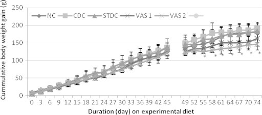
Although body weights were similar at onset of the experiment (54.82 ± 3.25 g), 6-week initial feeding of cafeteria diet (CD) increased body weight in the five experimental groups by 25–45% over the NC (Fig. 1.1). But 4-week intervention with VA supplemented diet at 5% and 15% dose-dependently decreased body weight gain (12.78% and 38.51%), compared to CDC. Oral orlistat also decreased body weight gain by 17.45%, relative to CDC, at end of the study (Fig. 1.2). However, there was no significant difference among groups in food consumption before and after intervention, implying that neither VA nor orlistat, adversely affected dietary intake (Fig. 1.3). Cumulative body weight gain calculated over the 10 weeks, showed a sequential increase which was not significantly different among groups, until 10 days after the commencement of VA supplemented feeding. The gain was increasingly lower and lower in the VA diet fed rats compared to the CDCs (P < 0.05) until end of the study, in a dose-dependent manner (Fig. 1.4).
Figure 1.1.

Body weight gain after 6-week cafeteria diet feeding (BWG1) and body weight gain to total diet ratio (G1/TD1) within the period. Values are the mean ± SD (n = 6); NC, normal pellet; CD 1–4 cafeteria diet only.
Figure 1.2.

Body weight gain after 4-week intervention with VA supplemented diet or orlistat (BWG 2) and body weight gain to total diet ratio (G2/TD2) within this period. Values are the mean ± SD (n = 6). NC, normal pellet; CDC, cafeteria diet only; STDC, orlistat treated; VAS 1, 5% VA and VAS 2, 15% VA, aP < 0.05 vs. NC, bP < 0.05 vs. CDC.
Figure 1.3.

Total diet (TD) consumed before 6 weeks (6WK) and after 4 weeks (4WK) VA supplemented feeding.
Figure 1.4.

Cummulative body weight gain of rats fed cafeteria diet alone and with VA supplemented diet or orlistat. Data represent the mean ± SD (n = 4–6 per group); ∗P < 0.05 vs. CDC.
3.2. Effect of VA supplemented diet/orlistat on % body fat and organs weights
The percentage body fat at end of the study was lower in animals fed 5% and 15% VA diet than in the CDCs by 28.04% and 30.02% respectively (P < 0.05). Body fat of orlistat-treated rats was intermediate, and did not differ significantly from either CD fed control or VA supplemented groups (Fig. 2). Weights of liver, kidneys and brain were lower in CDC compared to NC (P < 0.05). However, orlistat or VA diet restored brain weight (P < 0.05), but slightly increased the liver and kidney weights (Fig. 3).
Figure 2.

Body fat measured at end of study by dissection. %TWAT, total white adipose tissue/body weight; %PWAT, perirenal white adipose tissue/body weight; %EWAT, epididymal white adipose tissue/body weight. Data represent the mean ± SD (n = 4–6); aP < 0.05 vs. NC.
Figure 3.

Organ weights to body weight ratio at end of study (10 weeks). Values are the mean ± SD (n = 4–6). KWT, kidney weight; BWT, brain weight; LWT, liver weight; aP < 0.05 vs. NC, bP < 0.05 vs. CDC.
3.3. Effect of VA supplemented diet/orlistat on serum glucose
Serum glucose was higher (46.26%) in CDC than NC (P < 0.05), indicating glucose intolerance. However, 4-week oral orlistat, or dietary feeding with 5% and 15% VA diet decreased the glucose respectively by 31.80%, 38.75% and 34.65% (P < 0.05), to levels similar to the NCs (Fig. 4).
Figure 4.

Effect of VA supplemented diet/orlistat on serum glucose concentration at end of study. Values are the mean ± SD (n = 6); aP < 0.05 vs. NC; bP < 0.05 vs. CDC.
3.4. Effect of VA supplemented diet/orlistat on lipid components
3.4.1. Serum
Serum TG was higher in CDC than NC rats fed pellets only (P < 0.05). However in the VA supplemented groups, the TG was lower than the CDC (P < 0.05) (Fig. 5.1). Serum TCHOL was down regulated by VA supplemented feeding at 15% (P < 0.05) relative to CD. HDL-C was however not significantly impacted by the treatments.
Figure 5.1.

Effect of VA supplemented diet/orlistat on selected serum lipid components. Values are the mean ± SD (n = 4–6). HDL-C, high density lipoprotein cholesterol; TCHOL, total cholesterol; TG, triacylglycerol; aP < 0.05 vs. NC; bP < 0.05 vs. CDC.
3.4.2. Brain tissue
There was significant increase in brain TCHOL (P < 0.05) of CDC compared to NC. A 4-week intervention with oral orlistat and 15% VA diet caused a decrease in the levels (P < 0.05) (Fig. 5.2). Brain TG and HDL-C were not significantly impacted by intervention.
Figure 5.2.

Effect of VA supplemented diet/orlistat on lipid components measured in brain tissue. Values are the mean ± SD (n = 4–6). HDL-C, high density lipoprotein cholesterol; TCHOL, total cholesterol; TG, triacylglycerol; aP < 0.05 vs. NC; bP < 0.05 vs. CDC.
3.4.3. Liver tissue
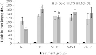
Hepatic TCHOL was increased (P < 0.05) in CDC compared to NC. Intervention with orlistat or VA diet decreased the cholesterol concentration relative to CDC (Fig. 5.3). However, this decrease was only significant in orlistat treated group (P < 0.05). Hepatic TG was also modulated by VA diet while HDL-C was not significantly impacted.
Figure 5.3.

Effect of VA supplemented diet/orlistat on lipid components measured in liver tissue. Values are the mean ± SD (n = 4–6). HDL-C, high density lipoprotein cholesterol; TCHOL, total cholesterol; TG, triacylglycerol; aP < 0.05 vs. NC; bP < 0.05 vs. CDC.
3.5. Effect of VA supplemented diet/orlistat on ALT and AST activities
3.5.1. Serum
Compared with the CDC, 5% and 15% VA diets respectively lowered serum ALT (41.35% and 27.13%) and AST (17.09% and 43.21%) activities (Fig. 6.1). Oral orlistat was found to raise ALT activity over CDC by 15.29%.
Figure 6.1.

Effect of VA supplemented diet/orlistat on serum amino transaminase activities. Values are the mean ± SD (n = 4–6); SALT = serum alanine amino transaminase; SAST = serum asparate amino transaminase; bP < 0.05 vs. CDC.
3.5.2. Liver tissue
The observed change in hepatic ALT activity was the opposite of that in serum. Four-week feeding of VA diets at 5% and 15% respectively increased ALT activity by 47.52% and 22.95% (P < 0.05). Oral orlistat also had no effect on hepatic ALT activity. Hepatic AST activity was not significantly changed at end of the study (Fig. 6.2).
Figure 6.2.

Effect of VA supplemented diet/orlistat on liver enzyme activities measured in liver tissue. values are the mean ± SD (n = 4–6); LALT = liver alanine amino transaminase; LAST = liver aspartate amino transaminase; bP < 0.05 vs. CDC.
3.6. Effect of VA supplemented diet/orlistat on adipose tissue histology
Photomicrographs of the histology of white adipose tissue of the control and treated rats at the end of the study are shown in Fig. 7 (Plates 1–5). The histology of WAT showed numerous adipocytes tightly packed and clumped together in the obese rats (Plate 2), compared to the NCs which showed normal adipocytes distribution and cells of regular sizes (Plate 1). However, 15% VA supplemented diet indicated histology similar to the NCs, suggesting inhibition of the hyperplasic growth of the adipocytes (Plate 5). The effect of orlistat and 5% VA supplemented diet were mild on the histological architecture of the adipose tissue (Plates 3 and 4). These observations of the adipose tissue histology correlate to % body weight results.
Figure 7.

Photomicrographs of white adipose tissue histology (400×) of rats at end of the study. Numerous deposits of adipocytes tightly packed and clumped together of plate 2, indicate hyperplasic obesity in untreated obese rats, compared to the NCs with normal adipocytes distribution and cells of regular sizes (Plate 1). Obese rats fed VA supplemented diet at 15% indicate histology similar to NC, suggesting an inhibition of the hyperplasic growth of the adipocytes (Plate 5). The effect of orlistat and 5% supplemented feeding were mild on the histological architecture of the adipose tissue (Plates 3 and 4).
4. Discussion
The potential of VA leaves as medication for dietary obesity was studied. Cafeteria diet, so-named by Kumar et al. (2011) compounded with processed and fast foods up to 45–54% of main course diet fed for 6 weeks, raised animals’ body weight by 25–45% above rats fed pellets only. On the basis of this, obesity model was successfully produced in conformity with the definition of Pi-Sunyer (1991): weight >20% above optimal, taking normal control rats as the optimum. Intervention with VA supplemented diet down regulated the animals’ body weights consistent with our earlier report (Atangwho et al., 2007a) and other researchers (Igile et al., 1995), though these earlier studies were carried out in normal animal subjects. Despite the characteristic bitter taste of VA leaves, food intake was not affected. An earlier investigation with broilers had also shown that feeding VA leaves as a substitute to maize-based diet up to 300 g/kg did not affect food intake and efficiency (Aregheore et al., 1998). This null effect on food intake, and the successive decrease in body weight gain, which became statistically significant only about 10 days after commencement of supplemented feeding; and continued to the end of experiment, strengthens the believe that weight reduction was indeed a consequence of the ingested VA leaves.
Measurement of adipose tissue weight has been used as a valid index in obesity related studies. For the first time, the present study showed a significant reduction in total body fat by feeding VA supplemented diet. WAT weight reduction appears as valid mechanism by which many anti-obesity medicinal plants exert their effects (Xia et al., 2010; Kumar et al., 2011; Kim et al., 2011). Although, we do not understand for now, how this WAT weight reduction is achieved; it is plausible that VA with reported hormone modulatory effect in diabetes, a metabolic condition akin to obesity, may also influence the actions of the several adipose tissue hormones such as the adiponectins. This is however, a subject for further research. The intermediate action of orlistat on WAT weight was expected, since orlistat exerts its action by sparing fat absorption in the GIT, rather than direct effect on adipose tissues (Ellrichmann et al., 2008). A longer period is required for overt effect on adipose cells, which probably explains why soon after withdrawal from clinical treatment with orlistat, weight rebounds. In this light alone, VA may be preferred to orlistat as a management for obesity.
Glucose intolerance, the link between obesity and diabetes (Kannel et al., 1979; Akiyama et al., 1996) was observed in the CDCs (46.26% above normal threshold). This agrees with Akiyama et al. (1996) who in their work, reported increased blood glucose in mice made obese by feeding a high fat diet for 28 days. VA supplemented feeding was shown for the first time to restore this intolerant condition. In related studies chloroform and ethanol extracts of VA respectively lowered serum glucose in normoglycemic and diabetic rats (Gyang et al., 2004; Atangwho et al., 2007b). This implies that VA may possess some prophylactic action against diabetes mellitus, in dietary feeding – a subject for further research.
Serum TG and TCHOL levels were decreased in VA fed rats in agreement with a previous report (Adaramoye et al., 2008). Although in the earlier report, VA extract was given pharmacologically, it is clear that this can be reproduced using dietary protocols, the most tolerable route of therapy. As in our previous studies with diabetic subjects (Atangwho et al., 2007a), serum HDL-C was not impacted by VA supplemented feeding, probably requiring a longer period for redistribution of the high protein component of the molecule. Hepatic lipid profile result paralleled that in serum. Cafeteria diet produced fatty liver, evident in increased TG and TCHOL levels, similar to the obesity model of Kumar et al. (2011). Four-weeks feeding with VA supplemented diet, decreased lipids but to a lower extent compared to orlistat. In the brain cells where TCHOL was also high following the 6-week CD, 15% VA supplemented feeding produced attenuation effect on TCHOL similar to orlistat. V. amygdalina may therefore share some mechanism with orlistat in its anti-obesity effect.
The activities of ALT and AST were significantly raised in CD fed rats, indicating a hepatotoxic tendency, imposed by obesity. Kim et al. (2011) have also observed significant increase in serum ALT activity in animal model of obesity. Supplemented feeding with VA but not orlistat, restored the enzyme activities to levels even lower than normal control. This may be an indication that VA has added advantage of hepato-protection, when used to manage obesity. The hepato-protective effect of VA extract in alloxan-induced diabetic subjects was earlier reported in our laboratory (Atangwho et al., 2007c). Decrease in hepatic ALT activity with CD feeding, was also restored by supplemented VA feeding. The hepatic fat deposition could have disrupted the cell integrity thereby depleting the enzyme protein in the liver; hence the observed decrease in obese rats. It was only necessary that VA supplemented feeding which modulated the fatty liver, also reversed the seeming damage by restoring the hepatic enzyme activity.
Numerous deposits of tightly packed and clumped adipocytes were seen in obese WAT histology depicting as increase in number of adipocytes – hyperplasia. Adipose tissue development in young mice and rats is a combination of two phases in which the first involves stem cell differentiation and in the second phase, the differentiated small cells gradually fill up with triacylglycerol (MacKellar et al., 2010). The animals in this study may yet have been in the cell differentiation stage particularly that at onset of experiment, the animals were just about 3 weeks. Moreover, the entire duration of study was relatively short. However VA supplementation at 15% indicated histology similar to normal control, suggesting an inhibition of the hyperplasic growth of the adipocytes. This opined adipocyte growth inhibition by VA supplemented feeding may be similar to its reported action on certain tumor cell, through which it has found relevance in treatment of breast cancers (Izevbigie, 2003).
5. Conclusion
VA fed as percentage dietary replacement can effectively attenuate obesity of dietary origin, via modulation of body and adipose tissue weights, cellular architecture and lipid metabolism, without affecting food intake. Additionally, the potential risks of hepatotoxicity and intolerance in glucose are alongside ameliorated. Further research is however needed to elucidate detail mechanism(s) by which this action is exerted.
Authors’ contributions
I.J.A. and E.E.E. conceived the study, designed the protocol and coordinated the experiment. The animal feeding and laboratory procedures were carried out by D.E.U., while A.U.O. performed the histological procedures. M.Z.A., M.A. and I.J.A. made significant contributions in data analysis, data interpretation, writing of the manuscript and designing the illustrations. All authors approved the final manuscript.
Acknowledgements
The authors sincerely thank the Academy of Science for the Developing World (TWAS) and the Universiti Sains Malaysia (USM) for the award of the TWAS-USM Fellowship to Item Justin Atangwho. The fellowship was responsible for the completion of this work and its eventual publication in this form.
Footnotes
Peer review under responsibility of King Saud University.
Contributor Information
Item J. Atangwho, Email: aijustyno@yahoo.com, dratangwho@gmail.com.
Emmanuel E. Edet, Email: emmanueleedet@yahoo.com.
Daniel E. Uti, Email: danuti4000@yahoo.com.
Augustine U. Obi, Email: austinhealth@yahoo.com.
Mohd. Z. Asmawi, Email: amzaini@usm.my.
Mariam Ahmad, Email: mariam@usm.my.
References
- Adaramoye O.A., Akintayo O., Achem J., Fafunso M.A. Lipid-lowering effect of methanolic extract of Vernonia amygdalina leaves in rats fed on high cholesterol diet. Vasc. Health Risk Manag. 2008;4:235–241. doi: 10.2147/vhrm.2008.04.01.235. [DOI] [PMC free article] [PubMed] [Google Scholar]
- Akiyama T., Tachibana I., Shirohara H., Watanabe N., Otsuki M. High-fat hypercaloric diet induces obesity, glucose intolerance and hyperlipidemia in normal adult male Wister rats. Diabet. Res. Clin. Pract. 1996;31:27–31. doi: 10.1016/0168-8227(96)01205-3. [DOI] [PubMed] [Google Scholar]
- Aregheore E.M.K., Makkar H.P.S., Becker K. Feed value of some browse plants from the central zone of Delta State. Nigeria Nig. Trop. Sci. 1998;38:97–104. [Google Scholar]
- Atangwho I.J., Ebong P.E., Egbung G.E., Eteng M.U., Eyong E.U. Effect of Vernonia amygdalina Del. on liver function in alloxan-induced hyper glycemic rats. J. Pharm. Bioresour. 2007;4:25–31. [Google Scholar]
- Atangwho I.J., Ebong P.E., Eteng M.U., Eyong E.U., Obi A.U. Effects of Vernonia amygdalina Del. leaf on kidney function of diabetic rats. Int. J. Pharmacol. 2007;3:143–148. [Google Scholar]
- Atangwho I.J., Ebong P.E., Eyong M.U., Eteng M.U., Uboh F.E. Vernonia amygdalina Del.: a potential prophylactic Antidiabetic agent in lipids complication. Glob. J. Pure Appl. Sci. 2007;13:103–106. [Google Scholar]
- Ekpo A., Eseyin O.A., Ikpeme A.O., Edoho E.J. Studies on some biochemical effect of Vernonia amygdalina in rats. Asian J. Biochem. 2007;2:193–197. [Google Scholar]
- Ellrichmann M., Kapelle M., Ritter P.R., Holst J.J., Herzig K.H., Schmidt W.E., Schimitz F., Meier J.J. Orlistat inhibition of intestinal lipase accurately increase appetite and attenuate post prandial glucagons – like peptide – 7 (7–36) – amide-1 cholesytokinin and peptide yy concentrations. J. Clin. Endocrinol. Metabol. 2008;93:3995–3998. doi: 10.1210/jc.2008-0924. [DOI] [PubMed] [Google Scholar]
- Ezekwe C.I., Obidia O. Biochemical effect of Vernonia amygdalina on rats liver microsomes. Nig. J. Biochem. Mol. Biol. 2001;16:174S–179S. [Google Scholar]
- Farombi E.O., Owoeye O. Antioxidant and chemopreventive properties of Vernonia amygdalina and Garcinia biflavonoid. Int. J. Environ. Res. Public Health. 2011;8:2533–2555. doi: 10.3390/ijerph8062533. [DOI] [PMC free article] [PubMed] [Google Scholar]
- Gyang S.S., Nyam D.D., Sokomba E.N. Hypoglycemic activity of Vernonia amygdalina (chloroform extract) in normglycamic and alloxan-induced hyperglucemic rats. J. Pharm. Bioresour. 2004;1:61–66. [Google Scholar]
- Igile G.O., Oleszek W., Jurzysta M., Burda S., Fafunso M., Fasanmade A.A. Nutritional assesment of Vernonia amygdalina leaves in growing mice. J. Agric. Food Chem. 1995;43:2162–2166. [Google Scholar]
- Ijeh I.I., Ejike C.E.C.C. Current perspectives on the medicinal potentials of Vernonia amygdalina Del. J. Med. Plant Res. 2010;5:1051–1061. [Google Scholar]
- Izevbigie E.B. Discovery of water-soluble anticancer agent (edotide) from a vegetable found in Benin City. Nigeria Exp. Biol. Med. (Maywood N.J.) 2003;228:293–298. doi: 10.1177/153537020322800308. [DOI] [PubMed] [Google Scholar]
- Kannel W.B., Gordon T., Castelli W.P. Obesity, lipids and glucose intolerance. The Framingham Study 1. Am. J. Clin. Nutr. 1979;32:1238–1245. doi: 10.1093/ajcn/32.6.1238. [DOI] [PubMed] [Google Scholar]
- Kim H.J., Kang H.J., Seo J.Y., Lee C.H., Kim L., Kim J. Antiobesity effect of oil extract of Ginseng. J. Med. Food. 2011;14:573–583. doi: 10.1089/jmf.2010.1313. [DOI] [PubMed] [Google Scholar]
- Kumar S., Alagawadi K.R., Rao M.R. Effect of Argyreia speciosa root extract on cafeteria diet-induced obesity in rats. Ind. J. Pharmacol. 2011;43:163–167. doi: 10.4103/0253-7613.77353. [DOI] [PMC free article] [PubMed] [Google Scholar]
- MacKellar J., Cushman S.N., Periwal V. Waves of adipose tissue growth in genetically obese zucker fatty rat. PLos ONE. 2010;5:e8197. doi: 10.1371/journal.pone.0008197. [DOI] [PMC free article] [PubMed] [Google Scholar]
- NIH. Principles of laboratory animal care guidelines. National Institute of Health, Bethesda, MD, Revised 1985, NIH Publication No. 85–23.
- Pi-Sunyer F.X. Health implications of obesity. Am. J. Clin. Nutr. 1991;53:1595S–1603S. doi: 10.1093/ajcn/53.6.1595S. [DOI] [PubMed] [Google Scholar]
- Shrestha S., Bhattarai B.R., Lee K.H., Cho H. Mono- and disalicylic acid derivatives: PTP1B inhibitors as potential anti-obesity drugs. Bioorg. Med. Chem. 2007;15:6535–6548. doi: 10.1016/j.bmc.2007.07.010. [DOI] [PubMed] [Google Scholar]
- Thompson A.K., Minihane A.-M., William C.M. Trans fatty acids and weight gain. Int. J. Obes. 2011;35:315–324. doi: 10.1038/ijo.2010.141. [DOI] [PubMed] [Google Scholar]
- Ugwu C.E., Alumana E.O., Ezeanyika L.U.S. Comparative effect of the leaves of Gongronema latifolium and Vernonia amygdalina incorporated diet on the lipid profile of rats. Biochemistry. 2009;21:59–65. [Google Scholar]
- Ugwu C.E., Olajide J.E., Alumana E.O., Ezeanyika L.U.S. Comparative effect of the leaves of Vernonia amygdalina and Telfairia occidentalis incorporated diet on the lipid profile of rats. Afr. J. Biochem. Res. 2011;5:28–32. [Google Scholar]
- Xia D., Wu X., Yang Q., Gong J., Zhang Y. Anti-obesity and hypolipidemic effects of a functional formula containing Prumus mume in mice fed high-fat diet. Afr. J. Biotechnol. 2010;9:2463–2467. [Google Scholar]
- Zhou J., Keenan M.J., Losso J.N., Raggio A.M., Shen L., McCutecheon K.L., Tulley R.T., Blackman M.R., Martin R.J. Dietary whey protein decreases food intake and body fat in rats. Obesity. 2011;19:1568–1573. doi: 10.1038/oby.2011.14. [DOI] [PMC free article] [PubMed] [Google Scholar]


