Abstract
DNA double-strand break (DSB) repair is essential for maintenance of genome stability. Recent work has implicated a host of chromatin regulators in the DNA damage response, and although several functional roles have been defined, the mechanisms that control their recruitment to DNA lesions remain unclear. Here, we find that efficient DSB recruitment of the INO80, SWR-C, NuA4, SWI/SNF, and RSC enzymes is inhibited by the non-homologous end joining machinery, and that their recruitment is controlled by early steps of homologous recombination. Strikingly, we find no significant role for H2A.X phosphorylation (γH2AX) in the recruitment of chromatin regulators, but rather their recruitment coincides with reduced levels of γH2AX. Our work indicates that cell cycle position plays a key role in DNA repair pathway choice and that recruitment of chromatin regulators is tightly coupled to homologous recombination.
Introduction
Cell viability and genomic stability are frequently threatened by chromosomal DNA double strand breaks (DSBs). DSBs can be induced by endogenous free oxygen radicals, collapsed replication forks, or by exposure to DNA damaging agents, such as ionizing radiation (IR), UV light, and chemicals1. The failure or improper repair of DSBs can result in cell death or gross chromosomal changes, including deletions, translocations, and fusions that promote genome instability and tumorigenesis2. Consequently, cells have developed complex signaling networks that sense DSBs, arrest the cell cycle, and activate repair pathways.
Eukaryotic cells have evolved two major mechanisms that repair chromosomal DSBs, non-homologous end joining (NHEJ) and homologous recombination (HR). NHEJ is the predominant DSB repair mechanism in the G1 phase of the cell cycle, whereas HR predominates in the S and G2 phases 3–7. In the case of NHEJ, the broken DNA ends are recognized and bound by the Ku70/Ku80 heterodimer which subsequently recruits other factors to facilitate ligation of the ends 8–10. In contrast, DSB repair by HR relies on sequence homology from an undamaged sister chromatid or a homologous DNA sequence to use as a template for copying the missing information. The first step of HR involves extensive processing of the DSB such that the 5’ ends of the DNA duplex that flank the DSB are resected to generate long, 3' single-stranded tails 11. Notably, extensive processing of the DSB ends is inhibited in G1 phase cells by the Ku70/80 complex 7, and increased CDK activity at the G1/S boundary activates DSB processing during later cell cycle phases 4,5,12.
DSB processing regulates the differential recruitment of two functionally related, checkpoint kinases ATM and ATR (Tel1 and Mec1, respectively in budding yeast). ATM recruitment does not require extensive DSB processing, while recruitment of the ATR/ATRIP (scMec1/Ddc2) checkpoint kinase complex requires the binding of the single stranded binding protein RPA to the processed DNA 13,14. One of the most intensively studied targets for checkpoint kinases is the histone variant H2A.X, which is phosphorylated at a C-terminal serine residue (H2A S129 in yeast or H2A.X S139 in higher eukaryotes; termed γH2AX). The formation of γH2AX is one of the earliest events at a DSB, and this mark spreads over at least a megabase of chromatin adjacent to each DSB in mammalian cells, and up to 50 kb on each side of a DSB in budding yeast 15,16. Although γH2AX is not essential for the initial recruitment of DSB response factors, it plays a role in stabilizing the binding of checkpoint factors to DSB chromatin 17. Besides its role in the DNA damage checkpoint, γH2AX has also been proposed to recruit chromatin regulatory factors, namely the ATP-dependent chromatin remodeling complexes INO80 and SWR-C18,19. These results have established γH2AX as both a ubiquitous hallmark and regulator of the chromatin response to DSBs.
In budding yeast, the DSB recruitment of chromatin regulators has been monitored primarily in asynchronous cell populations, and thus it is unclear if these events are linked to NHEJ or HR. In order to investigate whether the chromatin response to DNA damage is defined by a specific DSB repair pathway, we induced a single DSB within yeast cells synchronized in either G1 or G2/M cell cycle phases, and chromatin immunoprecipitation (ChIP) assays were performed to follow recruitment of many chromatin regulators. We surprisingly find that subunits of the INO80, SWR-C, NuA4, SWI/SNF, and RSC enzymes are primarily recruited outside of G1 phase, with the key NHEJ factor Ku70 inhibiting the recruitment of each of these enzymes in G1 cells. Furthermore, we find that recruitment of all chromatin regulators requires DSB processing and the Rad51 recombinase. In contrast to previous reports, we find that γH2AX plays no significant role in the recruitment of chromatin regulators to DSBs in either G2/M or asynchronous cells, though our data do suggest that chromatin regulators may enhance γH2AX dynamics during the HR process.
Results
Recruitment of chromatin regulators is cell cycle regulated
We use an established yeast system that has proven invaluable for monitoring the DSB recruitment of repair factors and chromatin regulators by chromatin immunoprecipitation (ChIP) analyses. This system allows for a single, persistent DSB to be induced on chromosome III by galactose-dependent expression of the HO endonuclease in a yeast strain that lacks homologous donor sequences 20 (hmlΔ hmrΔ; Fig. 1a). To investigate whether recruitment of chromatin regulators might be linked to the NHEJ or HR repair pathways, cells were first synchronized in G1 phase with alpha-factor mating pheromone (αF), and then released into three different media conditions: (1) galactose and alpha factor to induce a DSB in G1 cells; (2) galactose and hydroxyurea to induce a DSB as cells exit G1 phase and arrest in S phase; and (3) galactose and fresh media to induce a DSB as cells exit G1 and subsequently arrest at the G2/M DNA damage checkpoint. Cell cycle arrest was confirmed by flow cytometry analysis (Supplementary Fig. S1a). In this initial study, we followed recruitment of the Arp5 subunit of the INO80 chromatin remodeling enzyme. Surprisingly, recruitment of Arp5 was very low in G1 cells and in cells arrested in S phase. In contrast, Arp5 recruitment was robust in cells that had received a DSB outside of G1 phase and accumulated at the G2/M cell cycle checkpoint (Fig. 1b). To further investigate these results, cells were arrested in either G1 phase with alpha factor or in G2/M with nocodazole, followed by galactose addition to induce a DSB. Initial cell cycle arrest was confirmed by flow cytometry (Supplementary Fig. S2a). Once again, recruitment of INO80, monitored by both Arp5 and the catalytic Ino80 subunit, was robust only when a DSB was induced in G2/M cells, with low levels of recruitment observed at a DSB induced in G1 cells (Fig. 1c and Supplementary Fig. S2c). Consistent with previous findings in asynchronous cultures 18,19,21, recruitment of INO80 in G2/M-arrested cultures as well as asynchronous cultures was observed within a 10 kb chromatin domain adjacent to the DSB, and recruitment continued for at least 4 hours after DSB formation (Fig. 1c and Supplementary Fig. S2g). Importantly, recruitment of the NHEJ factor yKu70 was also monitored, and in this case DSB recruitment was equal in both G1 and G2/M cells, similar to previous studies 7,12 (Supplementary Fig. S2f).
Figure 1. Cell cycle regulated recruitment of chromatin modifying enzymes to an induced DSB.
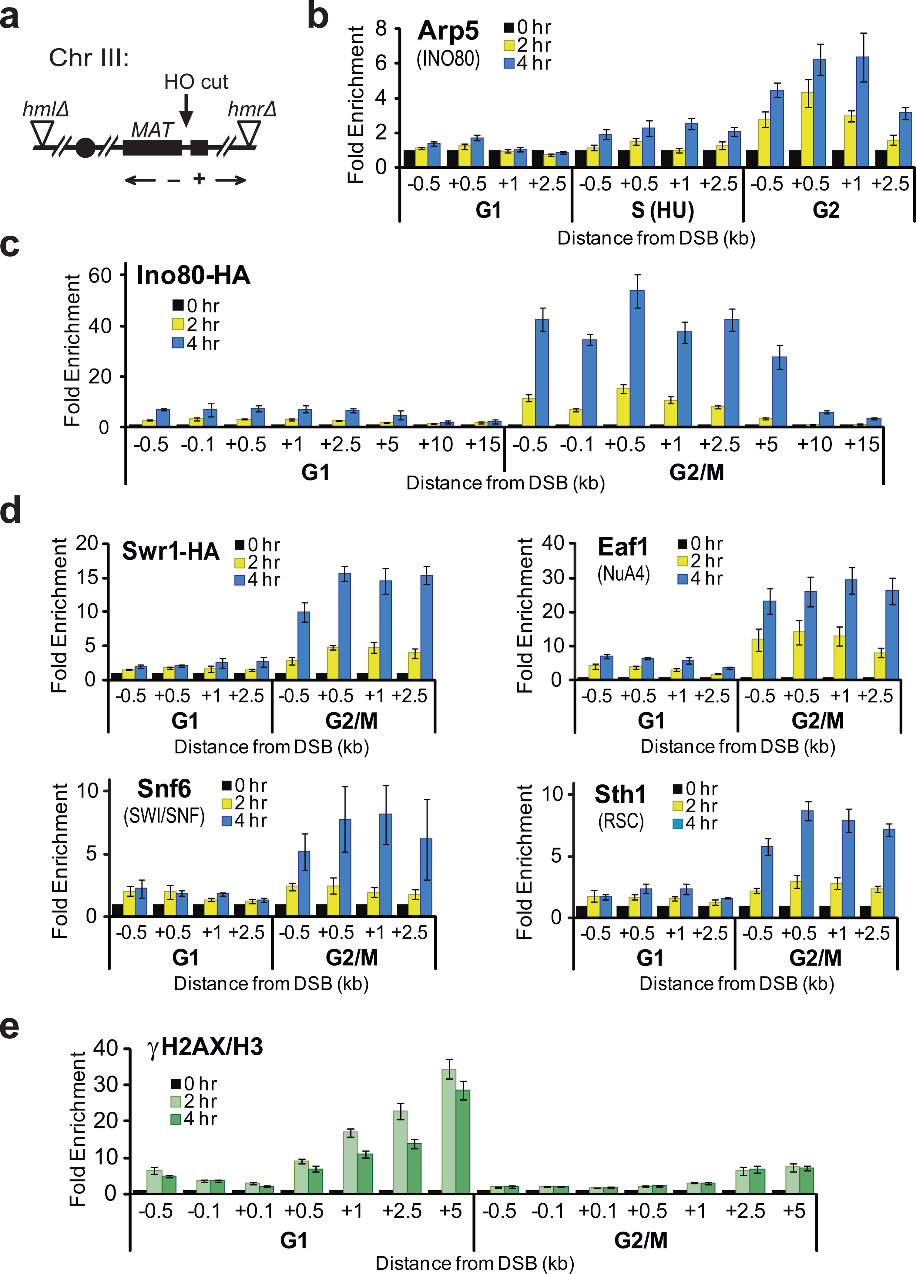
(a) Schematic of chromosome III of a donorless yeast strain harboring a galactose inducible HO endonuclease. Primers used during ChIP analyses are indicated according to their distance from the DSB, and designated with a “−” for centrosomal-proximal and “+” for centrosomal-distal.
(b) A wild-type, donorless strain was arrested in G1 using αF, and then split into three cultures: maintained in αF-arrest (“G1”), released into fresh media containing 0.2M hydroxyurea (“S(HU)”), or released into fresh media alone (“G2”). Galactose was also added at this time to induce a single DSB. Arp5 recruitment to areas surrounding the HO cut site was monitored by ChIP.
(c,d) A wild-type, donorless strain was arrested with either αF (“G1”) or nocodazole (“G2/M”), after which a DSB was induced by addition of galactose for the indicated times. Recruitment of various chromatin remodeling complexes to the DSB region was monitored by ChIP using antibodies to the indicated enzyme subunit. Fold enrichment reflects the %IP values normalized to the ACT1 locus, relative to time zero values.
(e) H2A phosphorylation (γH2AX) is cell cycle regulated. Cells were treated as in panel (c) and levels of γH2AX were determined and normalized to levels of histone H3 also determined by ChIP.
Data shown represent at least two biological replicates; error bars represent s.e.m.
Given the unanticipated result of differential recruitment of INO80 during the cell cycle, we conducted further ChIP assays to monitor recruitment of several other chromatin regulators, including subunits of the SWR-C, SWI/SNF, and RSC remodeling enzymes, as well as the NuA4 histone acetyltransferase complex. Interestingly, the recruitment of each of these chromatin regulators was much more robust outside of G1 phase, compared to G1 arrested cells (Fig. 1d and Supplementary Fig. S2d). These data suggest that there may be a common, cell-cycle regulated mechanism for recruitment of multiple chromatin regulators to a DSB.
γH2AX is dispensable for recruitment of chromatin regulators
Previous ChIP studies have indicated that formation of γH2AX is required for efficient DSB recruitment of INO80 and SWR-C within asynchronous cell populations 18,19. To understand how this mechanism interfaces with cell cycle regulation, we monitored the levels of γH2AX in chromatin surrounding DSBs formed in our experiments. Surprisingly, the levels of γH2AX surrounding the DSB were much lower in cells outside of G1 compared to those arrested in G1 (Fig. 1e, and Supplementary Figs. S1b and S3a). These contrasting levels of γH2AX are not due to changes in nucleosome density, as levels of H3 and H2B were reduced only ~2-fold in G2/M samples compared to G1, presumably due to DSB processing (Supplementary Fig. S3d, see below). Levels of γH2AX were also reduced in G2/M samples at early time points after DSB induction, when end processing has not progressed significantly (e.g. 30’), and when ChIP samples were processed in buffers containing 0.5M NaCl (Supplementary Fig. S3b,c). Furthermore, we monitored formation of γH2AX following exposure of synchronized cells to the DSB-inducing agent phleomycin and again observed more robust γH2AX formation in G1 cells compared to G2/M cells, indicating that these cell cycle differences are not unique to an HO-induced DSB (Supplementary Fig. S3e,f). The data suggest that γH2AX levels or dynamics may be dramatically altered in chromatin surrounding DSBs formed within G2/M cells. Furthermore, these results imply that the levels of γH2AX and chromatin regulators are anti-correlated, indicating that γH2AX may not be involved in their recruitment.
In order to re-examine the role of γH2AX in recruitment of chromatin regulators, we monitored recruitment events in two different strains that lack γH2AX: a strain expressing a derivative of H2A (bulk yeast H2A is the equivalent to mammalian H2A.X) where serine 129 has been changed to an alanine residue (hta1,2-S129A) 22, and a strain expressing a truncated H2A derivative that removes the final four C-terminal amino acids, including the Mec1/Tel1 phosphorylation site (hta1,2-S129Δ4) 23. Importantly, both of these strains exhibited similar sensitivity to the DNA damaging agent methyl methanesulfonate (MMS), as expected from previous studies 24 (Supplementary Fig. S4a). Surprisingly, neither H2A-S129A nor H2A-S129Δ4 reduced INO80 recruitment, irrespective of whether a DSB was induced in asynchronous or G2/M arrested cells (Fig. 2a and Supplementary Fig. S4c). Indeed, recruitment of the Arp5 subunit of INO80 was slightly elevated in the strain expressing the C-terminal H2A truncation (Fig. 2a). Similar results were found for Sth1 (RSC), Eaf1 (NuA4), Eaf3 (NuA4/Rpd3), Swi2 (SWI/SNF), and Yaf9 (NuA4/SWR-C) (Fig. 2b,c and Supplementary Fig. S4d,e). Interestingly, however, recruitment of the Snf6 subunit of SWI/SNF complex was markedly decreased in the absence the H2A C-terminus, even though its recruitment is not affected by the H2A-S129A substitution (compare Fig. 2b and Supplementary Fig. S4e), implicating other residues within the H2A C-terminus. Why recruitment of the Swi2 and Snf6 subunits of SWI/SNF differentially respond to the H2A C-terminus remains unclear. However, when taken together, the data indicate that γH2AX does not regulate recruitment of chromatin regulators.
Figure 2. γH2AX is not essential for recruitment of chromatin regulators to a DSB.
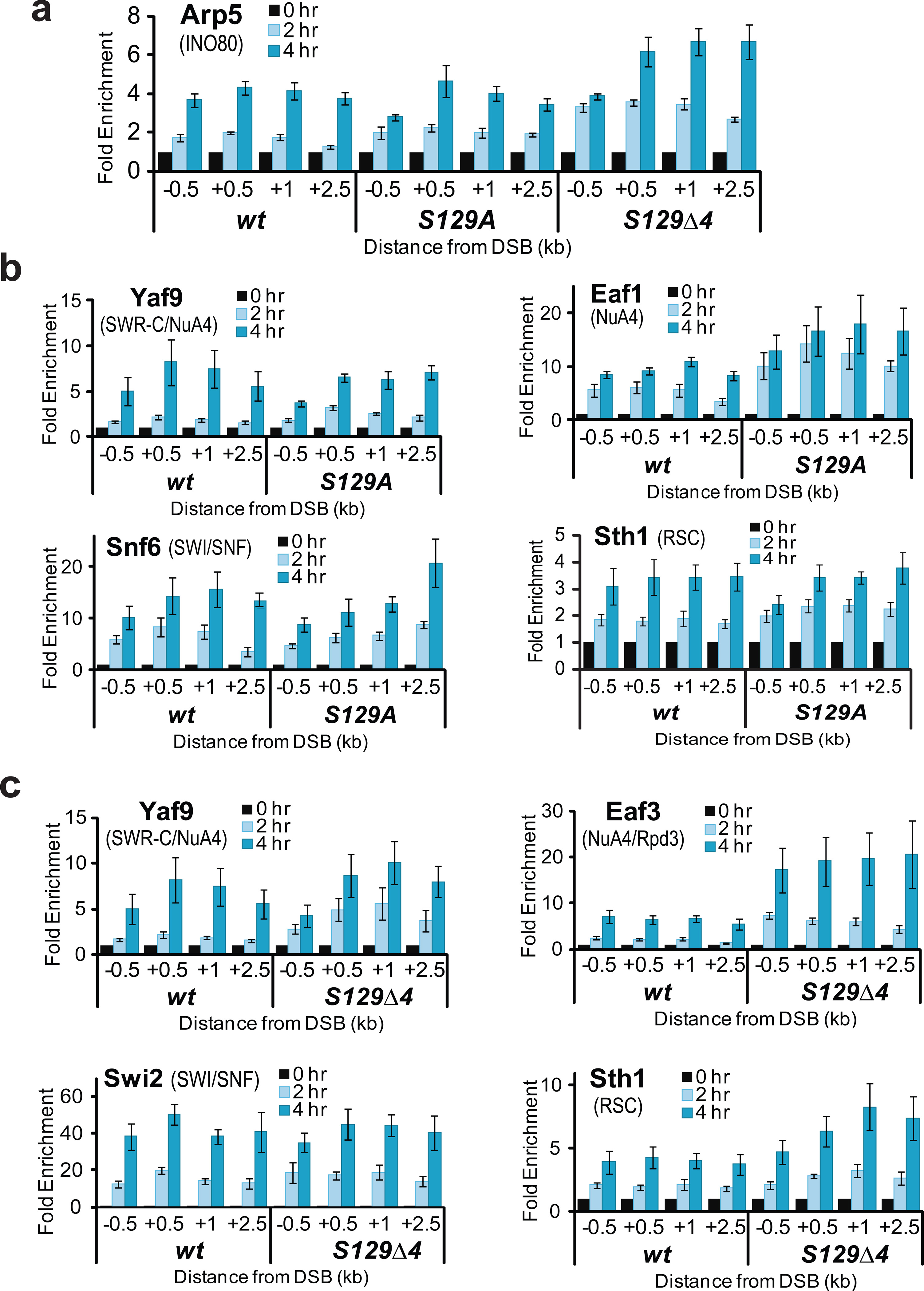
(a,b,c) Isogenic, donorless wild-type (wt), hta1,2-S129A (S129A), and hta1,2-S129Δ4 (S129Δ4) strains were arrested in G2/M using nocodazole, and analyzed by ChIP for recruitment of the indicated chromatin modifying enzyme subunits to the DSB region at the indicated time points after DSB induction. Data shown represent at least two biological replicates; error bars represent s.e.m.
Although our hta1,2-S129Δ4 and hta1,2-S129A alleles were created within the same JKM strain background as two previously published studies, our ChIP data are contradictory 18,19. We obtained the previously published hta1,2-S129* strain (also a four residue truncation; GA282418) and found that this strain shows similar sensitivity to DNA damaging agents as our hta1,2-129Δ4 strain (Supplementary Fig. S4a). However, strain GA2824 also exhibits an unexpected, severe growth defect in raffinose or lactate media, and liquid cultures arrested growth at low cell densities (e.g. OD600=0.4). Flow cytometry analysis also demonstrates that asynchronous cultures of GA2824 grown in raffinose media accumulate in the G1 phase of the cell cycle, and furthermore, this cell cycle distribution does not change following galactose addition to induce the HO endonuclease (Supplementary Fig. S4b). These growth defects precluded our ability to obtain high quality, reproducible ChIP data with this strain. Previous studies with the GA2824 strain have also indicated that γH2AX is required for efficient DSB processing 18. However, a recent study shows that γH2AX inhibits DSB processing 25, and we also observe increased levels of RPA adjacent to a DSB in our hta1,2-S129Δ4 and hta1,2-S129A strains (Supplementary Fig. S4f), consistent with a negative role for γH2AX in DSB processing. Since DSB processing is restricted in G1 cells, and INO80 and SWR-C are also poorly recruited in G1 cells, it seems likely that the aberrant slow growth and G1 accumulation phenotypes of the GA2824 strain were the cause of the previously observed defects in both DSB end processing and chromatin regulator recruitment, rather than a lack of γH2AX18.
Chromatin regulator recruitment requires DNA end processing
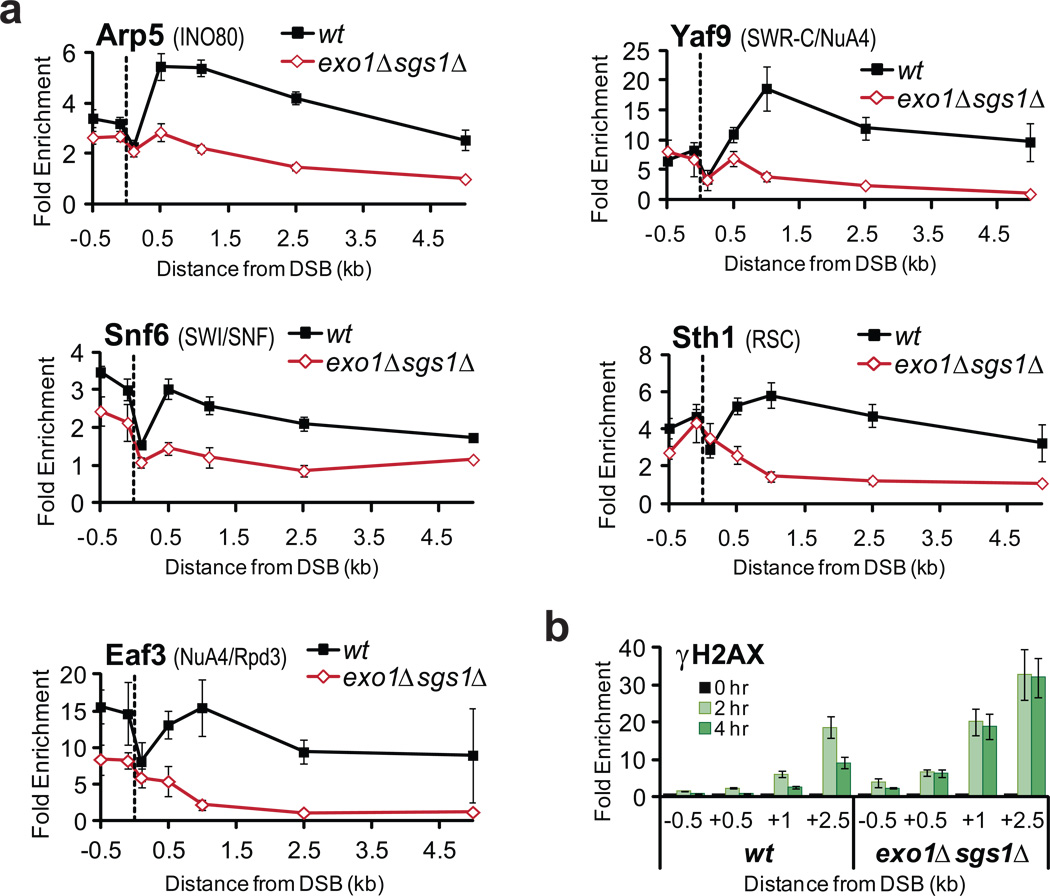
Our results indicate that the cell cycle regulation of the DSB response plays a key role in the recruitment of chromatin regulators to the DSB. Recruitment of chromatin factors outside of G1 coincides with the binding of RPA to ssDNA that is formed by the extensive processing of the DSB by the redundant Sgs1/Dna2 and Exo1 resection pathways 5,26–28 (Supplementary Fig. S2d). Notably, this relationship is also consistent with the poor recruitment of Arp5 (INO80) to a DSB induced within HU treated cells (Fig. 1b), as HU activates cell cycle checkpoints that inhibit DSB processing 29. To examine the possibility that DSB recruitment of chromatin regulators requires resection, we monitored recruitment in isogenic sgs1Δ, exo1Δ, and sgs1Δ exo1Δ strains. Strikingly, recruitment of Arp5 (INO80), Snf6 (SWI/SNF), Sth1 (RSC), Eaf3 (NuA4/Rpd3), or Yaf9 (NuA4/SWR-C) was greatly reduced in the sgs1Δ exo1Δ double mutant, with significant reductions occurring 500 bp distal to the DSB and reducing to basal levels by 2.5 kb distal (Fig. 3a). Importantly, the sgs1Δ exo1Δ strain showed a cell cycle profile identical to the wild-type strain (Supplementary Fig. S5a). Consistent with the functional redundancy of these processing enzymes, only a minor defect in recruitment of INO80 to a DSB was observed in exo1Δ or sgs1Δ single mutants (Supplementary Fig. S5b). Interestingly, γH2AX levels were also increased in the double mutant, most notably distal from the break (Fig. 3b). These results suggest that DSB processing is required for optimal recruitment of multiple chromatin regulators and that their recruitment correlates with decreased γH2AX levels.
Figure 3. DNA end processing is required for recruitment to a DSB.
Isogenic, donorless wild-type (wt) and exo1Δsgs1Δ strains were grown asynchronously, and analyzed by ChIP in the region surrounding the DSB for (a) recruitment of the indicated chromatin modifying enzyme subunits 4 hours after induction (dotted line indicates the HO cut site), and (b) levels of γH2AX at the indicated time points after induction. Data shown represent at least two biological replicates; error bars represent s.e.m.
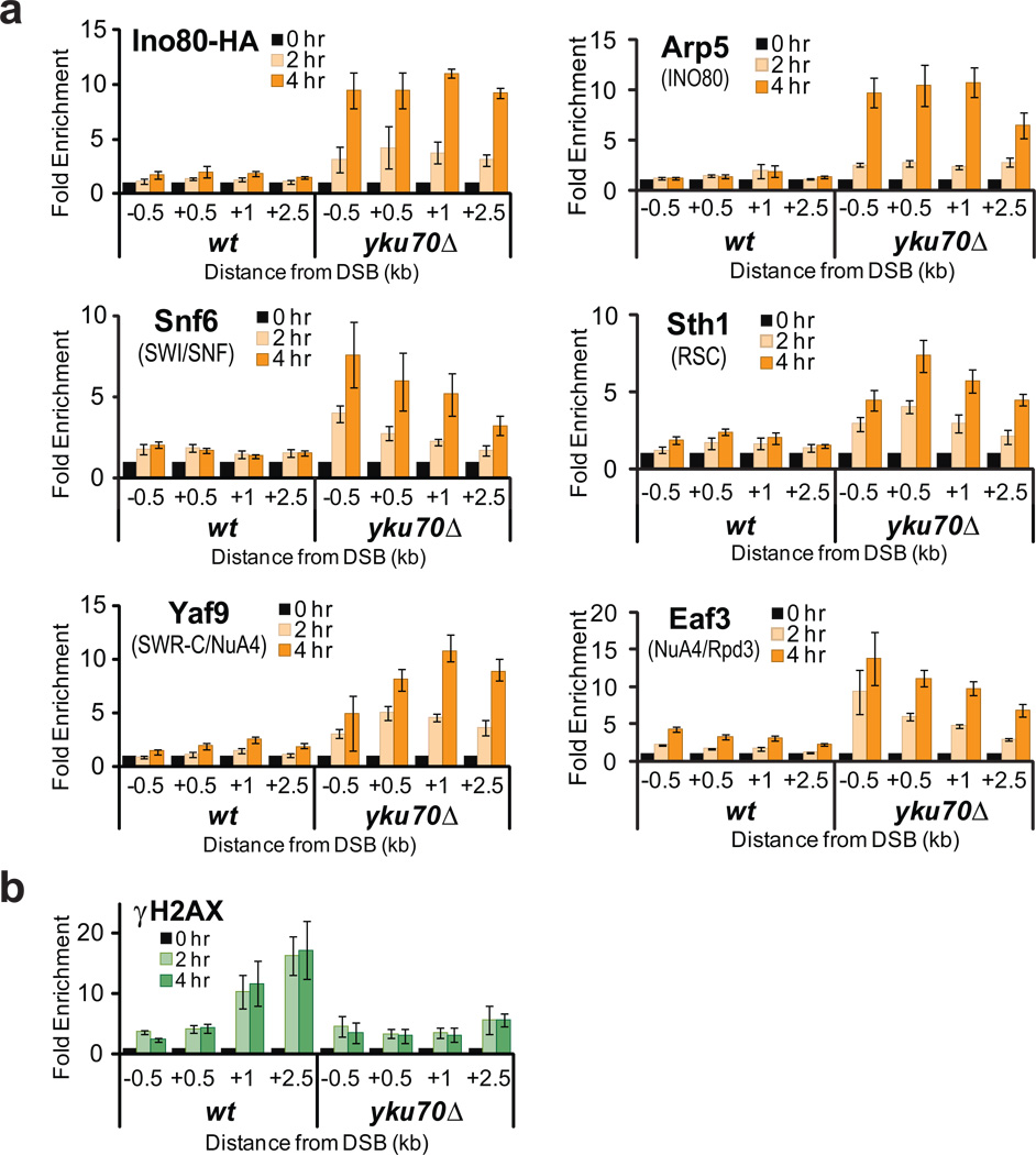
Previous studies have shown that the Ku70/80 heterodimer inhibits DSB processing in G1 cells, limiting DNA-end resection and promoting NHEJ 7. In addition, loss of Ku70 allows Rad52- and Rad51-dependent recombination events to occur efficiently in G1 arrested cells 30. To test whether the decreased recruitment of chromatin factors in G1 is due to limited DSB processing, an yku70Δ strain was arrested in G1, and ChIP assays were performed at an HO-induced DSB. Strikingly, recruitment of all chromatin regulators was restored to high levels in the G1-arrested yku70Δ cells (Fig. 4a). In contrast, γH2AX levels were reduced in the G1-arrested yku70Δ strain to levels previously seen in G2/M cultures, once again displaying an inverse relationship to the recruitment of chromatin regulators (Fig. 4b). These results suggest that yKu70/80 inhibits recruitment of chromatin regulators in G1 cells, and furthermore, that recruitment is independent of cell cycle position. Notably, inactivation of Ku70 does not restore the recruitment of INO80 and SWI/SNF in the absence of Sgs1 and Exo1 (Supplementary Fig. S5d), strongly supporting the idea that DSB processing facilitates the recruitment of chromatin regulatory factors at the DSB.
Figure 4. Ku inhibits recruitment of chromatin regulators in G1.
Isogenic, donorless wild-type (wt) and yku70Δ strains were arrested in G1with αF and analyzed by ChIP for (a) recruitment of the indicated chromatin modifying enzyme subunits, and (b) levels of γH2AX in the DSB region at the indicated time points after DSB induction. Data shown represent at least two biological replicates; error bars represent s.e.m.
Previous work has shown that the yeast Mre11/Rad50/Xrs2 (MRX) complex directs processing of the initial ~100 bp of DNA proximal to the DSB 28. Indeed, we find high levels of the ssDNA binding protein RPA proximal to the DSB in sgs1Δ exo1Δ cells (Supplementary Fig. S5c), a result consistent with MRX-dependent resection. To test whether MRX-dependent processing might be responsible for the residual recruitment of chromatin regulators observed in the sgs1Δ exo1Δ double mutant, Arp5 (INO80) recruitment was monitored in wild-type and mre11Δ cells arrested in G2/M. Note that a sgs1Δ exo1Δ mre11Δ strain was not constructed due to the expected growth defects of this strain. As shown in Supplementary figure S5e, Arp5 (INO80) recruitment was lost from DSB proximal chromatin in the absence of Mre11, and overall levels are similar to those found in G1 cells. Taken together, these data suggest a model in which DSB processing controls the recruitment of chromatin regulators, either through direct interactions with ssDNA or by subsequent events of the HR or the DNA damage checkpoint signaling pathway.
Rad51 is required for recruitment of chromatin regulators
Following DSB processing, the ssDNA ends are initially bound by the single strand DNA binding protein RPA, which is subsequently replaced by the key recombinase, Rad51 31. Therefore, we tested whether Rad51 is key for recruitment of chromatin regulators. Strikingly, recruitment of Arp5 (INO80) was nearly eliminated in the G2/M arrested rad51Δ strain, with a reduction to the two-fold recruitment level seen in G1 cells (Fig. 5a). Furthermore, recruitment of Swi2 (SWI/SNF), Snf6 (SWI/SNF), Sth1 (RSC), Eaf3 (NuA4/Rpd3), and Yaf9 (NuA4/SWR-C) were also nearly abolished (Fig. 5a and Supplementary Fig. S6a). In addition, levels of γH2AX were also increased in the absence of Rad51, consistent with one or more chromatin regulators controlling γH2AX dynamics (Fig. 5c). Importantly, Rad51 is not required for DSB processing or for establishing the checkpoint response 28,31,32 (Supplementary Fig. S6b), indicating that formation of ssDNA is not sufficient for recruitment of chromatin regulators or for decreased levels of γH2AX.
Figure 5. Rad51 is necessary for recruitment of chromatin regulators.
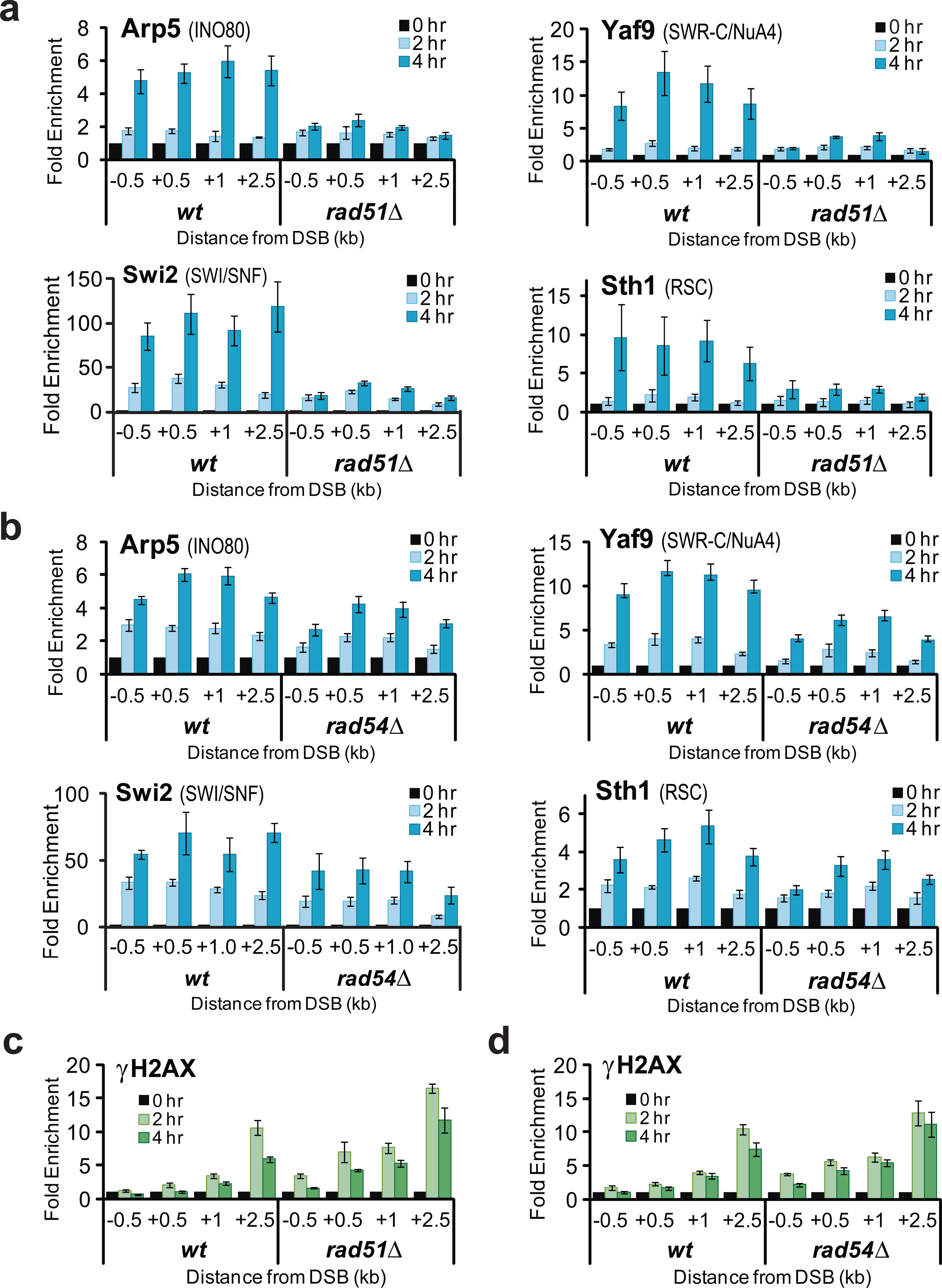
Isogenic, donorless wild-type (wt), and (a) rad51Δ or (b) rad54Δ strains were arrested in G2 with nocodazole, and analyzed by ChIP for recruitment of the indicated chromatin modifying enzyme subunits to the region the DSB region at the indicated time points after DSB induction. A dotted line indicates the HO cut site. (c,d) Levels of γH2AX determined by ChIP in the DSB region of experiments described in panels (a) and (b), repsectively. Data shown represent at least two biological replicates; error bars represent s.e.m.
Formation of the Rad51-ssDNA nucleoprotein filament plays a key role in the subsequent search and capture of a homologous DNA duplex. Rad51 also recruits Rad54 which is a member of the Snf2/Swi2 family of ATPases and exhibits weak chromatin remodeling activity in vitro 33. Rad54 plays at least two roles during HR. First, Rad54 has an ATP-independent activity that facilitates Rad51 loading onto DNA proximal to the DSB 34,35, and second, Rad54 plays an ATP-dependent role to convert the initial joint molecule into a stable, strand invasion product that can be extended by DNA polymerases 35,36. To investigate possible roles for Rad54 in the recruitment of chromatin regulators, a DSB was induced in G2/M arrested rad54Δ or rad54 K341R strains, the latter of which contains an allele of RAD54 that inactivates its ATPase activity 37. ChIP assays for Arp5 (INO80), Swi2 (SWI/SNF), Sth1 (RSC), Eaf3 (NuA4/Rpd3), or Yaf9 (NuA4/SWR-C), indicate a small but reproducible role for Rad54. In all of these cases, there is a defect in recruitment at locations proximal to the DSB, but less of an effect at distal locations (Fig. 5b and Supplementary Fig. S7a). However, very few recruitment defects were observed in the strain harboring the ATPase defective version of Rad54 (Supplementary Fig. S7b). In contrast, recruitment of Snf6 (SWI/SNF) was nearly abolished in the absence of Rad54, or when the ATPase activity of Rad54 was inactivated (Supplementary Fig. S7). Thus, recruitment of the Snf6 subunit of SWI/SNF is distinct from both the Swi2 catalytic subunit and from other chromatin regulators, requiring both Rad51 and the ATPase activity of Rad54.
Discussion
We have shown here that the recruitment of at least five chromatin regulatory enzymes – INO80, SWR-C, SWI/SNF, RSC, and NuA4 – are recruited to a DNA double strand break in a cell cycle-dependent manner, with at least five fold higher levels observed in G2/M cells compared to G1 cells. Our results indicate that recruitment is inhibited in G1 cells by the Ku70/80 complex, and that robust recruitment outside G1 is promoted by early steps of the HR process that lead to formation of the Rad51 nucleoprotein filament. Our data are not inconsistent with roles for chromatin regulators during NHEJ, as recruitment of chromatin regulators is low but not entirely abolished in G1. Indeed, recruitment of the INO80 complex in G1 cells is not affected by loss of Rad51, suggesting an independent mode for recruitment of chromatin regulators at this cell cycle phase. However, Rad51 is at least partially required for recruitment of INO80 in G1 cells that lack Ku70 (Supplementary Fig. S6c). Our data strongly support the view that chromatin regulators primarily impact repair events such as HR that occur following S phase. This idea is consistent with the known roles for the RSC, SWI/SNF, INO80, and SWR-C remodeling enzymes in distinct steps of HR and in cell cycle checkpoint control 22,38–41.
Whereas recruitment of human INO80 to DSBs does not require γH2AX 42, three studies previously implicated γH2AX in the recruitment of the yeast INO80 and SWR-C remodeling enzymes 18,19,21. This conclusion was based primarily on three results: (1) ChIP assays using a strain harboring an H2A C-terminal truncation allele (hta1,2-S129Δ4); (2) ChIP assays in a mec1 tel1 double mutant; and (3) co-purification of INO80 with γH2AX from cells treated with DNA damaging agents. Our current data indicate that the interpretation of previous ChIP data were confounded by the cell cycle distribution of the strain used: the previously employed H2A-S129Δ4 strain exhibits an aberrant accumulation of cells in G1, conditions where recruitment of INO80 and SWR-C is poor. Likewise, we envision that the lack of G2 checkpoint arrest in the mec1 tel1 double mutant led to a similar issue. Furthermore, we note that purification of INO80 in low salt buffers leads to co-purification of all four core histones 21, so it is expected that some level of γH2AX will be associated with INO80 under DNA damage conditions. It is perhaps not surprising that γH2AX does not control recruitment of INO80 or SWR-C, since their recruitment requires hours, whereas formation of the γH2AX domain occurs within minutes. In addition, loss of γH2AX leads to relatively little sensitivity to DNA damaging agents, whereas inactivation of INO80 causes a strong impact on the DNA damage response 18,24. Furthermore, as shown here and previously, the chromatin distribution of γH2AX and INO80 do not coincide at DSBs 16,19, and furthermore, our ChIP data show an anti-correlation in the recruitment of chromatin regulators and γH2AX signal. Although it remains a possibility that γH2AX may play a role within G1 cells, our data do not support a dominant role of γH2AX in recruitment of chromatin regulators to DSBs.
Previous studies in budding yeast have demonstrated high levels of γH2AX in both asynchronous cell populations and in cells arrested in G1. However, no previous studies have reported γH2AX levels for DSBs induced in cells synchronized outside of G1 phase. We were quite surprised to find a dramatic decrease in γH2AX levels for DSBs induced with G2/M cells. This decrease does not appear to be due solely to DSB processing, as γH2AX levels remain high in rad51Δ cells where DSB resection occurs normally. We envision that γH2AX may be established at normal levels in G2/M cells, but that it is subject to enhanced dynamics, likely catalyzed by one or more chromatin regulators. One possibility is that the low levels of γH2AX reflect dynamic exchange of H2A for H2A.Z by the SWR-C complex 22,43,44, although we find that γH2AX levels are not increased in a G2/M-arrested swr1Δ strain (Supplementary Fig. S8). Removal of γH2AX, particularly in G2/M cells, is consistent with recent studies, and our own data, indicating a negative impact of γH2AX on DSB processing 25 (Supplementary Fig. S4f).
We note that, while all the chromatin modifying complexes examined share a common set of requirements for recruitment to a DSB, only the Snf6 subunit of SWI/SNF shows a strong requirement for both the H2A C-terminus and Rad54, though neither was needed to recruit the Swi2 catalytic subunit. Our previous study indicated that Snf6 is uniquely associated with SWI/SNF 45, so it seems unlikely that it is recruited to DSBs by an independent mechanism. We favor a model in which the Snf6 subunit does not directly associate with nucleosomal DNA, and thus its crosslinking during the ChIP procedure is highly sensitive to small changes in SWI/SNF chromatin interactions.
How might Rad51 control the recruitment of chromatin regulators? One simple possibility is that the Rad51-ssDNA filament functions as an assembly platform for chromatin regulators and that each of these enzymes may directly interact with Rad51. Although there are no subunits held in common among all of the chromatin regulators that we have monitored, we note that each enzyme does harbor a member of the Actin-Related Protein (ARP) family that may provide a common interaction surface 46. Alternatively, it is possible that only a limited number of regulators interact directly with Rad51, and the activity of these few enzymes control recruitment of other complexes. Testing this latter possibility may require the development of strains where multiple essential regulators can be removed simultaneously.
Recently, studies in Drosophila and yeast have demonstrated that DSB processing and the Rad51 recombinase are required for long-range, intra-nuclear movements of DSB chromatin during the homology search step of HR 40,47, and to regulate repair of heterochromatic DSBs by HR 48. Interestingly, yeast studies indicate that the ATPase activity of Rad54 is also essential for enhanced DSB mobility 47. Although the INO80 enzyme has been suggested as a candidate factor that catalyzes DSB mobility 41, our recruitment data suggests that other remodeling enzymes may also contribute, as all enzymes tested require Rad51 for their recruitment to DSBs. How ATP-dependent remodeling might promote chromosome dynamics is currently unclear, though the orchestration of such a complex event may provide one explanation for why so many chromatin regulators are recruited to a DSB.
Methods
Yeast strains
All strains are derivatives of JKM139 or JKM17920, and were generated by the one-step PCR disruption method. Disruptions were confirmed by PCR analysis. Full genotypes are available in Supplementary Table S1. All strains were grown at 30 °C in lactate media (1% yeast extract, 2% bactopeptone, 2% lactic acid, 3% glycerol, 0.05% glucose, pH6.6) or YPR (1% yeast extract, 2% bactopeptone, 2% raffinose) pre-induction. HO induction was achieved by adding 2% galactose to each culture. Cultures were arrested in G1 using 1 µM alpha-factor (αF) treatment for 4 hours (bar1Δ strains). G2/M arrest was achieved using 30 µg/ml nocodazole for 4–5 hours. Arrests were confirmed by visual microscopy, followed by budding indices.
Chromatin Immunoprecipitation
Chromatin immunoprecipitations were performed with some modifications as previously described49: 50 mL of mid-log phase cells were cross-linked by adding 1% (final) formaldehyde for 15 minutes at room temperature, followed by neutralization with 150 mM (final) glycine for 5 minutes. Cell pellets were washed twice in cold TBS (50 mM Tris-Cl pH 7.5, 150 mM NaCl), and then resuspended in cold 400 µl FA-lysis buffer (50 mM HEPES-KOH pH 7.5, 140 mM NaCl, 1 mM EDTA, 1% Triton X-100, 0.1% sodium deoxycholate) plus 1× fresh “cOmplete” protease inhibitor cocktail (PIC; Roche). Cells were lysed with an equal volume of glass beads at 4 °C. After glass bead removal, samples were sonicated to shear DNA to an average size of 500 bp. An additional 1 mL FA-lysis buffer plus PIC was added and the chromatin lysate was purified by centrifugation at 14000 rpm for a total of 1.5 hours at 4 °C.
For most immunoprecipitations (IPs), 100–200 µl of the purified chromatin lysate was diluted up to 400ul with FA-lysis buffer plus PIC, and 1–2 µl antibody added. For SWI/SNF IPs, 1% (final) sarkosyl was also added. For high salt γH2AX IPs, FA-lysis buffer was replaced by FA-500 buffer (50 mM HEPES-KOH pH7.5, 500 mM NaCl, 1 mM EDTA, 1% Triton X-100, 0.1% sodium deoxycholate) All IPs were incubated overnight at 4 °C, followed by incubation with 15 µl equilibrated sepharose protein A beads (50% slurry; Rockland) for 2 hours at 4 °C. Pelleted beads were washed at room temperature, for 5–10 minutes each, sequentially with FA-lysis buffer (except high salt γH2AX IPs), FA-500 buffer, LiCl wash buffer (10 mM Tris-Cl pH 8.0, 250 mM LiCl, 1 mM EDTA, 0.5% NP-40, 0.5% sodium deoxycholate), and TE (10 mM Tris-Cl pH 7.5, 1 mM EDTA), followed by elution in Elution buffer (50 mM Tris-Cl pH 7.5, 10mM EDTA, 1% sodium dodecylsulfate) shaking for 10 minutes at 65 °C. For input samples, 10 µl purified chromatin lysate was diluted in 450 µl TE. All samples (IPs and inputs) were treated with proteinase K (0.2 mg/mL final) at 42 °C for 2 hours, cross-links reversed by incubation at 65 °C for ≥ 5 hours, and purified by phenol-chloroform extraction and ethanol precipitation. Input samples were diluted 20× over IP samples during DNA purification.
The IP and input DNA was analyzed by quantitative real-time PCR with iTaq SYBR Green Supermix with ROX (Biorad). Primer sequences are available in Supplementary Table S2. Fold enrichment represents the ratio of recovered DNA to input DNA of the break region, normalized to the same ratio obtained for the ACT1 ORF. These ratios were additionally normalized to pre-induction (0hr) values and corrected for DSB induction. Percent IP (for anti-RPA only) represents the ratio of IP DNA to input DNA corrected for dilution, and is not normalized to a control region, because those values approached zero. Error bars indicate standard error of the mean from at least 2 independent biological replicas and four PCR reactions.
Western blotting
Whole cell extracts were prepared by trichloroacetic acid precipitation and proteins were separated by sodium dodecyl sulfate-polyacrylamide electrophoresis in 18% acrylamide gels. Samples were blotted onto polyvinylidene difluoride membranes and probed with antibodies using standard methods.
Antibodies
Rabbit polyclonal antibodies to HA tag (ab9110), Arp5 (ab12099), Yaf9 (ab4468), Eaf3 (ab4467), and H2A-S129phos (γH2AX; ab15083) are commercially available from Abcam. Anti-H2B (39237) is available from Active Motif. Anti-Snf6 and anti-Swi2, Anti-RPA, Anti-Sth1, Anti-Eaf1, and Anti-Ku antibodies were kind gifts from J. Reese (Pennsylvania State University), V. Borde (Institut Curie), B. Cairns (University of Utah), J. Cote (Laval University Cancer Research Center), and S.E. Lee (University of Texas Health Science Center at San Antonio), respectively.
Flow cytometry
Approximately 1 mL of mid-log phase (~1–2 ×10⁷) cells were collected per sample, washed in water, fixed in 100% ethanol, and incubated at 4 °C rocking overnight. After fixation, cells were again washed in water, resuspended in 50 mM Tris pH 8.0, containing 200 µg/mL RNase A and incubated at 37 °C for 2–4 hours. Samples were then pelleted and resuspended in 50 mM Tris pH 7.5 containing 2 mg/mL Proteinase K and incubated at 50 °C for 30–60 minutes, followed by resuspension in 500 µl FACS buffer (200 mM Tris pH 7.5, 200 mM NaCl, 78 mM MgCl2). Approximately 100 µl of each sample was then incubated for 10 minutes at room temperature with 1 mL Sytox solution (50 mM Tris pH7.5, 1 µM Sytox Green (Molecular Probes S-7020)) and sonicated gently for approximately 30 seconds directly before analysis on a BD FACSCalibur flow cytometer. Data analysis and preparation completed with FlowJo.
Supplementary Material
Acknowledgements
We thank G. Ira for exo1 and sgs1 deletion strains; S. Gasser for providing strain GA2824; J. Cote for his H2A S129* strain; J. Reese, V. Borde, B. Cairns, J. Cote, and S.E. Lee for providing antibodies; and members of the Peterson lab for helpful discussion. This work was supported by a grant from the NIH (GM54096).
Footnotes
Author Contributions
G.B. designed and performed all experiments, and assisted in manuscript preparation; M.P-C. and C.L.P. played an equal role in project overview, data interpretation, and manuscript preparation.
Competing Financial Interests
The authors declare no competing financial interests.
References
- 1.Peterson C, Côté J. Cellular machineries for chromosomal DNA repair. Genes Dev. 2004;18:602–616. doi: 10.1101/gad.1182704. [DOI] [PubMed] [Google Scholar]
- 2.Khanna KK, Jackson SP. DNA double-strand breaks: signaling, repair and the cancer connection. Nat. Genet. 2001;27:247–254. doi: 10.1038/85798. [DOI] [PubMed] [Google Scholar]
- 3.Moore JK, Haber JE. Cell cycle and genetic requirements of two pathways of nonhomologous end-joining repair of double-strand breaks in Saccharomyces cerevisiae. Mol. Cell. Biol. 1996;16:2164. doi: 10.1128/mcb.16.5.2164. [DOI] [PMC free article] [PubMed] [Google Scholar]
- 4.Aylon Y, Liefshitz B, Kupiec M. The CDK regulates repair of double-strand breaks by homologous recombination during the cell cycle. EMBO J. 2004;23:4868–4875. doi: 10.1038/sj.emboj.7600469. [DOI] [PMC free article] [PubMed] [Google Scholar]
- 5.Ira G, et al. DNA end resection, homologous recombination and DNA damage checkpoint activation require CDK1. Nature. 2004;431:1011–1017. doi: 10.1038/nature02964. [DOI] [PMC free article] [PubMed] [Google Scholar]
- 6.Barlow JH, Lisby M, Rothstein R. Differential regulation of the cellular response to DNA double-strand breaks in G1. Mol. Cell. 2008;30:73–85. doi: 10.1016/j.molcel.2008.01.016. [DOI] [PMC free article] [PubMed] [Google Scholar]
- 7.Clerici M, Mantiero D, Guerini I, Lucchini G, Longhese MP. The Yku70–Yku80 complex contributes to regulate double-strand break processing and checkpoint activation during the cell cycle. EMBO Rep. 2008;9:810. doi: 10.1038/embor.2008.121. [DOI] [PMC free article] [PubMed] [Google Scholar]
- 8.Downs JA, Jackson SP. A means to a DNA end: the many roles of Ku. Nat. Rev. Mol. Cell Biol. 2004;5:367–378. doi: 10.1038/nrm1367. [DOI] [PubMed] [Google Scholar]
- 9.Wu D, Topper LM, Wilson TE. Recruitment and dissociation of nonhomologous end joining proteins at a DNA double-strand break in Saccharomyces cerevisiae. Genetics. 2008;178:1237. doi: 10.1534/genetics.107.083535. [DOI] [PMC free article] [PubMed] [Google Scholar]
- 10.Lieber MR. The mechanism of double-strand DNA break repair by the nonhomologous DNA end-joining pathway. Annu. Rev. Biochem. 2010;79:181–211. doi: 10.1146/annurev.biochem.052308.093131. [DOI] [PMC free article] [PubMed] [Google Scholar]
- 11.Bernstein KA, Rothstein R. At loose ends: resecting a double-strand break. Cell. 2009;137:807–810. doi: 10.1016/j.cell.2009.05.007. [DOI] [PMC free article] [PubMed] [Google Scholar]
- 12.Zhang Y, Shim EY, Davis M, Lee SE. Regulation of repair choice: Cdk1 suppresses recruitment of end joining factors at DNA breaks. DNA Repair. 2009;8:1235–1241. doi: 10.1016/j.dnarep.2009.07.007. [DOI] [PMC free article] [PubMed] [Google Scholar]
- 13.Schleker T, Nagai S, Gasser SM. Posttranslational modifications of repair factors and histones in the cellular response to stalled replication forks. DNA Repair. 2009;8:1089–1100. doi: 10.1016/j.dnarep.2009.04.010. [DOI] [PubMed] [Google Scholar]
- 14.Polo SE, Jackson SP. Dynamics of DNA damage response proteins at DNA breaks: a focus on protein modifications. Genes Dev. 2011;25:409–433. doi: 10.1101/gad.2021311. [DOI] [PMC free article] [PubMed] [Google Scholar]
- 15.Rogakou EP, Boone C, Redon C, Bonner WM. Megabase chromatin domains involved in DNA double-strand breaks in vivo. J. Cell Biol. 1999;146:905–916. doi: 10.1083/jcb.146.5.905. [DOI] [PMC free article] [PubMed] [Google Scholar]
- 16.Shroff R, et al. Distribution and dynamics of chromatin modification induced by a defined DNA double-strand break. Curr. Biol. 2004;14:1703–1711. doi: 10.1016/j.cub.2004.09.047. [DOI] [PMC free article] [PubMed] [Google Scholar]
- 17.Celeste A, et al. Histone H2AX phosphorylation is dispensable for the initial recognition of DNA breaks. Nat. Cell Biol. 2003;5:675–679. doi: 10.1038/ncb1004. [DOI] [PubMed] [Google Scholar]
- 18.Van Attikum H, Fritsch O, Hohn B, Gasser SM. Recruitment of the INO80 complex by H2A phosphorylation links ATP-dependent chromatin remodeling with DNA double-strand break repair. Cell. 2004;119:777–788. doi: 10.1016/j.cell.2004.11.033. [DOI] [PubMed] [Google Scholar]
- 19.Van Attikum H, Fritsch O, Gasser SM. Distinct roles for SWR1 and INO80 chromatin remodeling complexes at chromosomal double-strand breaks. EMBO J. 2007;26:4113. doi: 10.1038/sj.emboj.7601835. [DOI] [PMC free article] [PubMed] [Google Scholar]
- 20.Lee SE, et al. Saccharomyces Ku70, Mre11/Rad50, and RPA Proteins Regulate Adaptation to G2/M Arrest after DNA Damage. Cell. 1998;94:399–409. doi: 10.1016/s0092-8674(00)81482-8. [DOI] [PubMed] [Google Scholar]
- 21.Morrison AJ, et al. INO80 and γ-H2AX Interaction Links ATP-Dependent Chromatin Remodeling to DNA Damage Repair. Cell. 2004;119:767–775. doi: 10.1016/j.cell.2004.11.037. [DOI] [PubMed] [Google Scholar]
- 22.Papamichos-Chronakis M, Krebs JE, Peterson CL. Interplay between Ino80 and Swr1 chromatin remodeling enzymes regulates cell cycle checkpoint adaptation in response to DNA damage. Genes Dev. 2006;20:2437–2449. doi: 10.1101/gad.1440206. [DOI] [PMC free article] [PubMed] [Google Scholar]
- 23.Downs JA, et al. Binding of chromatin-modifying activities to phosphorylated histone H2A at DNA damage sites. Mol. Cell. 2004;16:979–990. doi: 10.1016/j.molcel.2004.12.003. [DOI] [PubMed] [Google Scholar]
- 24.Downs Ja, Lowndes NF, Jackson SP. A role for Saccharomyces cerevisiae histone H2A in DNA repair. Nature. 2000;408:1001–1004. doi: 10.1038/35050000. [DOI] [PubMed] [Google Scholar]
- 25.Chen X, et al. The Fun30 nucleosome remodeller promotes resection of DNA double-strand break ends. Nature. 2012;489:576–580. doi: 10.1038/nature11355. [DOI] [PMC free article] [PubMed] [Google Scholar]
- 26.Gravel S, Chapman JR, Magill C, Jackson SP. DNA helicases Sgs1 and BLM promote DNA double-strand break resection. Genes Dev. 2008;22:2767. doi: 10.1101/gad.503108. [DOI] [PMC free article] [PubMed] [Google Scholar]
- 27.Mimitou EP, Symington LS. Sae2, Exo1 and Sgs1 collaborate in DNA double-strand break processing. Nature. 2008;455:770–774. doi: 10.1038/nature07312. [DOI] [PMC free article] [PubMed] [Google Scholar]
- 28.Zhu Z, Chung W, Shim EY, Lee SE, Ira G. Sgs1 helicase and two nucleases Dna2 and Exo1 resect DNA double-strand break ends. Cell. 2008;134:981–994. doi: 10.1016/j.cell.2008.08.037. [DOI] [PMC free article] [PubMed] [Google Scholar]
- 29.Alabert C, Bianco JN, Pasero P. Differential regulation of homologous recombination at DNA breaks and replication forks by the Mrc1 branch of the S-phase checkpoint. EMBO J. 2009;28:1131–1141. doi: 10.1038/emboj.2009.75. [DOI] [PMC free article] [PubMed] [Google Scholar]
- 30.Trovesi C, Falcettoni M, Lucchini G, Clerici M, Longhese MP. Distinct Cdk1 requirements during single-strand annealing, noncrossover, and crossover recombination. PLoS Genet. 2011;7:e1002263. doi: 10.1371/journal.pgen.1002263. [DOI] [PMC free article] [PubMed] [Google Scholar]
- 31.Symington LS, Gautier J. Double-strand break end resection and repair pathway choice. Annu. Rev. Genet. 2011;45:247–271. doi: 10.1146/annurev-genet-110410-132435. [DOI] [PubMed] [Google Scholar]
- 32.Sugawara N, et al. DNA structure-dependent requirements for yeast RAD genes in gene conversion. Nature. 1995;373:84–86. doi: 10.1038/373084a0. [DOI] [PubMed] [Google Scholar]
- 33.Ceballos SJ, Heyer W-D. Functions of the Snf2/Swi2 family Rad54 motor protein in homologous recombination. Biochim. Biophys. Acta. 2011;1809:509–523. doi: 10.1016/j.bbagrm.2011.06.006. [DOI] [PMC free article] [PubMed] [Google Scholar]
- 34.Wolner B, et al. Recruitment of the recombinational repair machinery to a DNA double-strand break in yeast. Mol. Cell. 2003;12:221–232. doi: 10.1016/s1097-2765(03)00242-9. [DOI] [PubMed] [Google Scholar]
- 35.Wolner B, Peterson CL. ATP-dependent and ATP-independent roles for the Rad54 chromatin remodeling enzyme during recombinational repair of a DNA double strand break. J. Biol. Chem. 2005;280:10855–10860. doi: 10.1074/jbc.M414388200. [DOI] [PubMed] [Google Scholar]
- 36.Sugawara N, Wang X, Haber JE. In vivo roles of Rad52, Rad54, and Rad55 proteins in Rad51-mediated recombination. Mol. Cell. 2003;12:209–219. doi: 10.1016/s1097-2765(03)00269-7. [DOI] [PubMed] [Google Scholar]
- 37.Petukhova G, Van Komen S, Vergano S, Klein H, Sung P. Yeast Rad54 promotes Rad51-dependent homologous DNA pairing via ATP hydrolysis-driven change in DNA double helix conformation. J. Biol. Chem. 1999;274:29453–29462. doi: 10.1074/jbc.274.41.29453. [DOI] [PubMed] [Google Scholar]
- 38.Chai B, Huang J, Cairns BR, Laurent BC. Distinct roles for the RSC and Swi/Snf ATP-dependent chromatin remodelers in DNA double-strand break repair. Genes Dev. 2005;19:1656–1661. doi: 10.1101/gad.1273105. [DOI] [PMC free article] [PubMed] [Google Scholar]
- 39.Shim EY, et al. RSC mobilizes nucleosomes to improve accessibility of repair machinery to the damaged chromatin. Mol. Cell. Biol. 2007;27:1602–1613. doi: 10.1128/MCB.01956-06. [DOI] [PMC free article] [PubMed] [Google Scholar]
- 40.Miné-Hattab J, Rothstein R. Increased chromosome mobility facilitates homology search during recombination. Nat. Cell Biol. 2012;14:510–517. doi: 10.1038/ncb2472. [DOI] [PubMed] [Google Scholar]
- 41.Neumann FR, et al. Targeted INO80 enhances subnuclear chromatin movement and ectopic homologous recombination. Genes Dev. 2012;26:369–383. doi: 10.1101/gad.176156.111. [DOI] [PMC free article] [PubMed] [Google Scholar]
- 42.Kashiwaba S-I, Kitahashi K. The mammalian INO80 complex is recruited to DNA damage sites in an ARP8 dependent manner. Biochemical and bbophysical Research Communications. 2010;402:619–625. doi: 10.1016/j.bbrc.2010.10.066. [DOI] [PubMed] [Google Scholar]
- 43.Mizuguchi G, et al. ATP-driven exchange of histone H2AZ variant catalyzed by SWR1 chromatin remodeling complex. Science. 2004;303:343. doi: 10.1126/science.1090701. [DOI] [PubMed] [Google Scholar]
- 44.Luk E, et al. Stepwise Histone Replacement by SWR1 Requires Dual Activation with Histone H2A.Z and Canonical Nucleosome. Cell. 2010;143:725–736. doi: 10.1016/j.cell.2010.10.019. [DOI] [PMC free article] [PubMed] [Google Scholar]
- 45.Peterson CL, Dingwall A, Scott MP. Five SWI/SNF gene products are components of a large multisubunit complex required for transcriptional enhancement. Proc. Natl. Acad. Sci. U. S. A. 1994;91:2905–2908. doi: 10.1073/pnas.91.8.2905. [DOI] [PMC free article] [PubMed] [Google Scholar]
- 46.Boyer LA, Peterson CL. Actin-related proteins (Arps): conformational switches for chromatin-remodeling machines? Bioessays. 2000;22:666–672. doi: 10.1002/1521-1878(200007)22:7<666::AID-BIES9>3.0.CO;2-Y. [DOI] [PubMed] [Google Scholar]
- 47.Dion V, Kalck V, Horigome C, Towbin BD, Gasser SM. Increased mobility of double-strand breaks requires Mec1, Rad9 and the homologous recombination machinery. Nat. Cell Biol. 2012;14:502–509. doi: 10.1038/ncb2465. [DOI] [PubMed] [Google Scholar]
- 48.Chiolo I, et al. Double-Strand Breaks in Heterochromatin Move Outside of a Dynamic HP1a Domain to Complete Recombinational Repair. Cell. 2011;144:732–744. doi: 10.1016/j.cell.2011.02.012. [DOI] [PMC free article] [PubMed] [Google Scholar]
- 49.Papamichos-Chronakis M, Petrakis T, Ktistaki E, Topalidou I, Tzamarias D. Cti6, a PHD domain protein, bridges the Cyc8-Tup1 corepressor and the SAGA coactivator to overcome repression at GAL1. Mol. Cell. 2002;9:1297–1305. doi: 10.1016/s1097-2765(02)00545-2. [DOI] [PubMed] [Google Scholar]
Associated Data
This section collects any data citations, data availability statements, or supplementary materials included in this article.


