Abstract
Importance
The FKBP5 gene product regulates glucocorticoid receptor (GR) sensitivity and hypothalamic-pituitary-adrenal axis functioning and has been associated with many stress-related psychiatric disorders. The study of intermediate phenotypes, such as emotion-processing biases and their neural substrates, provides a way to clarify the mechanisms by which FKBP5 dysregulation mediates risk for psychiatric disorders.
Objective
To examine whether allelic variations for a putatively functional single-nucleotide polymorphism associated with FKBP5 gene regulation (rs1360780) would relate differentially to attention bias for threat. this was measured through behavioral response on a dot probe task and hippocampal activation during task performance. Morphologic substrates of differential hippocampal response were also measured.
Design
Cross-sectional study conducted from 2010 to 2012 examining associations between genotype, behavioral response, and neural response (using functional magnetic resonance imaging [fMRI]) on the dot probe; voxel-based morphometry and global and local shape analyses were used to measure structural differences in hippocampi between genotype groups.
Setting
Participants were recruited from primary care clinics of a publicly funded hospital in Atlanta, Georgia.
Participants
An African American cohort of adults (N=103) was separated into 2 groups by genotype: one genotype group included carriers of the rs1360780 T allele, which has been associated with increased risk for posttraumatic stress disorder and affective disorders; the other group did not carry this allele. Behavioral data included both sexes (N=103); the MRI cohort (n=36) included only women.
Main Outcome Measures
Behavioral and fMRI (blood oxygen level–dependent) response, voxel-based morphometry, and shape analyses.
Results
Carriers of the rs1360780 T allele showed an attention bias toward threat compared with individuals with-out this allele (F1,90=5.19, P=.02). Carriers of this allele demonstrated corresponding increases in hippocampal activation and differences in morphology; global and local shape analyses revealed alterations in hippocampal shape for TT/TC compared with CC genotype groups.
Conclusion
Genetic variants of FKBP5 may be associated with risk for stress-related psychiatric disorders via differential effects on hippocampal structure and function, resulting in altered attention response to perceived threat.
In the past decade, FKBP5 (OMIM 602623) has emerged as a promising genetic candidate for investigations of vulnerability to mood and anxiety disorders.1 The FKBP5 gene encodes for the protein FK506 binding protein 5–51, which regulates sensitivity of the glucocorticoid receptor (GR). The GR is a critical component of the body's stress response system and a primary binding site for cortisol, a corticosteroid released during acute and chronic stress. The FKBP5 protein interacts with heat shock protein 90 to reduce GR affinity, modulating cortisol activity and preventing translocation of the GR complex to the nucleus.2 Through this process, FKBP5 appears to decrease GR sensitivity (or increase GR resistance) to cortisol following stress. Dysregulated GR signaling is common in affective disorders1,3 and may be mediated by FKBP5 gene expression. Polymorphisms in FKBP5 have been associated with posttraumatic stress disorder (PTSD),4–10 depression,11 depressive episode recurrence,12 and bipolar disorder.13 Notably, associations between FKBP5 polymorphisms and psychiatric disorders have been somewhat variable between white and African American populations.6,14
Although these studies have outlined clear associations between FKBP5 and various affective disorders, mechanisms by which FKBP5 dysregulation may mediate symptom risk are still unknown. This relationship may be clarified through the study of intermediate phenotypes: discrete, heritable traits associated with genetic variance and disease phenotype.15 Given the role of FKBP5 in endocrine response to stress, this gene is likely to influence maladaptive behaviors that comprise intermediate phenotypes that underlie stress-related psychiatric disorders.16 One candidate intermediate phenotype is biased attention to emotionally valenced cues, which has been widely observed in anxiety17 and depression18 and linked to allelic variations in the serotonin transporter gene.19,20 There is some evidence to suggest the involvement of FKBP5 in this phenomenon. Multiple studies indicate that attention biases are closely associated with circulating levels of cortisol21–25 and that these biases may be influenced by cortisol administration.24 Given the role of the GR in modulating the body's response to cortisol, attention biases for emotional stimuli could be regulated through GR signaling and thus moderated by polymorphisms in genes that influence the GR receptor complex, such as FKBP5.
Furthermore, emerging evidence suggests that attention biases may be associated with alterations in brain function among individuals who hold increased genetic risk for psychiatric disorders. For example, Thomason and colleagues26 found that children who carried specific allelic variants of the serotonin transporter gene (related to depressive vulnerability) demonstrated an attention bias toward fearful facial expressions and increased blood oxygen level–dependent (BOLD) signal in parietal and paralimbic regions compared with participants without this allele.
For FKBP5 in particular, the hippocampus is an attractive target for brain-behavior phenotype investigations given that this region has been associated with high GR density27 and FKBP5 expression; furthermore, psychiatric disorders linked to FKBP5 genetic status, particularly PTSD, have been associated with abnormal hippocampal volume28–30 and function.31,32 The hippocampus also has been implicated in the processing of context during acquisition of conditioned fear responses and, in concert with prefrontal regions,33 participates in the appraisal of potential threat in new situations.34–36 These hippocampal functions may be mediated partly by FKBP5 given the potential role of this gene in the stress response.
Thus, the goal of the present study was to investigate the relationship between an FKBP5 polymorphism and a candidate cognitive phenotype, attention bias for threat cues, in highly traumatized individuals, some of whom met criteria for Axis I psychiatric disorders. We selected a putatively functional37FKBP5 single-nucleotide polymorphism (SNP) that has been associated with anxious and depressive psychiatric disorders: rs1360780. Recent experiments37 have shown that the rs1360780 risk allele is associated with increased transcription of FKBP5 following stimulation by glucocorticoids, possibly by increasing binding of the risk-allele containing DNA sequence to TATA-box binding protein, a transcriptional enhancer. Increased FKBP5 messenger RNA transcription in peripheral blood cells immediately after trauma exposure has been associated with a higher risk for subsequent PTSD.38
Attention biases were characterized through behavioral and neural responses during functional magnetic resonance imaging (fMRI) in a subset of these individuals. We tested the hypothesis that risk allele carriers would demonstrate significantly greater attention bias for threat and increased neural response in the hippocampus compared with individuals without this allele. In addition, we conducted structural analyses within this region of interest (ROI) to characterize the neural substrates of differential task response between individuals with and without the risk allele.
METHODS
PARTICIPANTS
A total of 110 adult (aged 20–62 years) African American men and women (sex and race self-identified) were enrolled as part of a larger study investigating genetic risk for stress-related disorders; study procedures were approved by the institutional review boards of Emory University School of Medicine and Georgia State University. Participants were recruited from the general medical clinics of a publicly funded hospital, as detailed previously.4,39 Individuals were deemed eligible for participation if they could give informed consent and understand English, as determined by a study researcher. Participants were administered the Traumatic Events Inventory (TEI) to detail the frequency and type of traumatic experiences, the PTSD Symptom Scale (PSS)40 to measure the presence and frequency of current PTSD symptoms, and the Structured Clinical Interview for DSM-IV disorders41 to detect comorbid psychiatric disorders such as major depressive disorder (MDD) or bipolar disorder. Demographic and clinical characteristics of this sample are detailed in eTable 1 (http://www.jamapsych.com).
A subset of these participants (47 women) completed magnetic resonance imaging (MRI); data from these (all traumatized) individuals were included in a prior study of PTSD42 and were selected from the larger sample on the basis of inclusion/exclusion criteria for the present study. Exclusion criteria for this study included current psychotropic medication use; medical or physical conditions that preclude MRI scanning; a diagnosis of bipolar disorder, schizophrenia, or other psychotic disorder; medical conditions that contribute significantly to psychiatric symptoms; history of head injury or loss of consciousness for more than 5 minutes; and a history of neurologic illness.
DNA EXTRACTION, GENOTYPING, AND QUALITY CONTROL
Samples for DNA were extracted from saliva (DNAdvance extraction kit; Beckman Coulter Genomics) collected in vials (Oragene; DNA Genotek Inc) or from whole blood (E.Z.N.A. Blood DNA Midi Kit; Omega Bio-tek) collected in EDTA tubes. All DNA for genotyping was quantified (PicoGreen; Invitrogen Corp, or NanoDrop; Thermo Fisher Scientific Inc) and normalized to a concentration of 5 ng/μL. The DNA was plated into 384 plates at 10 ng and dried prior to performing the genotyping reactions.
For genotyping, 1 mL of whole blood or 500 μL of Oragene saliva was used for extraction. Genotyping of FKBP5 SNP rs1360780 was conducted (TaqMan SNP Genotyping Assay with TaqMan Genotyping Master Mix; Applied Biosystems Inc) and alleles were discerned (7900HT Fast Real-Time PCR System; Applied Biosystems Inc). Negative controls and within- and across-plate duplicates were used for quality control. All negative controls passed quality control. Forty percent of the samples were genotyped in duplicate with no genotype discordants. The TaqMan genotype call rate was 97%. Alleles were in Hardy-Weinberg equilibrium (African Americans only, P=.19).
The FKBP5 SNP rs1360780 was selected on the basis of prior studies37,43 with FKBP5 polymorphisms within this population. We tested for associations under a risk=T allele carrier model. Participants were categorized into 2 groups on the basis of genotype: individuals with 1 or 2 copies of the T allele, previously associated with risk of psychiatric disorders, were categorized in the risk group (TC/TT), whereas individuals without this allele (CC) were categorized in the resilient group.
ATTENTION BIAS (DOT PROBE) TASK DESCRIPTION
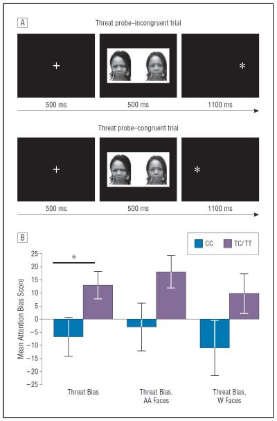
A dot probe task was used to measure attention biases for threat; this measure has reliably detected biases in prior studies17 of individuals with emotional disorders. Task procedures have been described44 (also detailed in eMethods), and the trial structure is illustrated in Figure 1A. Briefly, in each of 80 trials a central fixation cross appears for 500 ms, followed by a pair of face photographs (both of the same model) presented for 500 ms. Each pair comprises either 1 emotionally expressive (threatening/happy) and 1 neutral face or 2 neutral faces. After the offset of the faces, an asterisk replaces 1 of the faces for 1100 ms. Participants quickly indicate, with a forced-choice button press, whether the asterisk appeared on the left or right side of the screen. A modified version of this task, detailed elsewhere,42 was presented during neuroimaging.
Figure 1.
Attention bias (dot probe) task. A, Trial structure. Rows illustrate trials that were used to calculate threat bias and as functional magnetic resonance imaging contrast conditions. The top row displays trials in which the probe appears on the opposite side of the threatening expression (threat probe incongruent); the bottom row displays trials in which the probe appears on the same side of the threatening expression (threat probe congruent). B, Attention bias to threat as a function of FKBP5 genotype: TC/TT genotypes demonstrate attention bias toward threat, compared with CC genotype. Chart illustrates mean attention bias score (error bars indicate standard error of the mean), for threat faces (both races, combined) and separated by African American (AA) and white (W) race type, as a function of genotype group. *P<.03.
Emotion bias scores were first calculated by subtracting response time to emotion-congruent stimuli (probes that replace happy or angry/threatening pictures) from response time to emotion-incongruent stimuli (probes that replace neutral pictures); these scores were decomposed into threat bias and happy bias scores for all stimuli combined and separately for African American and white faces. Positive scores indicated attention capture by the threat or happy cue, and negative scores reflected attention avoidance of this cue.
STATISTICAL ANALYSIS OF BEHAVIORAL DATA
Univariate analyses of covariance were conducted to examine whether genotype status was related to attention bias for threatening faces in general (threat bias) or for threatening white (threat bias white) or African American (threat bias African American) faces after covarying for the presence of bipolar disorder, MDD, total trauma exposure (TEI total score), and PTSD symptoms (PSS total score). A similar analysis was conducted to examine potential associations with happy bias score. In addition, 3 separate factorial analyses of covariance were conducted to examine potential interactions of genotype, PTSD diagnosis, and trauma exposure (in childhood or adulthood) on threat bias score after covarying for depressive psychiatric disorders. A final analysis was conducted to compare bias scores for the CC, TC, and TT groups after covarying for psychiatric disorders. A threshold of P<.05 was used to determine statistical significance.
MRI ACQUISITION AND PREPROCESSING, fMRI DATA ANALYSIS
Scanning took place in a research-dedicated scanner (Siemens 3-Tesla; Siemens AG). A high-resolution T1-weighted structural scan was acquired using an MPRAGE sequence (176 slices, field of view [FOV]=256 mm cubic voxels; 1×1×1 mm slice; repetition time [TR]=2600 ms; echo time [TE]=3.02 ms; inversion time=900 ms; flip angle=8°). During task administration, a total of 26 contiguous echo-planar, T2-weighted images parallel to the anterior-posterior commissure line were acquired (TR=2530 ms; TE=30 ms; FOV=240 mm; 64×64 matrix; 3.75×3.75×4.0 mm voxel). Statistical Parametric Mapping, version 5 (SPM5, Wellcome Department of Neurology, London, England; http://www.fil.ion.ucl.ac.uk/spm/) was used for file conversion, image preprocessing, and fMRI statistical analyses. Functional images were slice-time corrected (with a high-pass filter applied) and realigned to the first image to correct for motion. The mean of the realigned undistorted images was then coregistered with the structural T1 volume, spatially normalized to standardized Montreal Neurological Institute (MNI) space45 based on the position of the anterior and posterior commissure and smoothed with an 8-mm full-width half-maximum gaussian kernel.
For each participant's data, a first-level, fixed-effects model was created with vectors for onset time of each condition, including threat/neutral, happy/neutral, and neutral/neutral trials; each trial included face-pair presentation and probe. Box-car functions using 1, −1 contrast conventions were used to indicate voxels that had a higher activation level for the primary contrast condition, which was activation to threat probe-incongruent trials (ie, the probe replaced the neutral face of a threat-neutral pair) minus threat probe-congruent trials (the probe replaced the threat face of a threat-neutral pair), the same events that were used to calculate threat bias score. Analysis using 2-tailed unpaired t tests was then conducted to examine between-group differences in FKBP5 genotype and BOLD signal change to threat within ROIs. Primary analyses were conducted with ROIs, and brainwide analyses were conducted for completeness. First, an uncorrected statistical threshold of P<.005 and an extent threshold of 5 or more voxels per cluster were applied to brainwide analyses. Region-of-interest analyses were conducted with masks of the parahippocampal gyrus created with the WFU PickAtlas Toolbox (Wake University School of Medicine)46; a P<.05 threshold, extent threshold of 5 or more voxels per cluster was used to determine statistical significance. A nonlinear transformation (http://bioimagesuite.yale.edu/mni2tal/index.aspx) was used to convert coordinates from MNI to Talairach space,47 and a Talairach daemon48 was used to localize anatomic coordinates of voxels associated with statistically significant patterns of BOLD activation.
VOXEL-BASED MORPHOMETRY ANALYSIS
With the brain extraction toolkit in the Oxford Centre for Functional MRI of the Brain (FMRIB) Software Library,49 all structural images were (1) skull-stripped (evaluated individually for errors), (2) segmented with FMRIB's automated segmentation tool,50 (3) aligned to MNI152 standard space using affine registration, and (4) underwent nonlinear registration using FMRIB's nonlinear image registration tool. Following alignment, a study-specific template was generated. The segmented images were then smoothed using an isotropic gaussian kernel with a sigma of 3 mm, and voxelwise general linear model was carried out using permutation-based, nonparametric testing51 to control for type I error. Statistical thresholding was set at uncorrected P<.001 for ROIs.
SHAPE ANALYSIS
Global shape analysis was performed using methods fully detailed in eTable 2. Following skull extraction, images were fed through a global shape analysis workflow that automatically extracted each brain into 56 distinct regions. Inhomogeneity correction of images was followed by affine registration, nonlinear registration to the Laboratory of Neuroimaging Probabilistic Brain Atlas (LPB40),52 and subsequent automatic volume parcellation, during which each voxel in the brain was labeled.53 The regional boundaries of the resulting labeled image were then converted into 56 distinct shapes,54 which underwent subsequent preprocessing, modeling, and analysis. A total of 5 shape metrics were used to perform group comparisons between the TC/TT and CC genotypes: volume, surface area, mean curvature, shape index, and curvedness. The resultant output of this analysis included sets of raw P values and false-discovery rate (FDR)-corrected P values.55 eTable 2 shows the standard intrinsic shape measures computed for each region.
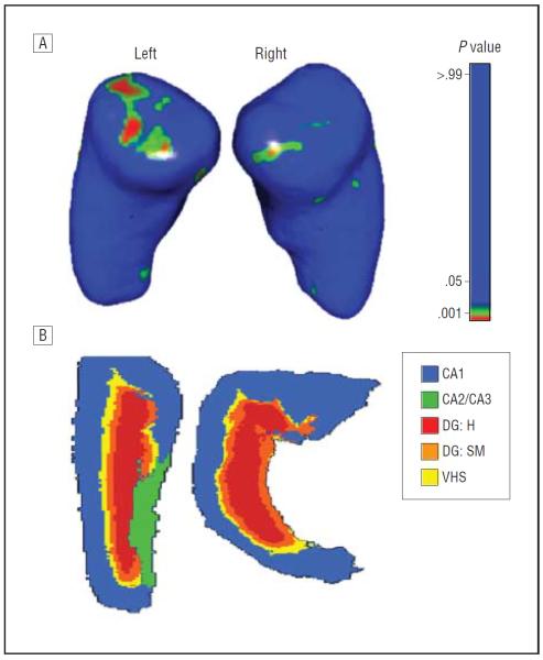
Local shape analyses of both hippocampi54 were conducted to identify the specific vertices/subregions that demonstrated significant radial distance and/or displacement vector field variations from the study-defined mean hippocampal shape template.56 In the local shape analysis protocol, the structural attributes and cortical measures are calculated per vertex on specific shape regions that are first coregistered across participants to establish homologous anatomic features before statistically analyzing them against various participant demographic, clinical, or phenotypic data.57 To control for multiple comparisons testing, FDR correction was applied. An atlas (Figure 2B) was used to identify anatomic locations of between-group differences.56
Figure 2.
FKBP5 polymorphism is associated with differences in hippocampal shape. A, Local shape analyses of the left and right hippocampus. Smaller P values (false-discovery rate corrected) reflect greater spatial displacement for TC/TT vs CC genotypes. B, Cross-sectional views of the 3-dimensional hippocampal atlas56 used for reference. CA indicatescornu ammonis; DG,dentate gyrus; H, hilum; SM, stratum moleculare; and VHS,vestigial hippocampal sulcus.
RESULTS
GROUP CHARACTERISTICS
Genotype data were not available for 5 participants because of insufficient quality of the DNA samples, leaving a total participant sample of 105. In this sample, participants reported experiencing a range of 0 to 10 traumatic experiences, with 7.7% of participants reporting no trauma exposure. Approximately 6.7% of participants met criteria for a lifetime history of bipolar disorder, 13.3% met the criteria for current MDD and, based on PSS items (in keeping with DSM-IV PTSD criteria), 41.2% met the criteria for PTSD. Within this sample, no significant differences (P>.05) emerged in demographic or clinical characteristics between CC and TC/TT genotype groups (eTable 1). Although significant differences were observed between genotype groups for psychotropic medication use (P=.006, χ2=7.51), no significant differences in threat or happy bias score were observed between participants who were or were not taking psychotropic medication (P>.05). Within the imaging cohort, age and level of current PTSD symptoms were similar for risk and resilient genotypes. However, because the incidence of trauma exposure differed significantly between groups (P<.05), this served as a covariate in subsequent analyses in addition to PTSD symptoms. A comparison of clinical and demographic characteristics between the entire sample and the MRI cohort revealed no significant differences in age, PTSD symptoms, trauma exposure, household monthly income, and educational level (all P>.05).
BEHAVIORAL DIFFERENCES IN ATTENTION BIAS TO THREAT BETWEEN FKBP5 (rs1360780) GENOTYPES
Two other participants were excluded from behavioral data analyses because of many skipped trials or inaccurate responses on trials (>20%), leaving a total of 103 participants with valid behavioral data. Univariate tests including trauma exposure and presence of PTSD, MDD, and bipolar disorder as covariates and 3 threat bias indices (threat bias score for the combined race types threat bias score for African American, or threat bias score for whitefaces) as dependent variables were conducted. After covarying for trauma exposure and the presence of PTSD, MDD, and bipolar disorder, risk allele (TC/TT) carriers demonstrated a statistically significant (F1,90=5.19, P=.02) bias toward threat (African American and white races combined: mean [SD] threat bias score, 13.1 [37.2]) compared with the CC genotype group (−6.8 [46.2]) (Figure 1B). These findings were verified with permutation testing, using PLINK software, version 1.07,58 to ensure that results did not depend on distributional assumptions; a total of 100 000 permutations were performed.
Separate univariate analyses revealed no significant genotype associations with happy bias (composite bias score or separated by face race; P>.05). Further univariate analyses (including the same covariates) revealed no significant genotype effects for threat bias for white faces (P=.10), although threat bias findings for African American faces approached statistical significance (F1,90=3.48, P=.06). Results from separate factorial analyses of covariance including psychiatric disorder covariates and threat bias score as the dependent variable indicated no significant interactions of genotype status and (1) childhood sexual or physical abuse (P=.85) or (2) adult trauma exposure (presence or absence, as reported in the TEI) (P=.44). A univariate analysis including genotype and PTSD status as factors did not reveal significant interactions of these variables on threat bias score (P=.69). Genotype contributed significantly to this model (P=.04), whereas PTSD status did not, although findings approached statistical significance (P=.17). A final factorial analysis of covariance revealed that, when CC, TC, and TT genotype groups were examined separately, no significant main effects were observed after covarying for psychiatric disorders (possibly because of limited sample size), though findings approached statistical significance (F1,90=2.47; P=.09); individuals in the CC genotype group demonstrated a slight bias away from threat (mean bias score,−6.78 [46.21]), whereas both the TC (13.25 [39.26]) and TT (12.71 [41.40]) groups demonstrated a bias toward threat.
fMRI DIFFERENCES IN HIPPOCAMPAL ACTIVATION TO THREAT BETWEEN FKBP5 GENOTYPES
Because of excessive motion (7) and/or brain parenchyma abnormalities (4), 11 participants were excluded from fMRI and MRI data analyses, leaving 36 participants (10 CC, 26 TC/TT); another (TC/TT) participant was excluded from fMRI (but not structural MRI) analyses, resulting from many skipped trials (>20%) on the task. Behavioral data from the cohort of participants who completed the fMRI version of the task (n=35) yielded no significant genotype effects on threat bias regardless of face race (P>.05). Correlations among attention bias scores from these 2 data sets are provided in the eResults.
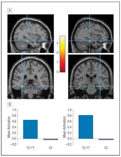
In a whole-brain analysis for the threat incongruent vs threat congruent trials contrast condition, participants with the TC/TT genotype demonstrated significantly greater BOLD signal in the right hippocampus and left parahippocampal gyrus compared with those having the CC genotype (uncorrected P<.005) (eTable 3 and Figure 3); these findings did not change significantly when hippocampal volume was entered as a covariate (eTable 4). The CC genotype, relative to TC/TT genotype, did not demonstrate greater BOLD signal in hippocampal regions (eTable 3).
Figure 3.
FKBP5 polymorphism is associated with differential hippocampal activation during attention to threat. A, Statistical parametric maps of left and right hippocampus activation during the processing of threat probe-incongruent vs threat probe-congruent faces in TC/TT>CC genotype. Activations are shown overlaid onto a canonical T1 magnetic resonance image. The colored bar represents t scores for activations. Maximally activated voxels from the left parahippocampal gyrus (x, y, z: −36, −35, −8) and right hippocampus (x, y, z: 36, −24, −12), P<.05 (small-volume correction, family-wise error). Data are reported using the coordinate system of Talairach and Tournoux. B, Genotype differences in averaged blood oxygen level–dependent signal (contrast time series extracted from 6-mm spherical regions of interest) to this contrast condition.
Subsequent ROI analyses confirmed these findings, with the TC/TT genotype group demonstrating increased activation in the right and left hippocampus relative to the CC group (P<.05) (eTable 3). The BOLD signal values were extracted from 6-mm spheres within these regions and entered into 2 separate, 3-level hierarchical regressions, with trauma incidence (TEI total; level 1), PTSD symptoms (PSS total; level 2), and genotype (level 3) serving as predictors of BOLD signal in these regions. Trauma exposure did not predict a significant amount of variance in BOLD signal within the right (β=−0.15, R2=0.02, P>.05) or left (β=−11, R2=0.01, P>.05) hippocampus; when added to this model, PTSD symptoms likewise did not predict a significant proportion of variance in BOLD signal within the right (β=0.24, R2=0.08, P>.05) or left hippocampus (β=0.03, R2=0.02, P>.05). However, when added in the third step of these models, FKBP5 genotype accounted for a significant amount of variance in BOLD signal for the right (β=−0.44, R2=0.28, P<.05) and left hippocampus (β=−0.46, R2=0.21, P<.05). There was no direct association between threat bias score and neural response within ROIs.
MORPHOLOGIC ANALYSIS OF DIFFERENCES IN HIPPOCAMPAL VOLUME AND SHAPE BETWEEN FKBP5 GENOTYPES
At the uncorrected P<.001 statistical threshold, voxel-based morphometry findings indicated no between-group differences in gray matter density of the hippocampus. The global shape analysis revealed some between-group differences in shape index of the left hippocampus (uncorrected P<.05). Additionally, local shape analysis results (Figure 2B) revealed significant between-group differences in displacement vector field measure in the left and right hippocampus (FDR corrected P<.001). Relative to the right hippocampus, a larger area of the left hippocampus displayed between-group spatial displacement vector metric differences, predominantly in the cornu ammonis 1 (CA1) region. The genotypic effect on hippocampal surface area was reduced (P>.05) when TEI score and/or hippocampal volume were included as covariates.
To ensure that these genotypic effects were not present in similar cortical and subcortical regions, these local shape analyses were conducted on the left and right insula and caudate. None of these regions showed statistically significant genotypic effects (P>.05).
COMMENT
The present study investigated associations among genotypic variants of a putatively functional FKBP5 SNP, rs1360780, and response to threat cues in an attention bias paradigm. In addition to investigating behavioral response, we examined corresponding differences in function and structure within the hippocampus, a brain region that appears to be affected by FKBP5 gene expression. Our findings from these parallel behavioral, functional, and structural neuroimaging data sets revealed that, relative to individuals without this allele, carriers of the T allele (which has been linked to incidence of psychiatric disorders37,43) demonstrated (1) an attention bias toward threat, (2) increased hippocampal activation to threat, and (3) differences in hippocampal shape.
Preferential attention processing of threat cues has purported relevance to the development and maintenance of pathologic anxiety.59 The findings from the present study document, for what we believe to be the first time, an association between FKBP5 polymorphism and threat bias. An increasing number of studies indicate that FKBP5 genetic variability is linked to behaviors that reflect atypical or dysregulated emotion processing, including aggression60 and suicidality.61 Furthermore, earlier findings4 reflected interactive effects of genetics and environmental stress (ie, childhood abuse), that is, the genetic effects were most pronounced in the presence of high levels of prior stress or trauma. Notably, we found that the relationship between FKBP5 allelic variance and attention bias for threat remained significant after controlling for variance associated with psychiatric disorders, as well as trauma exposure, and no interactive effects were observed among FKBP5 genotype, trauma exposure, and posttraumatic psychiatric disorders. Thus, the associations observed between FKBP5 genotypic variance and attention bias were not better accounted for by variance linked to environmental stress or psychiatric disorders. It is also possible that, when considering the relationship between FKBP5 genotype and attention bias for threat, the effects of maltreatment, exposure to traumatic events in adulthood, and a relatively high number of life stressors (eg, occupational stress, racial discrimination, bereavement, and divorce) may be additive and not interactive. Although the cross-sectional design of this study precludes definitive statements regarding risk, our findings suggest that FKBP5 risk allele carriers, compared with individuals without this allele, are more likely to demonstrate a heightened vigilance for ostensibly mild threat cues. An attentional preference for these mild threat cues, particularly among individuals with trait anxious characteristics, indicates increased susceptibility to psychiatric disorders under stressful conditions.
In this study, risk allele carriers (who are likely to demonstrate increased transcription of FKBP5 following stress exposure) also demonstrated heightened hippocampal activation in response to threat, specifically, to a condition that approximates threat bias. To our knowledge, no previous studies have examined associations between FKBP5 polymorphisms and hippocampal function, particularly response to threat. However, prior data16 identified FKBP5 as a putative modulator of physiologic stress response systems, suggesting that these associations merit attention. Our findings indicate that increased hippocampal responsiveness to threat cues was selectively associated with risk allele carrier status, implicating this gene in neural mechanisms of threat evaluation. Given the density of GRs in the hippocampus and the role of FKBP5 in regulating GR function, the increased hippocampal activation may represent, in part, FKBP5-mediated sensitization of neural systems to external threat signals. Risk allele carriers may have a lower threshold for perceiving threat in the environment compared with individuals without this allele, and thus may demonstrate heightened responsiveness in neural circuits mediating threat evaluation. Notably, earlier studies of pathologically anxious individuals have revealed enhanced activation in hippocampal regions,62 as well as frontal regions with limbic connections, during expression of attention biases.33,63 Thus, the heightened hippocampal response to mild threat cues observed in risk allele carriers could represent higher baseline responsiveness in neural networks engaged during threat evaluation in these individuals.
These functional differences also may be understood in the context of structural differences. Although no significant differences in hippocampal density were noted, risk allele carriers demonstrated differences in hippocampal shape compared with individuals without this allele, specifically in the CA1 region. Abnormalities in hippocampal structure, particularly the CA1 region,64,65 have been noted frequently in individuals with stress-related disorders.66,67 This region may be particularly susceptible to the effects of stress,68 and changes in CA1 plasticity could be partially mediated by FKBP5 mechanisms given previous observations69 of FKBP5 messenger RNA upregulation following stress exposure. Individuals within our study population had experienced a relatively high incidence of trauma exposure and psychosocial stress, which has been linked to suppressed neuronal proliferation in the hippocampus.70 These conditions are likely to interact with genetic vulnerability, and structural differences in the hippocampus may be particularly apparent among individuals who carry a risk genotype, one that has been associated37,43 with adverse behaviors and diagnostic outcomes.
In addition, there were no significant genotypic effects in other regions of the brain examined, providing evidence to support the specificity of FKBP5 genetic effects on the hippocampus. Thus, the structural differences observed here indicate a putative morphologic substrate for the differential response to threat observed between genotype groups. However, given that genotypic effects were no longer apparent after including trauma exposure in the statistical model, replication in a larger sample is needed to ascertain the robustness of these findings.
Other study limitations must be noted. First, because only female face stimuli were used in this version of the dot probe, it was impossible to investigate potential interactive effects of sex and attention biases. Similarly, the lack of white participants prohibited examination of stimulus×participant race interactions and their effects on attention biases. Also, our imaging cohort included only women, thus precluding investigation of sex-specific differences in hippocampal response or structure. In addition, some research71 suggests that FKBP5 may be a target of progesterone; unfortunately, endocrine and menstrual cycle data were unavailable in the present study to examine potential relationships with behavioral and neural response. These factors merit consideration in future investigations. Although hypothesis testing was limited to a particular ROI, the relatively small size of our imaging cohort (particularly the size of 1 genotype group) increases the likelihood of a type II error. Furthermore, mediational analyses would provide an ideal statistical approach for determining the indirect effects of hippocampal function and size on attention bias for threat; however, our limited sample size did not confer sufficient statistical power to detect these effects. Thus, these neuroimaging findings should be viewed as preliminary and warrant confirmation in genetic neuroimaging studies conducted with larger samples.
In summary, we observed that genotype status for a putatively functional FKBP5 SNP was associated with differential behavioral and hippocampal response during performance of an attention bias task. Structural differences in hippocampal formation served to further distinguish these genotype groups. Taken together, these data may represent behavioral and neural markers of FKBP5 function and merit further investigation in further studies of genetic risk for affective psychiatric disorders.
Acknowledgments
Funding/Support: This work was primarily supported by grants R01MH071537 (Dr Ressler) and F32MH095456 (Dr Fani) from the National Institutes of Mental Health. Support also was received from Emory and Grady Memorial Hospital General Clinical Research Center, grant M01RR00039 from the National Institutes of Health National Centers for Research Resources, and the Burroughs Wellcome Fund (Dr Ressler).
Footnotes
Conflict of Interest Disclosures: None reported.
Online-Only Material: The eTables, eMethods, and eResults are available at http://www.jamapsych.com.
Additional Contributions: Technical support was provided by Timothy Ely, BS; Kimberly Kerley, AA; Justine Phifer, BS; Asante Kamkwalala, BS; and Lauren Sands, MPH; Karin Mogg, PhD, and Brendan Bradley, PhD, assisted in developing the dot probe measure.
REFERENCES
- 1.Hauger RL, Olivares-Reyes JA, Dautzenberg FM, Lohr JB, Braun S, Oakley RH. Molecular and cell signaling targets for PTSD pathophysiology and pharmacotherapy. Neuropharmacology. 2012;62(2):705–714. doi: 10.1016/j.neuropharm.2011.11.007. [DOI] [PMC free article] [PubMed] [Google Scholar]
- 2.Denny WB, Valentine DL, Reynolds PD, Smith DF, Scammell JG. Squirrel monkey immunophilin FKBP51 is a potent inhibitor of glucocorticoid receptor binding. Endocrinology. 2000;141(11):4107–4113. doi: 10.1210/endo.141.11.7785. [DOI] [PubMed] [Google Scholar]
- 3.Anacker C, Zunszain PA, Carvalho LA, Pariante CM. The glucocorticoid receptor: pivot of depression and of antidepressant treatment? Psychoneuroendocrinology. 2011;36(3):415–425. doi: 10.1016/j.psyneuen.2010.03.007. [DOI] [PMC free article] [PubMed] [Google Scholar]
- 4.Binder EB, Bradley RG, Liu W, Epstein MP, Deveau TC, Mercer KB, Tang Y, Gillespie CF, Heim CM, Nemeroff CB, Schwartz AC, Cubells JF, Ressler KJ. Association of FKBP5 polymorphisms and childhood abuse with risk of post-traumatic stress disorder symptoms in adults. JAMA. 2008;299(11):1291–1305. doi: 10.1001/jama.299.11.1291. [DOI] [PMC free article] [PubMed] [Google Scholar]
- 5.Boscarino JA, Erlich PM, Hoffman SN, Rukstalis M, Stewart WF. Association of FKBP5, COMT and CHRNA5 polymorphisms with PTSD among outpatients at risk for PTSD. Psychiatry Res. 2011;188(1):173–174. doi: 10.1016/j.psychres.2011.03.002. [DOI] [PMC free article] [PubMed] [Google Scholar]
- 6.Xie P, Kranzler HR, Poling J, Stein MB, Anton RF, Farrer LA, Gelernter J. Interaction of FKBP5 with childhood adversity on risk for post-traumatic stress disorder. Neuropsychopharmacology. 2010;35(8):1684–1692. doi: 10.1038/npp.2010.37. [DOI] [PMC free article] [PubMed] [Google Scholar]
- 7.Sarapas C, Cai G, Bierer LM, Golier JA, Galea S, Ising M, Rein T, Schmeidler J, Müller-Myhsok B, Uhr M, Holsboer F, Buxbaum JD, Yehuda R. Genetic markers for PTSD risk and resilience among survivors of the World Trade Center attacks. Dis Markers. 2011;30(2–3):101–110. doi: 10.3233/DMA-2011-0764. [DOI] [PMC free article] [PubMed] [Google Scholar]
- 8.Mehta D, Gonik M, Klengel T, Rex-Haffner M, Menke A, Rubel J, Mercer KB, Pütz B, Bradley B, Holsboer F, Ressler KJ, Müller-Myhsok B, Binder EB. Using polymorphisms in FKBP5 to define biologically distinct subtypes of posttraumatic stress disorder: evidence from endocrine and gene expression studies. Arch Gen Psychiatry. 2011;68(9):901–910. doi: 10.1001/archgenpsychiatry.2011.50. [DOI] [PMC free article] [PubMed] [Google Scholar]
- 9.van Zuiden M, Geuze E, Willemen HL, et al. Glucocorticoid receptor pathway components predict posttraumatic stress disorder symptom development: a prospective study. Biol Psychiatry. 2012;71(4):309–316. doi: 10.1016/j.biopsych.2011.10.026. [DOI] [PubMed] [Google Scholar]
- 10.Koenen KC, Uddin M. FKBP5 polymorphisms modify the effects of childhood trauma. Neuropsychopharmacology. 2010;35(8):1623–1624. doi: 10.1038/npp.2010.60. [DOI] [PMC free article] [PubMed] [Google Scholar]
- 11.Zobel A, Schuhmacher A, Jessen F, Höfels S, von Widdern O, Metten M, Pfeiffer U, Hanses C, Becker T, Rietschel M, Scheef L, Block W, Schild HH, Maier W, Schwab SG. DNA sequence variants of the FKBP5 gene are associated with unipolar depression. Int J Neuropsychopharmacol. 2010;13(5):649–660. doi: 10.1017/S1461145709991155. [DOI] [PubMed] [Google Scholar]
- 12.Binder EB, Salyakina D, Lichtner P, Wochnik GM, Ising M, Pütz B, Papiol S, Seaman S, Lucae S, Kohli MA, Nickel T, Künzel HE, Fuchs B, Majer M, Pfennig A, Kern N, Brunner J, Modell S, Baghai T, Deiml T, Zill P, Bondy B, Rupprecht R, Messer T, Köhnlein O, Dabitz H, Brückl T, Müller N, Pfister H, Lieb R, Mueller JC, Lôhmussaar E, Strom TM, Bettecken T, Meitinger T, Uhr M, Rein T, Holsboer F, Muller-Myhsok B. Polymorphisms in FKBP5 are associated with increased recurrence of depressive episodes and rapid response to antidepressant treatment. Nat Genet. 2004;36(12):1319–1325. doi: 10.1038/ng1479. [DOI] [PubMed] [Google Scholar]
- 13.Willour VL, Chen H, Toolan J, Belmonte P, Cutler DJ, Goes FS, Zandi PP, Lee RS, MacKinnon DF, Mondimore FM, Schweizer B, DePaulo JR, Jr, Gershon ES, McMahon FJ, Potash JB; Bipolar Disorder Phenome Group; NIMH Genetics Initiative Bipolar Disorder Consortium. Family-based association of FKBP5 in bipolar disorder. Mol Psychiatry. 2009;14(3):261–268. doi: 10.1038/sj.mp.4002141. [DOI] [PMC free article] [PubMed] [Google Scholar]
- 14.Lekman M, Laje G, Charney D, Rush AJ, Wilson AF, Sorant AJ, Lipsky R, Wisniewski SR, Manji H, McMahon FJ, Paddock S. The FKBP5-gene in depression and treatment response—an association study in the Sequenced Treatment Alternatives to Relieve Depression (STAR*D) cohort. Biol Psychiatry. 2008;63(12):1103–1110. doi: 10.1016/j.biopsych.2007.10.026. [DOI] [PMC free article] [PubMed] [Google Scholar]
- 15.Rasetti R, Weinberger DR. Intermediate phenotypes in psychiatric disorders. Curr Opin Genet Dev. 2011;21(3):340–348. doi: 10.1016/j.gde.2011.02.003. [DOI] [PMC free article] [PubMed] [Google Scholar]
- 16.Touma C, Gassen NC, Herrmann L, Cheung-Flynn J, Büll DR, Ionescu IA, Heinzmann JM, Knapman A, Siebertz A, Depping AM, Hartmann J, Hausch F, Schmidt MV, Holsboer F, Ising M, Cox MB, Schmidt U, Rein T. FK506 binding protein 5 shapes stress responsiveness: modulation of neuroendocrine reactivity and coping behavior. Biol Psychiatry. 2011;70(10):928–936. doi: 10.1016/j.biopsych.2011.07.023. [DOI] [PubMed] [Google Scholar]
- 17.Bar-Haim Y, Lamy D, Pergamin L, Bakermans-Kranenburg MJ, van IJzendoorn MH. Threat-related attentional bias in anxious and nonanxious individuals: a meta-analytic study. Psychol Bull. 2007;133(1):1–24. doi: 10.1037/0033-2909.133.1.1. [DOI] [PubMed] [Google Scholar]
- 18.Bourke C, Douglas K, Porter R. Processing of facial emotion expression in major depression: a review. Aust N Z J Psychiatry. 2010;44(8):681–696. doi: 10.3109/00048674.2010.496359. [DOI] [PubMed] [Google Scholar]
- 19.Fox E, Ridgewell A, Ashwin C. Looking on the bright side: biased attention and the human serotonin transporter gene. Proc Biol Sci. 2009;276(1663):1747–1751. doi: 10.1098/rspb.2008.1788. [DOI] [PMC free article] [PubMed] [Google Scholar]
- 20.Pérez-Edgar K, Bar-Haim Y, McDermott JM, Gorodetsky E, Hodgkinson CA, Goldman D, Ernst M, Pine DS, Fox NA. Variations in the serotonin-transporter gene are associated with attention bias patterns to positive and negative emotion faces. Biol Psychol. 2010;83(3):269–271. doi: 10.1016/j.biopsycho.2009.08.009. [DOI] [PMC free article] [PubMed] [Google Scholar]
- 21.Dandeneau SD, Baldwin MW, Baccus JR, Sakellaropoulo M, Pruessner JC. Cutting stress off at the pass: reducing vigilance and responsiveness to social threat by manipulating attention. J Pers Soc Psychol. 2007;93(4):651–666. doi: 10.1037/0022-3514.93.4.651. [DOI] [PubMed] [Google Scholar]
- 22.Fox E, Cahill S, Zougkou K. Preconscious processing biases predict emotional reactivity to stress. Biol Psychiatry. 2010;67(4):371–377. doi: 10.1016/j.biopsych.2009.11.018. [DOI] [PMC free article] [PubMed] [Google Scholar]
- 23.McHugh RK, Behar E, Gutner CA, Geem D, Otto MW. Cortisol, stress, and attentional bias toward threat. Anxiety Stress Coping. 2010;23(5):529–545. doi: 10.1080/10615801003596969. [DOI] [PubMed] [Google Scholar]
- 24.Putman P, Hermans EJ, Koppeschaar H, van Schijndel A, van Honk J. A single administration of cortisol acutely reduces preconscious attention for fear in anxious young men. Psychoneuroendocrinology. 2007;32(7):793–802. doi: 10.1016/j.psyneuen.2007.05.009. [DOI] [PubMed] [Google Scholar]
- 25.Roelofs K, Bakvis P, Hermans EJ, van Pelt J, van Honk J. The effects of social stress and cortisol responses on the preconscious selective attention to social threat. Biol Psychol. 2007;75(1):1–7. doi: 10.1016/j.biopsycho.2006.09.002. [DOI] [PubMed] [Google Scholar]
- 26.Thomason ME, Henry ML, Paul Hamilton J, Joormann J, Pine DS, Ernst M, Goldman D, Mogg K, Bradley BP, Britton JC, Lindstrom KM, Monk CS, Sankin LS, Louro HM, Gotlib IH. Neural and behavioral responses to threatening emotion faces in children as a function of the short allele of the serotonin transporter gene. Biol Psychol. 2010;85(1):38–44. doi: 10.1016/j.biopsycho.2010.04.009. [DOI] [PMC free article] [PubMed] [Google Scholar]
- 27.Morimoto M, Morita N, Ozawa H, Yokoyama K, Kawata M. Distribution of glucocorticoid receptor immunoreactivity and mRNA in the rat brain: an immunohistochemical and in situ hybridization study. Neurosci Res. 1996;26(3):235–269. doi: 10.1016/s0168-0102(96)01105-4. [DOI] [PubMed] [Google Scholar]
- 28.Bremner JD, Randall P, Scott TM, Bronen RA, Seibyl JP, Southwick SM, Delaney RC, McCarthy G, Charney DS, Innis RB. MRI-based measurement of hippocampal volume in patients with combat-related posttraumatic stress disorder. Am J Psychiatry. 1995;152(7):973–981. doi: 10.1176/ajp.152.7.973. [DOI] [PMC free article] [PubMed] [Google Scholar]
- 29.Gurvits TV, Shenton ME, Hokama H, Ohta H, Lasko NB, Gilbertson MW, Orr SP, Kikinis R, Jolesz FA, McCarley RW, Pitman RK. Magnetic resonance imaging study of hippocampal volume in chronic, combat-related posttraumatic stress disorder. Biol Psychiatry. 1996;40(11):1091–1099. doi: 10.1016/S0006-3223(96)00229-6. [DOI] [PMC free article] [PubMed] [Google Scholar]
- 30.Bremner JD, Vythilingam M, Vermetten E, Southwick SM, McGlashan T, Nazeer A, Khan S, Vaccarino LV, Soufer R, Garg PK, Ng CK, Staib LH, Duncan JS, Charney DS. MRI and PET study of deficits in hippocampal structure and function in women with childhood sexual abuse and posttraumatic stress disorder. Am J Psychiatry. 2003;160(5):924–932. doi: 10.1176/appi.ajp.160.5.924. [DOI] [PubMed] [Google Scholar]
- 31.Acheson DT, Gresack JE, Risbrough VB. Hippocampal dysfunction effects on context memory: possible etiology for posttraumatic stress disorder. Neuropharmacology. 2012;62(2):674–685. doi: 10.1016/j.neuropharm.2011.04.029. [DOI] [PMC free article] [PubMed] [Google Scholar]
- 32.Shin LM, Liberzon I. The neurocircuitry of fear, stress, and anxiety disorders. Neuropsychopharmacology. 2010;35(1):169–191. doi: 10.1038/npp.2009.83. [DOI] [PMC free article] [PubMed] [Google Scholar]
- 33.Monk CS, Nelson EE, McClure EB, Mogg K, Bradley BP, Leibenluft E, Blair RJ, Chen G, Charney DS, Ernst M, Pine DS. Ventrolateral prefrontal cortex activation and attentional bias in response to angry faces in adolescents with generalized anxiety disorder. Am J Psychiatry. 2006;163(6):1091–1097. doi: 10.1176/ajp.2006.163.6.1091. [DOI] [PubMed] [Google Scholar]
- 34.Ji J, Maren S. Hippocampal involvement in contextual modulation of fear extinction. Hippocampus. 2007;17(9):749–758. doi: 10.1002/hipo.20331. [DOI] [PubMed] [Google Scholar]
- 35.Sanders MJ, Wiltgen BJ, Fanselow MS. The place of the hippocampus in fear conditioning. Eur J Pharmacol. 2003;463(1–3):217–223. doi: 10.1016/s0014-2999(03)01283-4. [DOI] [PubMed] [Google Scholar]
- 36.Fanselow MS. Contextual fear, gestalt memories, and the hippocampus. Behav Brain Res. 2000;110(1–2):73–81. doi: 10.1016/s0166-4328(99)00186-2. [DOI] [PubMed] [Google Scholar]
- 37.Klengel T, Mehta D, Anacker C, Rex-Haffner M, Pruessner JC, Pariante CM, Pace TW, Mercer K, Mayberg HS, Bradley B, Nemeroff CB, Holsboer F, Heim CM, Ressler KJ, Rein T, Binder EB. Allele-specific FKBP5 DNA demethylation mediates gene-childhood trauma interactions. Nat Neurosci. 2013;16(1):33–41. doi: 10.1038/nn.3275. [DOI] [PMC free article] [PubMed] [Google Scholar]
- 38.Segman RH, Shefi N, Goltser-Dubner T, Friedman N, Kaminski N, Shalev AY. Peripheral blood mononuclear cell gene expression profiles identify emergent post-traumatic stress disorder among trauma survivors. Mol Psychiatry. 2005;10(5):500–513. 425. doi: 10.1038/sj.mp.4001636. [DOI] [PubMed] [Google Scholar]
- 39.Bradley RG, Binder EB, Epstein MP, Tang Y, Nair HP, Liu W, Gillespie CF, Berg T, Evces M, Newport DJ, Stowe ZN, Heim CM, Nemeroff CB, Schwartz A, Cubells JF, Ressler KJ. Influence of child abuse on adult depression: moderation by the corticotropin-releasing hormone receptor gene. Arch Gen Psychiatry. 2008;65(2):190–200. doi: 10.1001/archgenpsychiatry.2007.26. [DOI] [PMC free article] [PubMed] [Google Scholar]
- 40.Falsetti SA, Resnick HS, Resick PA, Kilpatrick DG. The modified PTSD symptom scale: a brief self-report measure of posttraumatic stress disorder. Behav Ther. 1993;16(6):161–162. [Google Scholar]
- 41.First MB, Spitzer RL, Williams JBW, Gibbon M. Structured Clinical Interview for DSM-IV. American Psychiatric Press; Washington, DC: 1995. [Google Scholar]
- 42.Fani N, Jovanovic T, Ely TD, Bradley B, Gutman D, Tone EB, Ressler KJ. Neural correlates of attention bias to threat in post-traumatic stress disorder. Biol Psychol. 2012;90(2):134–142. doi: 10.1016/j.biopsycho.2012.03.001. [DOI] [PMC free article] [PubMed] [Google Scholar]
- 43.Binder EB, Bradley RG, Liu W, Epstein MP, Deveau TC, Mercer KB, Tang Y, Gillespie CF, Heim CM, Nemeroff CB, Schwartz AC, Cubells JF, Ressler KJ. Association of FKBP5 polymorphisms and childhood abuse with risk of post-traumatic stress disorder symptoms in adults. JAMA. 2008;299(11):1291–1305. doi: 10.1001/jama.299.11.1291. [DOI] [PMC free article] [PubMed] [Google Scholar]
- 44.Fani N, Tone EB, Phifer J, Norrholm SD, Bradley B, Ressler KJ, Kamkwalala A, Jovanovic T. Attention bias toward threat is associated with exaggerated fear expression and impaired extinction in PTSD. Psychol Med. 2012;42(3):533–543. doi: 10.1017/S0033291711001565. [DOI] [PMC free article] [PubMed] [Google Scholar]
- 45.Mazziotta J, Toga A, Evans A, Fox P, Lancaster J, Zilles K, Woods R, Paus T, Simpson G, Pike B, Holmes C, Collins L, Thompson P, MacDonald D, Iacoboni M, Schormann T, Amunts K, Palomero-Gallagher N, Geyer S, Parsons L, Narr K, Kabani N, Le Goualher G, Boomsma D, Cannon T, Kawashima R, Mazoyer B. A probabilistic atlas and reference system for the human brain: International Consortium for Brain Mapping (ICBM) Philos Trans R Soc Lond B Biol Sci. 2001;356(1412):1293–1322. doi: 10.1098/rstb.2001.0915. [DOI] [PMC free article] [PubMed] [Google Scholar]
- 46.Maldjian JA, Laurienti PJ, Kraft RA, Burdette JH. An automated method for neuroanatomic and cytoarchitectonic atlas–based interrogation of fMRI data sets. Neuroimage. 2003;19(3):1233–1239. doi: 10.1016/s1053-8119(03)00169-1. [DOI] [PubMed] [Google Scholar]
- 47.Talairach J, Tournoux P. Co-planar Stereotaxic Atlas of the Human Brain: Three-Dimensional Proportional System. Georg Thieme; Stuttgart, Germany: 1988. [Google Scholar]
- 48.Lancaster JL, Woldorff MG, Parsons LM, Liotti M, Freitas CS, Rainey L, Kochunov PV, Nickerson D, Mikiten SA, Fox PT. Automated Talairach atlas labels for functional brain mapping. Hum Brain Mapp. 2000;10(3):120–131. doi: 10.1002/1097-0193(200007)10:3<120::AID-HBM30>3.0.CO;2-8. [DOI] [PMC free article] [PubMed] [Google Scholar]
- 49.Smith SM. Fast robust automated brain extraction. Hum Brain Mapp. 2002;17(3):143–155. doi: 10.1002/hbm.10062. [DOI] [PMC free article] [PubMed] [Google Scholar]
- 50.Zhang Y, Brady M, Smith S. Segmentation of brain MR images through a hidden Markov random field model and the expectation-maximization algorithm. IEEE Trans Med Imaging. 2001;20(1):45–57. doi: 10.1109/42.906424. [DOI] [PubMed] [Google Scholar]
- 51.Nichols TE, Holmes AP. Nonparametric permutation tests for functional neuroimaging: a primer with examples. Hum Brain Mapp. 2002;15(1):1–25. doi: 10.1002/hbm.1058. [DOI] [PMC free article] [PubMed] [Google Scholar]
- 52.Shattuck DW, Mirza M, Adisetiyo V, Hojatkashani C, Salamon G, Narr KL, Poldrack RA, Bilder RM, Toga AW. Construction of a 3D probabilistic atlas of human cortical structures. Neuroimage. 2008;39(3):1064–1080. doi: 10.1016/j.neuroimage.2007.09.031. [DOI] [PMC free article] [PubMed] [Google Scholar]
- 53.Zhuowen T, Narr KL, Dollar P, Dinov I. Brain anatomical structure segmentation by hybrid discriminative/generative models. IEEE Trans Med Imaging. 2008;27(4):495–508. doi: 10.1109/TMI.2007.908121. [DOI] [PMC free article] [PubMed] [Google Scholar]
- 54.Dinov I, Lozev K, Petrosyan P, Liu Z, Eggert P, Pierce J, Zamanyan A, Chakrapani S, Van Horn J, Parker DS, Magsipoc R, Leung K, Gutman B, Woods R, Toga A. Neuroimaging study designs, computational analyses and data provenance using the LONI pipeline. PLoS One. 2010;5(9):e13070. doi: 10.1371/journal.pone.0013070. doi:10.1371/journal.pone.0013070. [DOI] [PMC free article] [PubMed] [Google Scholar]
- 55.Chu A, Cui J, Dinov ID. SOCR analyses: implementation and demonstration of a new graphical statistics educational toolkit. J Stat Softw. 2009;30(3):1–19. doi: 10.18637/jss.v030.i03. [DOI] [PMC free article] [PubMed] [Google Scholar]
- 56.Yushkevich PA, Avants BB, Pluta J, Das S, Minkoff D, Mechanic-Hamilton D, Glynn S, Pickup S, Liu W, Gee JC, Grossman M, Detre JA. A high-resolution computational atlas of the human hippocampus from postmortem magnetic resonance imaging at 9.4 T. Neuroimage. 2009;44(2):385–398. doi: 10.1016/j.neuroimage.2008.08.04. [DOI] [PMC free article] [PubMed] [Google Scholar]
- 57.Dinov ID, Torri F, Macciardi F, Petrosyan P, Liu Z, Zamanyan A, Eggert P, Pierce J, Genco A, Knowles JA, Clark AP, Van Horn JD, Ames J, Kesselman C, Toga AW. Applications of the pipeline environment for visual informatics and genomics computations. BMC Bioinformatics. 2011;12:304. doi: 10.1186/1471-2105-12-304. doi:10.1186/1471-2105-12-304. [DOI] [PMC free article] [PubMed] [Google Scholar]
- 58.Purcell S, Neale B, Todd-Brown K, Thomas L, Ferreira MA, Bender D, Maller J, Sklar P, de Bakker PI, Daly MJ, Sham PC. PLINK: a tool set for whole-genome association and population-based linkage analyses. Am J Hum Genet. 2007;81(3):559–575. doi: 10.1086/519795. [DOI] [PMC free article] [PubMed] [Google Scholar]
- 59.Mogg K, Bradley BP. A cognitive-motivational analysis of anxiety. Behav Res Ther. 1998;36(9):809–848. doi: 10.1016/s0005-7967(98)00063-1. [DOI] [PubMed] [Google Scholar]
- 60.Bevilacqua L, Carli V, Sarchiapone M, George DK, Goldman D, Roy A, Enoch MA. Interaction between FKBP5 and childhood trauma and risk of aggressive behavior. Arch Gen Psychiatry. 2012;69(1):62–70. doi: 10.1001/archgenpsychiatry.2011.152. [DOI] [PMC free article] [PubMed] [Google Scholar]
- 61.Roy A, Gorodetsky E, Yuan Q, Goldman D, Enoch MA. Interaction of FKBP5, a stress-related gene, with childhood trauma increases the risk for attempting suicide. Neuropsychopharmacology. 2010;35(8):1674–1683. doi: 10.1038/npp.2009.236. [DOI] [PMC free article] [PubMed] [Google Scholar]
- 62.van den Heuvel OA, Veltman DJ, Groenewegen HJ, Witter MP, Merkelbach J, Cath DC, van Balkom AJ, van Oppen P, van Dyck R. Disorder-specific neuroanatomical correlates of attentional bias in obsessive-compulsive disorder, panic disorder, and hypochondriasis. Arch Gen Psychiatry. 2005;62(8):922–933. doi: 10.1001/archpsyc.62.8.922. [DOI] [PubMed] [Google Scholar]
- 63.Britton JC, Lissek S, Grillon C, Norcross MA, Pine DS. Development of anxiety: the role of threat appraisal and fear learning. Depress Anxiety. 2011;28(1):5–17. doi: 10.1002/da.20733. [DOI] [PMC free article] [PubMed] [Google Scholar]
- 64.Cole J, Toga AW, Hojatkashani C, Thompson P, Costafreda SG, Cleare AJ, Williams SC, Bullmore ET, Scott JL, Mitterschiffthaler MT, Walsh ND, Donaldson C, Mirza M, Marquand A, Nosarti C, McGuffin P, Fu CH. Subregional hippocampal deformations in major depressive disorder. J Affect Disord. 2010;126(1–2):272–277. doi: 10.1016/j.jad.2010.03.004. [DOI] [PMC free article] [PubMed] [Google Scholar]
- 65.Liu L, Schulz SC, Lee S, Reutiman TJ, Fatemi SH. Hippocampal CA1 pyramidal cell size is reduced in bipolar disorder. Cell Mol Neurobiol. 2007;27(3):351–358. doi: 10.1007/s10571-006-9128-7. [DOI] [PubMed] [Google Scholar]
- 66.Woon FL, Sood S, Hedges DW. Hippocampal volume deficits associated with exposure to psychological trauma and posttraumatic stress disorder in adults: a meta-analysis. Prog Neuropsychopharmacol Biol Psychiatry. 2010;34(7):1181–1188. doi: 10.1016/j.pnpbp.2010.06.016. [DOI] [PubMed] [Google Scholar]
- 67.Kempton MJ, Salvador Z, Munafò MR, Geddes JR, Simmons A, Frangou S, Williams SC. Structural neuroimaging studies in major depressive disorder: meta-analysis and comparison with bipolar disorder. Arch Gen Psychiatry. 2011;68(7):675–690. doi: 10.1001/archgenpsychiatry.2011.60. [DOI] [PubMed] [Google Scholar]
- 68.Kozlovsky N, Matar MA, Kaplan Z, Kotler M, Zohar J, Cohen H. Long-term down-regulation of BDNF mRNA in rat hippocampal CA1 subregion correlates with PTSD-like behavioural stress response. Int J Neuropsychopharmacol. 2007;10(6):741–758. doi: 10.1017/S1461145707007560. [DOI] [PubMed] [Google Scholar]
- 69.Scharf SH, Liebl C, Binder EB, Schmidt MV, Müller MB. Expression and regulation of the Fkbp5 gene in the adult mouse brain. PLoS One. 2011;6(2):e16883. doi: 10.1371/journal.pone.0016883. doi:10.1371/journal.pone.0016883. [DOI] [PMC free article] [PubMed] [Google Scholar]
- 70.Leuner B, Gould E. Structural plasticity and hippocampal function. Annu Rev Psychol. 2010;61:111–140. C1–C3. doi: 10.1146/annurev.psych.093008.100359. doi:10.1146/annurev.psych.093008.100359. [DOI] [PMC free article] [PubMed] [Google Scholar]
- 71.Hubler TR, Denny WB, Valentine DL, Cheung-Flynn J, Smith DF, Scammell JG. The FK506-binding immunophilin FKBP51 is transcriptionally regulated by progestin and attenuates progestin responsiveness. Endocrinology. 2003;144(6):2380–2387. doi: 10.1210/en.2003-0092. [DOI] [PubMed] [Google Scholar]


