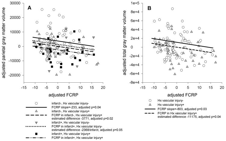
Figure 1.
Scatterplots showing the relationships between Freesurfer regional volumes and FCRP. FCRP and volumes were regressed on age, gender, magnet strength, intracranial volume, and cognitive status, and the residuals were plotted against each other. The slope of the line of best fit is the same as the regression coefficient for FCRP in the linear models that included history and evidence of vascular injury. When there are significant differences due to evidence and/or history of vascular injury, regression lines offset by the estimated difference are plotted. A: FCRP and parietal gray matter volume, illustrating effect of history of vascular injury, evidence of vascular injury, and combined effect of history and evidence of vascular injury. B: FCRP and total gray matter volume, illustrating effect of history of vascular injury.

