Abstract
The exact replication of natural tooth color in a metal ceramic restoration is a challenging fact as its affected by enumerable factors. Research revealing the influence of base metal alloys with different porcelain systems on the color of the restorations have shown minimal interest. The aim of the study was to evaluate the optical influence of different alloys (mainly base metal) and ceramic systems affecting the final color of metal–ceramic restorations. Four commercial ceramic alloys, two Ni–Cr, one Co–Cr and a high-noble alloy were combined with two porcelains in metal–ceramic specimens with a standardized thickness of layers. Ten disc-shaped specimens were prepared for each alloy/porcelain combination. High-noble was used as control group. Only opaque and dentin layers were applied. The specimens were analysed with Spectrophotometer, and data were obtained in the CIELAB color system. The recorded data were analysed with a one-way ANOVA and multiple range test by tukey—HSD procedure to identify the significant groups at 5 % level. The final color of the metal–ceramic specimens were significantly affected by both type of the alloy and the porcelain systems used (P < 0.0001). The Co-Cr alloy-porcelain combination produced least color difference when compared with the high-noble control group. There was significant difference (P < 0.0001) between both the brands of Ni–Cr alloy–porcelain systems. For all the alloy-porcelain combinations VMK 95 porcelain showed minimum color difference compared to the d-SIGN porcelain group. Conclusion: The final color of metal ceramic specimens was influenced both from the type of base metal alloy substructure and from the type of overlying porcelain.
Keywords: Metal–ceramic restorations, Ceramic alloys, Color matching, CIE lab
Introduction
The eventual success of any prosthesis is dependent on the morphological outline form, surface translucency, and color of the restoration [1]. Aesthetically superior restorations are now possible as a result of improvements in materials and fabrication techniques [2]. However, one of the most challenging aspects of aesthetic dentistry is color reproduction in restorations. Metal ceramic crowns are the most commonly prescribed fixed prostheses for both anterior and posterior teeth restorations [3]. The science of color is a complex specialty, which involves the principles of physics, chemistry and psychology [4]. Inadequacies related to color matching arise from structural differences that exist between metal–ceramic crowns and natural teeth, the limited range of available ceramic shades, inadequate shade guides and different composition of ceramic materials [5, 6]. The most significant difference between natural teeth and metal ceramic restorations affecting the color is the presence of metal framework. The presence of metal though increases the strength of restoration, however markedly detracts the aesthetic result. The final resultant color of metal ceramic restorations depends upon the type of ceramic used [7], the thickness of the ceramic layer, the particle size of ceramic material, refractive index of ceramic, the number of firings [8, 9]. The firing parameters, the applied stains, the type of metal alloy [9–15], composition of the alloy and its thickness have a definite effect on the color of the restoration. Color mismatch have been found between fired porcelain and shade guides [16], between shade guides [17, 18] and porcelain from different batches [19]. Color differences are also associated with specific metal ions contained in the dental alloys used for metal ceramic restorations [20–22]. Inconsistencies in a person’s ability to reliably select color matches are well documented [23, 24].
Various analyses with Spectrophotometer to compare the influence of different alloy substrates on the color of metal–ceramic restorations have been done [10, 11, 13]. Among all the alloys used for fabrication of porcelain fused to metal restorations, the base metal alloys are commonly utilized in contemporary clinical practice owing to superior physical properties, ease of oxidation, better bonding with ceramic compared to high-noble alloys and are economic [8, 11, 13]. The selection of right combination of alloy and porcelain material to produce the most clinically significant changes still remains unclear. To date, minimal research has been done to document the effects of only the base metal alloys on the color of metal-ceramic restorations. It is very well authenticated that high-noble alloys produce clinically acceptable color replication compared to other ceramic alloys [4], thus gold-platinum alloy was used as a control group.
Hence this study was attempted with the aim of investigating the optical influence of three different base metal alloys and two popular porcelains systems on the color of metal ceramic restorations when compared against high-noble alloy.
Materials and Methods
Preparation of Metal Substructure
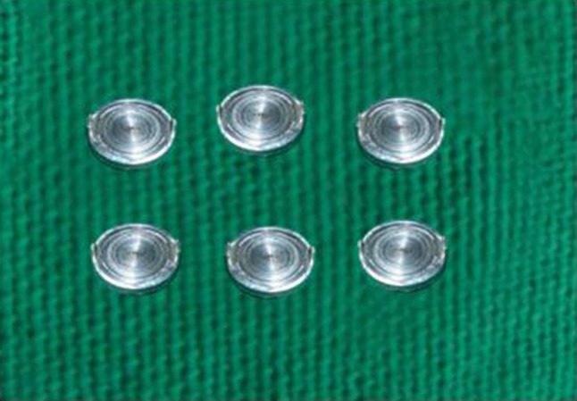
Three commercially available base metal alloys, two nickel–chromium (Ni–Cr) and one cobalt–chromium (Co–Cr) were combined with two porcelains systems in metal ceramic specimens with a standardized thickness of layer. High-noble alloy was used as a control group. The brand names, manufacture information, composition of the alloys as provided by the manufactures and the classification according to ADA are shown in Table 1. A standard circular stainless steel metal disc measuring 10 mm diameter and 5 mm in thickness was designed with two small metal extensions of 1.2 mm for application of ceramic layer (Fig. 1). Ten discs were made for each alloy group. The standard metal disc was duplicated using addition silicones (Aquasil Soft Putty—Regular Set, Dentsply, DeTrey, Germany), wax patterns (Inlay Wax Medium, GC Fuji, Japan) were made and then casted according to the manufacturers recommendations.. The extensions for ceramic was not enclosed but was of an open type in order to avoid the influence of alloy color over ceramic material. The flat porcelain bearing surface on each disc were adjusted with stones (Shofu, Co, Kyoto, Japan), sand blasted with 50 μm aluminium oxide particles, were cleaned with ultrasonic digital cleanser (Unikleen, Imperial Product). They were oxidized according to the manufacturer’s recommendation. (Fig. 2)
Table 1.
Composition of metal alloys as purported by the manufacturers and classification according to ADA
| Alloys | Composition | ADA classification | Manufacturer |
|---|---|---|---|
| Bellabond plus (subgroup A) | Ni—65.2 % Cr—22.5 % Mo, Fe, Si, Mn, Nb—9.5 % | Predominately base metal | BEGO Co, Bremer, Germany |
| Heraenium S (subgroup B) | Ni—62.9 % Cr—23 % Si—2 % Ce—<1 % Fe—1.5 % Mo—10 % | Predominately base metal | Heraus Kulzer GmbH, Germany |
| d-SIGN 30 (subgroup C) | Co—60.2 % Cr—30.1 % Ga—3.9 % Nb—3.2 % Si, Mo, Fe, B, Al, Li <1 % | Predominately base metal | Williams, Ivoclar Vivadent, Schaan, Liechtenstein |
| d.SIGN 98 (control group) | Au—85.9 % Pt—12.1 % Zn—1.5 % In—<1.0 % Ir—<1.0 % Ta, Fe, Mn—<1.0 % | High-noble alloy | Williams, the ceramic systems Group Ivoclar Vivadent, Schaan, Liechtenstein |
Fig. 1.

Standard metal disc
Fig. 2.

Alloy specimen
Porcelain Application
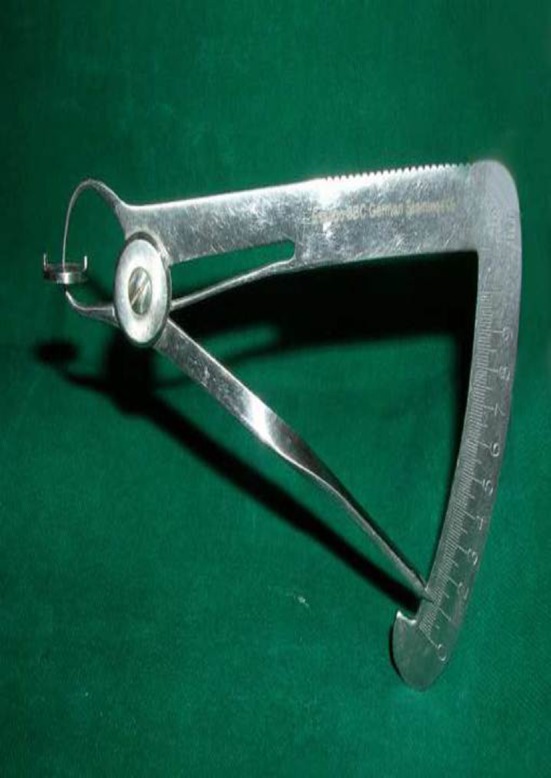
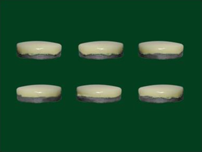
Two accepted porcelain systems were used. Shade A2 (Vita Classical Shade Guide) of d-SIGN ceramic material and shade 2M2 (Vitapan 3D Master Shade Guide) of VMK 95 ceramic material were employed. Two layers of opaque (powder form—Vita & paste form—d-SIGN) and dentin porcelain were applied until the desired range of thickness was achieved. The first layer was applied as slurry followed by normal paint on layer technique. Excess material was applied if any corrections were required. The thickness of opaque was 0.2 ± 0.05 mm and dentin layers were 1 ± 0.05 mm. A total thickness of ceramic layer was 1.2 mm. The layer thickness was evaluated with calliper (Iwanson gauge) having (accuracy of 0.1 mm) after each firing. There was no application of enamel porcelain. Glazing of the specimens were not done, they were finished to a uniform gloss using waterproof abrasive paper. The discs were fired separately under vita Vacumat 40 T furnace (Vita Zahnfabrik, Bad Sackingen, Germany) for both the porcelain systems. The furnace was calibrated before each firing and the settings were changed according to the porcelains system. (Fig. 3)
Fig. 3.

Metal–ceramic specimen
Color Determination
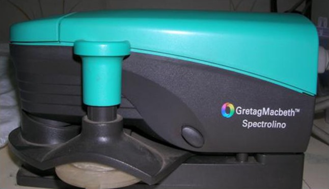
The color coordinates of each specimen was measured with a Spectrophotometer (Gretag Macbeth TM Spectrolino Reflectance Spectrophotometer Central Leather Research Institute, Chennai, India) set to the standard illumination source D65 with a 2° observation angle according to the 1931 CIE recommendation (Fig. 4) The data were based according to the CIELAB recommendation [25]. The data were displayed in L*, a*, b* values according to the CIELAB system. Each set of recorded data represented the mean value of 3 measurements. The device was calibrated before measurements of each specimen. The direct comparison of L*, a*, b*and ΔE values were done. ΔE Color difference was calculated using the formula ΔE = [(ΔL*) 2 + (Δa*) 2 + (Δb*) 2] ½ Mean color differences between alloy porcelain combinations were calculated and compared with control group combination values, to verify if there was any significant color differences noted. The recorded data were analysed with a one-way ANOVA and multiple range test by tukey—HSD procedure to identify the significant groups at 5 % level.
Fig. 4.

Spectrophotometer
Group Analysis
There were two main groups based upon the ceramic systems Group I—d-SIGN (Ivoclar Vivadent, Schaan, Liechtenstein) Group II—VMK 95(Vita Zahnfabrik, Bad Sackingen, Germany) Each main group had three subgroups A, B, and C based upon the base metal alloy and a control group I and group II respectively for high-noble alloy. Subgroup A—Ni–Cr alloy (Bellabond plus, BEGO Co, Bremer, Germany) Subgroup B—Ni–Cr alloy (Heraenium S, Heraus Kulzer GmbH, Germany) Subgroup C—Co-Cr alloy (d SIGN 30 Williams, Ivoclar Vivadent, Schaan Liechtenstein) high-noble alloy–porcelain group served as control. Control CI—High-noble alloy and d-SIGN ceramic combination Control CII—High-noble alloy and VMK 95 ceramic combination
Results
The final color of the metal-ceramic specimens were significantly affected by both type of the alloy and the porcelain systems used (P < 0.0001). The group II C, Co-Cr alloy porcelain combination produced least color difference ΔE = 1.347 when compared with the high-noble control group. There was significant difference (P < 0.0001) between both the brands of Ni–Cr alloy–porcelain systems ΔE > 3.3(group I A, B & II A, B) which was considered visually discernable as shown in Table 2. For the entire alloy-porcelain combinations VMK 95 porcelain group II showed minimum color difference compared to the d-SIGN porcelain group I. The descriptive statistics of L*, a*, b* values are furnished in Table 3. The mean “L” value was highest for subgroup II A (72.218 ± 1.470) and lowest for subgroup I B (62.572 ± 0.723). The Mean “L” values for group I was low compared to group II. The mean “a” value was more towards green axis for all the alloy- porcelain combinations. The mean “a” value was highest for subgroup I C (−2.598 ± 0.137) and lowest for subgroup II A (−1.512 ± 0.052). The mean “a” values were less for group II compared to group I. The mean “b” values were towards the yellow axis and the highest was for subgroup II C (12.184 ± 0.648) and lowest for subgroup I C (10.059 ± 0.416). The mean “b” values were less for group I compared to group II.
Table 2.
Mean ∆E values for each subgroup
| Variable | Group & subgroup | Mean ± SD | SE | Median range |
|---|---|---|---|---|
| Control I | 1.006 ± 0.634 | 0.204 | 1.127 (0.154 to 1.989) | |
| ∆E | I A | 4.316 ± 0.686 | 0.217 | 4.328 (3.515 to 5.312) |
| I B | 6.460 ± 1.872 | 0.592 | 5.832 (5.440 to 11.635) | |
| I C | 2.943 ± 0.469 | 0.148 | 3.118 (2.198 to 3.476) | |
| Control II | 0.941 ± 0.741 | 0.199 | 0.899 (1.174 to 0.963) | |
| II A | 3.004 ± 1.383 | 0.437 | 2.902 (0.858 to 5.561) | |
| II B | 4.634 ± 1.969 | 0.623 | 5.709 (1.814 to 6.685) | |
| II C | 1.347 ± 0.741 | 0.234 | 1.447 (0.134 to 2.063) |
Table 3.
Descriptive Statistics
| Variable | Group & subgroup | Mean ± SD | SE | Median range |
|---|---|---|---|---|
| L | I A | 63.081 ± 0.660 | 0.209 | 63.023 (62.052 to 64.401) |
| I B | 62.572 ± 0.723 | 0.229 | 62.880 (61.278 to 63.451) | |
| I C | 62.817 ± 0.431 | 0.136 | 62.708 (62.340 to 63.630) | |
| II A | 72.218 ± 1.470 | 0.465 | 72.519 (70.001 to 74.832) | |
| II B | 67.545 ± 2.140 | 0.677 | 68.663 (64.321 to 70.024) | |
| II C | 69.617 ± 0.542 | 0.172 | 69.785 (68.262 to 70.154) |
| Variable | Group & subgroup | Mean ± SD | SE | Median range |
|---|---|---|---|---|
| a | I A | −2.364 ± 0.088 | 0.028 | −2.386 (−2.165 to −2.451) |
| I B | −2.552 ± 0.117 | 0.037 | −2.527 (−2.710 to −2.384) | |
| I C | −2.598 ± 0.137 | 0.043 | −2.579 (−2.785 to −2.381) | |
| II A | −1.512 ± 0.052 | 0.016 | −1.515 (−1.583 to −1.443) | |
| II B | −1.877 ± 0.380 | 0.120 | −1.804 (−2.449 to −1.403) | |
| II C | −1.549 ± 0.149 | 0.047 | −1.585 (−1.780 to − 1.347) |
| Variable | Group & subgroup | Mean ± SD | SE | Median range |
|---|---|---|---|---|
| b | I A | 11.383 ± 0.934 | 0.295 | 11.360 (10.156 to 12.530) |
| I B | 10.073 ± 1.006 | 0.318 | 10.472 (7.585 to 10.883) | |
| I C | 10..059 ± 0.416 | 0.132 | 9.979 (9.546 to 10.780) | |
| II A | 12.037 ± 0.924 | 0.292 | 12.045 (10.362 to 13.192) | |
| II B | 11.409 ± 0.547 | 0.173 | 11.575 (10.018 to 11.815) | |
| II C | 12.184 ± 0.648 | 0.205 | 12.119 (10.974 to 13.142) |
Discussion
One of the major challenges encountered by the clinician in the field of Prosthodontics is to replicate the natural color of teeth in metal ceramic restorations. The inconsistencies in color with such restorations have been reported by various authors [16–22] like type of alloy and ceramic used, thickness of ceramic, particle size of ceramic, number of firing cycles, firing temperature, composition of alloy. Base metals among the ceramic alloys are extensively used in clinical practice because of many factual advantages over other noble alloys [8, 11, 13]. Thus this study destined to evaluate the influence of base metal alloys over color of porcelain fused to metal restorations. Having known high-noble alloy produces enhanced esthetic with porcelain [4], Au–Pt was used as a control group instead of shade guides. Most of the shade tabs are made of high fusing porcelain giving an unrealistic representation with presence of characterization, absence of metal backing difference of structure and applied ceramic layer [26]. With multiple etiological factors affecting the color of metal ceramic restorations, two well accepted low-fusing feldspathic Ivoclar and Vita porcelain systems were employed for fabrication metal-ceramic specimens. Since it’s idealistic to study all the shades, two commonly advocated shades A2 for d-SIGN and 2M2 for Vita in clinical practice in South India were opted.
Direct comparison of shades A2 and 2M2 under Reflectance Spectrophotometer revealed minimal color difference with ∆E value of 0.5 was obtained to exclude the variability between both the shades. Instrumental color measurement using Spectrophotometer with CIELAB has the advantages of obviating the subjective aspects of color assessment and of expediting the determination of color [3]. In this study a Reflectance Spectrophotometer based upon the CIE metrics was used to describe the color coordinates of the samples to produce the most accurate color measurements [27, 28]. The specimens were fabricated based upon average thickness of anterior metal–ceramic restorations [29]. Only opaque and dentin porcelain were applied as the opacifiers in the opaque material and certain metal oxides in the dentin porcelain aid in masking the color of the metal. Enamel porcelain was not applied as the color imparting pigments are negligible in enamel powders, they produce translucency of the restoration. Finishing of the discs was done without glazing as the study was done in vitro, the chances of ingress of oral fluids or bacteria are minimal [30].
The results of the present study indicate a strong influence of the base metal alloy and porcelain system on the final color of the metal-ceramic complex. The cobalt chromium alloy -porcelain combination produced least color difference when compared with the high-noble control group. There was significant difference between both the brands of nickel–chromium alloy–porcelain systems also. For all the alloy–porcelain combinations Vita porcelain showed minimum color difference compared to the Ivoclar porcelain group. The present study results confirm the results of previous studies, in which significant color differences were found with base metal alloys when, compared with high-noble alloy and also confirm that porcelain system had a significant influence on the final color of metal-ceramic restorations [4, 10, 11, 13] and in which significant color differences between different brands of porcelain of the same nominal shade in both opaque and layered porcelain samples were detected [1]. The present study color differences between Ni–Cr alloys (Heranium) showed visually detectable color difference with ∆E > 3.7 (∆E = 4.634). According to Johnston WM et al. [28] color difference of ∆ > 3.7 is considered to be a poor match.
The result of this study show that the necessary thickness of opaque and body porcelain to match the shade sample varies from shade to shade and also from one system to another. The thickness of body porcelain controls the amount of color pigments. The thicker the porcelain, the more concentrated the color [15]. The general mechanisms by which various agents react with the overlying porcelain, responsible for the discoloration have been proposed in the dental literature. Bulk transfer involves the diffusion of an element from the interior of the alloy into the porcelain. Surface diffusion occurs when an element from the very thin oxide layer on the alloy passes along the metal–ceramic interface and into the porcelain. The third mechanism of vapor deposition involves the elevated-temperature vaporization of different component elements from the alloy composition and their subsequent deposition onto the porcelain surface, followed by diffusion into the interior of the porcelain which results in discoloration [4]. The results show higher color difference in nickel–chromium alloy than the cobalt–chromium alloy. The Ni ions are colorants that have been shown to produce a neutral grey color in sodium silicate glasses and are probably associated with color changes in porcelain [28]. The presence of Molybdenum which is very less at a level of <1 % and so very less oxide was formed. Color differences in the present study have been shown differences between both brands of Ni–Cr alloys also. The Ni–Cr alloy (Heraenium) had higher color differences compared to Ni–Cr alloy (Bellabond Plus). The compositional difference in Ni–Cr alloy (Heraenium) shows that more oxide layer is formed because of the presence of molybdenum Mo—10 % and Chromium—23 % compared to Ni–Cr alloy (Bellabond Plus) where presence of Mo and Cr are less and hence formation of less oxide layer [31]. The color of a porcelain restoration is the result of diffuse reflectance from a translucent layer covering an underlying opaque layer. The double-layer color effects in porcelain restorations resulting from body and opaque layers have been described by O’Brien et al. according to Kubelka–Munk theory. The minimum color differences found with Vita porcelain system compared to the Ivoclar system may be attributed to scattering coefficients and transmission, which may be affected by variations of different refractive index of the particle size as studied by Lund et al. [31]. The particle size and shape of the porcelain powder have an effect on the scattering coefficient [7, 9]. Thus the present study provides more scope for further research where additional alloys and different shades of porcelain could be studied. Variations in visual perception could be studied with different operators at different age intervals, different lighting conditions, and varying distances. Advanced imaging techniques in metallurgy like X-ray diffraction, mass emission spectroscopy to evaluate ionic diffusion and transfer at molecular level influencing oxide formation of base metal alloys has to be explored in detail further.
Conclusion
Within the limitations of this study, it can be concluded that
Minimum color difference was found with Co-Cr alloy porcelain combination than the Ni–Cr alloy-porcelain combinations in all the subgroups
When comparing the efficacy of the various porcelain systems influencing color, the Vita porcelain had minimum color differences than the Ivoclar porcelain systems
Clinical Implication
Varying color combinations result with different alloy porcelain systems which are not concurrent with their respective parent shade guides. An exact choice of alloy and specific porcelain system is mandatory while communicating to the laboratory. Also, a customized metal-ceramic shade guide could drastically improve color matching with metal-ceramic restorations.
Acknowledgments
Source of support
The color of the specimens in this article were studied at “Department of Tanning, Central Leather Research Institute of India, Adyar, Chennai, Tamilnadu, India”.
References
- 1.Seghi RR, Johnston WM, O’Brien WJ. Spectrophotometric analysis of color differences between porcelain systems. J Prosthet Dent. 1986;56(1):35–40. doi: 10.1016/0022-3913(86)90279-9. [DOI] [PubMed] [Google Scholar]
- 2.Tung FF, Goldstein GR, Jang S, Hittelman E. The repeatability of an intra-oral dental colorimeter. J Prosthet Dent. 2002;88(6):585–590. doi: 10.1067/mpr.2002.129803. [DOI] [PubMed] [Google Scholar]
- 3.Douglas RD, Przybylska M. Predicting porcelain thickness required for dental shade matches. J Prosthet Dent. 1999;82(2):143–149. doi: 10.1016/S0022-3913(99)70147-2. [DOI] [PubMed] [Google Scholar]
- 4.Stavridakis MM, Papazoglou E, Seghi RR, Johnston WM, Brantley WA (2004) Effect of different high palladium metal-ceramic alloys on the color of opaque and dentin porcelain. J Prosthet Dent 92(2):170–178 [DOI] [PubMed]
- 5.Preston JD. Current status of shade selection and color matching. Quintessence Int. 1985;16(1):47–58. [PubMed] [Google Scholar]
- 6.Miller LL. Shade matching. J Esthet Dent. 1993;5(4):143–153. doi: 10.1111/j.1708-8240.1993.tb00771.x. [DOI] [PubMed] [Google Scholar]
- 7.Barghi N, Richardson JT. A study of various factors influencing shade of bonded porcelain. J Prosthet Dent. 1978;39(3):282–284. doi: 10.1016/S0022-3913(78)80096-1. [DOI] [PubMed] [Google Scholar]
- 8.Barghi N. Color and glaze: effect of repeated firings. J Prosthet Dent. 1982;47(4):393–395. doi: 10.1016/S0022-3913(82)80088-7. [DOI] [PubMed] [Google Scholar]
- 9.Jorgenson MW, Good Kind RJ. Spectrophotometric study of five porcelain shades relative to the dimension of color, porcelain thickness, and repeated firings. J Prothet Dent. 1979;42(1):96–105. doi: 10.1016/0022-3913(79)90335-4. [DOI] [PubMed] [Google Scholar]
- 10.Brewer JD, DA Akersck G, Scrensen SE. Spectrophotometric analysis of the influence of metal substrates on the color of metal–ceramic restorations. J Dent Res. 1985;64(1):74–77. doi: 10.1177/00220345850640011501. [DOI] [PubMed] [Google Scholar]
- 11.Jacobs SH, Good Acre CJ, Moore BK, Dykema RW. Effect of porcelain thickness and type of metal–ceramic alloy on color. J Prosthet Dent. 1987;57(2):138–145. doi: 10.1016/0022-3913(87)90135-1. [DOI] [PubMed] [Google Scholar]
- 12.Lund PS, Piotrowski TJ. Color changes of porcelain surface colorants resulting from firing. Int J Prosthodont. 1992;5(1):22–27. [PubMed] [Google Scholar]
- 13.Crispin BJ, Seghi RR, Globe H. Effect of different metal ceramic alloys on the color of opaque and dentin porcelain. J Prosthet Dent. 1991;65(3):351–356. doi: 10.1016/0022-3913(91)90224-K. [DOI] [PubMed] [Google Scholar]
- 14.Crispin BJ, Okamolosk GH. Effect of porcelain crown substructure on visually perceivable value. J Prosthet Dent. 1991;66(2):209–212. doi: 10.1016/S0022-3913(05)80049-6. [DOI] [PubMed] [Google Scholar]
- 15.Barghi N, Lorenzana RE. Optimum thickness of opaque and body porcelain. J Prosthet Dent. 1982;48(4):429–431. doi: 10.1016/0022-3913(82)90080-4. [DOI] [PubMed] [Google Scholar]
- 16.Groh CL, O’Brien WJ, Boenke KM. Difference in color between fired porcelain and shade guides. Int J Prosthodont. 1992;5(6):510–514. [PubMed] [Google Scholar]
- 17.O’Brien WJ, Boenke KM, Groh CL. Coverage errors of two shade guides. Int J Prosthodont. 1991;4(1):45–50. [PubMed] [Google Scholar]
- 18.Paravina RD, Powers JM, Fay RM. Color comparison of two shade guide. Int J Prosthodont. 2002;15(1):73–78. [PubMed] [Google Scholar]
- 19.Barghi N, Pedreo JAE, Bosch RR. Effects of batch variation on shade of dental porcelain. J Prosthet Dent. 1985;54(5):625–627. doi: 10.1016/0022-3913(85)90235-5. [DOI] [PubMed] [Google Scholar]
- 20.Bertolloti RL. Alloys for porcelain fused to metal restorations. In: O’Brien W, editor. Dental materials and their selection. 3. Chicago: Quintessence; 2002. pp. 200–209. [Google Scholar]
- 21.Fanderlik L. Glass science and technology: 5 optical properties of glass. St. Louis: Elsevier; 1983. pp. 320–330. [Google Scholar]
- 22.Yamamoto M. Metal ceramics: principles and methods of Makoto Yamamoto. Chicago: Quintessence; 1985. pp. 483–494. [Google Scholar]
- 23.Culpepper WD. A comparative study of shade—matching procedure. J Prosthet Dent. 1970;24(2):166–173. doi: 10.1016/0022-3913(70)90140-X. [DOI] [PubMed] [Google Scholar]
- 24.Davison SP, Myslinski NR. Shade selection by color vision—defective dental personnel. J Prosthet Dent. 1990;63(1):97–101. doi: 10.1016/0022-3913(90)90276-I. [DOI] [PubMed] [Google Scholar]
- 25.Sharma A. Understanding color management. Clifton Park: Thomson Delmar Learning; 1985. [Google Scholar]
- 26.Sorensen JA, Torres TJ. Improved color matching of metal-ceramic restorations part III: innovations in porcelain application. J Prosthet Dent. 1988;59(1):1–7. doi: 10.1016/0022-3913(88)90096-0. [DOI] [PubMed] [Google Scholar]
- 27.Kourtis SG, Tripodakis AP, Doukoudakis AA. Spectrophotometric evaluation of the optical influence of different metal alloys and porcelains in the metal–ceramic complex. J Prosthet Dent. 2004;92(5):477–485. doi: 10.1016/j.prosdent.2004.08.012. [DOI] [PubMed] [Google Scholar]
- 28.Johnston WM, Kao EC (1989) Assessment of appearance match by visual observation and clinical colorimetry. J Dent Res 68(5):819–822 [DOI] [PubMed]
- 29.Shillingburg HT. Fundamentals of fixed Prosthodontics. 3. Chicago: Quintessence; 2006. pp. 142–145. [Google Scholar]
- 30.Rhoads JE, Rudd KD, Morrow RM (1985) Dental laboratory procedures: fixed partial dentures, 2nd edn. Mosby, St. Louis, pp 282–284
- 31.Lund TW, Sahwabacher WB, Goodkind RJ. Spectrophotometric study of the relationship between body porcelain color and applied metallic oxide pigments. J Prosthet Dent. 1985;53(6):790–796. doi: 10.1016/0022-3913(85)90158-1. [DOI] [PubMed] [Google Scholar]


