Abstract
A rare case of ascaris coming out through the anterior abdominal wall is reported here. A 40-year-old female had undergone dilatation and curettage by a quack. On the second day she presented with presented with features of peritonitis. She was explored. Resection anastomosis of the ileum was done for multiple perforations of the ileum. Patient developed a fistula in the anterior abdominal wall which was draining bile-colored fluid. On the 12th postoperative day a 10-cm-long worm was seen coming out through the fistulous tract which was found to be Ascaris lumbricoids. Ascaris lumbricoids can lead to many complications ranging from worm colic to intestinal obstruction, volvulus, peritonitis, pancreatitis, cholangiohepatitis, liver abscess and many more. Worm has been reported to come out through mouth, nostrils, abdominal drains, T-tubes etc. But ascaris coming out through the anterior abdominal wall is very rare hence reported here.
Keywords: Ascaris, bile, perforation
INTRODUCTION
Ascariasis is an important medical, social and economic problem in many underdeveloped countries where public health, sanitation and personal hygiene are at the lowest level (1-3). It can cause a variety of complications like biliary obstruction, pancreatitis, small bowel obstruction, and gangrene. Ascaris has been reported to come out through chest tube. Nasogastric tube, T tube etc. We report this case in which ascaris came out through anterior abdominal wall.
CASE REPORT
A 40-year-old female had undergone dilatation and curettage for missed abortion by a quack. On the second day she developed abdominal pain in the periumblical area which later on involved the whole abdomen. Pain was followed by abdominal distension and fever. There was no history of nausea, vomiting, hemetemesis, malena. There was history of anorexia.
Physical examination revealed an ill-looking female with pulse of 100 bpm, blood pressure of 100/60 and temperature of 100°F. Abdominal examination showed guarding and rebound tenderness all over. Chest and cardiovascular examination was essentially normal.
Chest X-ray of both domes of the diaphragm showed gas under the diaphragm. Abdominal sonography showed free fluid in the peritoneal cavity. A diagnosis of peritonitis was made and patient was explored. Operative findings were:
Four perforations in the ileum in a segment of about 15 cm.
About 300 ml of dirty bile-stained fluid in the peritoneal cavity.
Pus flakes were present all over.
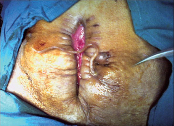
Resection anastomosis of the ileum was done and patient was put on Ryle's tube, IV fluids, and IV antibiotics (ceftrioxone salbactum and tinidazole). Ryle's tube was removed on the third postoperative day. Patient was shifted to liquid orals on the fifth postoperative day. Patient developed pus discharge from wound. Swab culture sensitivity was sterile. Skin sutures were removed and pus was drained. Wound was dressed daily and healing was achieved by secondary intention. On the 10th postoperative day patient developed a fistula just below the umbilicus which drained a small amount of bile-stained fluid, about 20 ml/day. On the 12th postoperative day a 10-cm-long worm was seen coming out through the fistulous tract. Discharge from the fistula decreased subsequently. Fistula closed of its own after three days. Wound healed completely by secondary intention. Patient was administered 400 mg of Albandazole for three consecutive days. Patient is on follow-up for the last six months.
DISCUSSION
Ascaris lumbricoids is the most common intestinal parasite.[1] It affects about 25% of the world's population in third-world countries. Maximal incidence is in the preschool and school-age groups.[2,3,4] Males and females are equally affected.[2,3] Humans are affected by ingestion of eggs from contaminated food, water and infected soil. Eggs reach the duodenum where shell is dissolved and larva comes out. Larvae penetrate the intestinal wall and reach the bloodstream. They are filtered out by capillaries of the lung where they break alveoli. They ascend the trachea and pass over the epiglottis by cough. They are later swallowed and reach back to the intestinal lumen.[2,3,4] The adult worm lives in the jejunal lumen. Adult worm can cause mechanical intestinal obstruction,[2,3,4,5,6,7,8] or it can migrate to some other sites causing variety of complications like pancreatitis,[3,4] biliary obstruction,[2,4,8] cholangiohepatitis,[4] liver abscess,[3,9] appendicitis,[4,8] intestinal perforation,[3,4] granulomatous peritonitis,[5,8,10] and ascaris empyema.[11,12] Agarwal et al.,[13] reported a case of ascaris coming out through a ventriculoperitoneal shunt. Kar et al.,[14] reported a case of ascaris coming out through a T-tube tract. All these complications can lead to significant morbidity and mortality. Ascaris coming out through the anterior abdominal wall is very rare, hence reported here [Figure 1].
Figure 1.

Ascaris coming out through the anterior abdominal wall
Footnotes
Source of Support: Nil
Conflict of Interest: None declared
REFERENCES
- 1.Rao PL, Sharma AK, Yadav K, Mitra SK, Pathak IC. Acute intestinal obstruction in children as seen in North West India. Indian Pediatr. 1978;15:1017–20. [PubMed] [Google Scholar]
- 2.Mahmoud AA. Intestinal nematodes. In: Mandell GI, Douglas RG, Bennet JE, editors. Principles and practice of infectious diseases. Edinburg: Churchill Livingstone; 1990. pp. 2137–8. [Google Scholar]
- 3.Surrendran N, Paulose MO. Intestinal complications of round-worms in children. J Paediatr Surg. 1998;23:931–5. doi: 10.1016/s0022-3468(88)80388-9. [DOI] [PubMed] [Google Scholar]
- 4.Markell EK, Voge M, John DT. The intestinal nematodes. In: Ozmat S, editor. Medical parasitology. Philadelphia: WB Saunders; 1992. pp. 261–93. [Google Scholar]
- 5.De Sa AE. Surgical Ascariasis. Indian J Surg. 1996;28:182–90. [Google Scholar]
- 6.Cole GJ. Surgical manifestations of Ascaris lumbricoides in the intestine. Br J Surg. 1965;52:444–7. doi: 10.1002/bjs.1800520611. [DOI] [PubMed] [Google Scholar]
- 7.Louw JH. Abdominal complications of Ascaris lumbricoide infestation in children. Br J Surg. 1966;53:510–21. doi: 10.1002/bjs.1800530606. [DOI] [PubMed] [Google Scholar]
- 8.Ochoa B. Surgical complications of ascariasis. World J Surg. 1991;15:222–7. doi: 10.1007/BF01659056. [DOI] [PubMed] [Google Scholar]
- 9.Saw HS, Somasundram K, Kamath R. Hepatic ascariasis. A case report and review of the literature. Arch Surg. 1974;108:733–5. doi: 10.1001/archsurg.1974.01350290095017. [DOI] [PubMed] [Google Scholar]
- 10.Mylvaganam C, Pannabokke RG. Extra-intestinal ascaris granuloma. J Trop Med Hyg. 1969;72:98–100. [PubMed] [Google Scholar]
- 11.Sen MK, Chakrabarti S, Ojha UC, Daima SR, Gupta R, Suri JC. Ectopic ascariasis: An unusual case of pyopneumothorax. Indian J Chest Dis Allied Sci. 1998;40:131–2. [PubMed] [Google Scholar]
- 12.Zamora Almeida O. Localization of Ascaris lumbricoides in the thoracic cavity. Report of a case. Rev Cubana Med Trop. 1976;28:71–5. [PubMed] [Google Scholar]
- 13.Agarwal P. Round worm migration along ventriculoperitoneal shunt tract: A rare complication. J Postgrad Med. 2000;46:37–8. [PubMed] [Google Scholar]
- 14.Kar M, Saha I, Kar JK, Mukhopadhyay M. Wandering ascaris coming out through the T-tube tract: Rare occurrence. J Indian Med Assoc. 2004;102:225. [PubMed] [Google Scholar]


