Abstract
Sporadic occurrence of transformed tumor cells is under the surveillance of the host immune system and such cells are effectively eliminated by immune-mediated cell death. During tumor progression, the antitumor effects of the tumor microenvironment are suppressed by diverse immunosuppressive mechanisms. In this research, we suggest novel immune evasion strategy of tumor cells through a transforming growth factor (TGF)-β1- and hypoxia-dependent mechanism. Experimental results showed that TGF-β1 and hypoxia induced mitogen-activated protein kinase phosphatase (MKP)-1 expression within 1 h, resulting in attenuation of c-Jun N-terminal kinase (JNK) phosphorylation and subsequent death receptor-mediated cell death. In addition, analysis of microarray data and immunostaining of MKP-1 in hepatocellular carcinoma (HCC) patient samples revealed that expression of MKP-1 is notably higher in tumors than in normal tissues, implying that MKP-1-dependent suppression of immune-mediated cell death takes place only in the tumor. To prove that MKP-1 can act as a mediator of immune escape by tumors, we determined whether chemo-resistance against several anticancer drugs could be overcome by knockdown of MKP-1. Cytotoxic assays showed that chemotherapy with siRNA targeting MKP-1 was significantly more effective than chemotherapy in the presence of MKP-1. Thus, we conclude that TGF-β1 and hypoxia ensure tumor cell survival and growth through expression of MKP-1.
Keywords: cell death, hypoxia, mitogen-activated protein kinase, mitogen-activated protein kinase phosphatase, transforming growth factor, tumor resistance
Recent progress in defining the cellular and molecular networks that regulate the immune responses in the tumor microenvironment has renewed the cancer-immune interaction paradigm.1 Tumors foster a tolerant microenvironment and activate diverse immunosuppressive mechanisms, eventually counteracting effective immune responses.2 These immunosuppressive strategies can pre-exist, arise through outgrowth of escape mutants, or take place during tumor-sculpting immune system activity.3, 4 These dual effects of the immune system on developing tumors facilitated refinement of the cancer immunosurveillance hypothesis into the ‘cancer immunoediting' hypothesis proposed by Schreiber et al.5 In this project, we report novel findings relating to tumor-specific evasion of immune-mediated cell death by transforming growth factor (TGF)-β1- and hypoxia-dependent mechanisms.
TGF-β is a mitogenic factor that regulates a wide variety of cellular responses, including migration, immune activity, apoptosis, differentiation, and proliferation.6 Many evidences support a strong correlation between TGF-β and cancer. Cancer cells are able to produce and secrete TGF-β1, which causes immunosuppression in cancer patients in the absence of cytotoxic treatment.7, 8 Also, elevated levels of TGF-β1 have been found in a number of experimental models and in specimens from patients with cancer.9 TGF-β1 has the potential to function as a tumor suppressor through effects on proliferation, replication potential, and apoptosis. However, paradoxically, TGF-β1 also modulates cell migration, invasion, angiogenesis, and the microenvironment in ways that cancer cells may exploit to their advantage.10 Consequently, the output of a TGF-β1 response is highly contextual throughout development, across different tissues and in cancer. This dual role of TGF-β1 in cancer has long been noted, but its mechanistic basis, operating logic, and clinical relevance have remained elusive.11
Mitogen-activated protein kinase phosphatase (MKP)-1, also known as dual specificity phosphatase1 (DUSP1), is one of the many target proteins of TGF-β1, and is known to specifically dephosphorylates c-Jun N-terminal kinase (JNK) and p38.12, 13, 14, 15 We postulated that immune-mediated cell death is remarkably affected by pre-existence of TGF-β, because MKP-1 augmentation may result in suppression of JNK phosphorylation, which is a major requirement of the death-receptor mediated cell death pathway.16
The major death receptor is part of the TNF superfamily, which is characterized by an intracellular death domain. Tumor necrosis factor (TNF)-α, TNF-related apoptosis-inducing ligand (TRAIL), and Fas ligand (FasL) can activate death receptor-mediated cell death.17, 18, 19 We dissected crosstalk between TNF-α and TGF-β1 signal transduction components. Many roles of TNF-α are mediated by the transcription factor nuclear factor-κB (NF-κB). In addition to acting through NF-κB, TNF-α also exerts its function through activation of JNK, p38, and p42/44 mitogen-activated protein kinase (MAPK) cascades, which participate in various cellular responses.20, 21 Notably, JNK contributes to caspase activation and apoptosis by multiple mechanisms.22, 23, 24, 25 Despite environmental dependence, sustained activation of JNK induces cell death, and many cellular components are involved in crosstalk with JNK signaling.26, 27 On the basis of these reports, we investigated the crosstalk between TGF-β1 signaling and the death receptor-mediated cell death pathway to unravel a novel regulatory mechanism of cell death. We identified MKP-1-JNK as a regulatory intersection, and further investigated the tumor-specific nature of this control of cell death and its contribution to tumor resistance to the host immune system.
Results
Block of immune-mediated cell death by TGF-β1 pretreatment
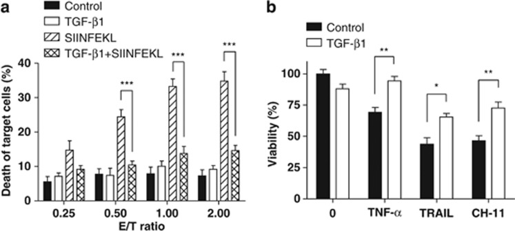
To unravel how TGF-β1 changes the sensitivity to CD8+ T cells, we cocultured OT-1 mouse (ovalbumin (OVA)-specific CD8+ T cell receptor transgenic mouse)-derived T cells and OVA peptide (SIINFEKL)-loaded MC38 cells with or without TGF-β1 pretreatment. In the absence of TGF-β1 pretreatment, an increase in effector (OT-1 mouse-derived CD8+ T cell)-to-target ratio increased cell-mediated cytotoxicity in immunized MC38 cells. When TGF-β1 was applied in MC38 cells 1 h before coculture, however, most cells were intact, indicating that TGF-β1 blocked CD8+ T cell-mediated cell death (Figure 1a). Because CD8+ T cells release diverse cytokines, such as TNF-α, TRAIL, and FasL, we applied each cytokine to HepG2 cells with or without TGF-β1 1 h before pretreatment to mimic the cytotoxic function of CD8+ T cells. We measured cell viability by WST-1 assay, and the results showed a protective role for TGF-β1, even though TGF-β1 alone did not exert proliferative effects (Figure 1b). These results suggest that there is a crosstalk between TGF-β1 and death receptor-mediated cell death pathways. Among the many types of death receptor-mediated cell death pathways activated by CD8+ T cells, we focused on TNF-α, a major cytokine in inflammation, cell survival, proliferation, and apoptosis.
Figure 1.
Block of immune-mediated cell death by TGF-β1 pretreatment. (a) MC38 cells were immunized with SIINFEKL peptide. Six hours after coculture of effector (OT-1 mouse-derived CD8+ T cells) and target (MC38 cells) cells, cytotoxicity was evaluated by lactate dehydrogenase (LDH) release assay. TGF-β1 (10 ng/ml) was applied in MC38 cells 1 h before coculture (mean±S.E.M., n=4; Bonferroni's post hoc test was applied for multiple comparisons in two-way ANOVA, ***P<0.001). (b) HepG2 cells were incubated in the presence or absence of TGF-β1 (10 ng/ml) for 1 h, and then treated with TNF-α (10 ng/ml), TRAIL (500 ng/ml), and CH-11 (500 ng/ml) for 24 h. Cell viability was measured by WST-1 assay (mean±S.E.M., n=4; *P<0.05, **P<0.01 by Student's t-test)
Attenuation of TNF-α-induced JNK signaling by TGF-β1 pretreatment
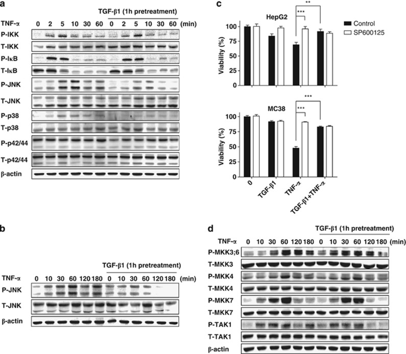
To investigate the mechanism of this crosstalk, we evaluated the effect of TGF-β1 pretreatment on internal transduction of the TNF-α signal. In the absence of TGF-β1, TNF-α treatment induced phosphorylation of MAPKs, such as JNK, which peaked at about 10 min. It also led to IκB kinase (IKK) phosphorylation, followed by IκB degradation and NF-κB translocation. However, pretreatment with TGF-β1 for 1 h suppressed phosphorylation of JNK, but did not affect NF-κB signaling (Figure 2a). Figure 2b shows that TGF-β1 pretreatment also suppressed prolonged phosphorylation of JNK. Treatment with the JNK inhibitor SP600125 largely blocked TNF-α-induced cell death, while TGF-β1 did not further contribute to the viability of HepG2 and MC38 cells (Figure 2c). These data show that TNF-α-induced cell death is JNK-dependent, and that attenuation of JNK phosphorylation by TGF-β1 pretreatment protects cells from TNF-α-induced cytotoxicity. To determine whether signaling components acting upstream of JNK show similar behavior, phosphorylation of MKK4/7, MKK 3/6, and TAK1 was measured by immunoblot analysis. Their signals were not significantly altered by TGF-β1 pretreatment (Figure 2d). These data suggest that this crosstalk between TNF-α and TGF-β1 signaling pathways originates directly from JNK.
Figure 2.
Attenuation of TNF-α-induced JNK phosphorylation by TGF-β1 pretreatment. (a–b) Intracellular signaling components were subjected to immunoblot analysis. Huh-7 cells were incubated in the presence or absence of TGF-β1 (10 ng/ml) for 1 h, and then treated with TNF-α (10 ng/ml) for up to 60 (a) or 180 min (b). Cell lysates were subjected to immunoblot analysis of IKK, IκB, JNK, p38, and p42/44. β-actin was used as the loading control. (c) Cell viability was measured by WST-1 assay. The JNK inhibitor SP600125 (10 μM) was applied 1 h before treatment with TNF-α or TGF-β1. WST-1 reagent was added 24 h after cytokine treatment (mean±S.E.M., n=4; **P<0.01, ***P<0.001 by Student's t-test). (d) Upstream signaling components of JNK and p38 were evaluated by immunoblotting, as in (a)
JNK and MKP-1 mediate crosstalk between TGF-β1 and TNF-α signaling pathway
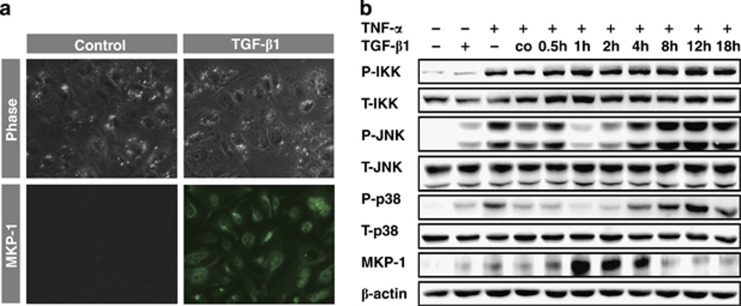
One of the most convincing candidates that may directly dephosphorylate JNK is MKPs. To elucidate the detailed mechanism of crosstalk between TGF-β1 and TNF-α signaling pathway, we evaluated the involvement of MKP-1. Immunocytochemistry clearly showed TGF-β1-dependent expression of MKP-1 in Huh-7 cells within 1 h (Figure 3a). We also performed immunoblot analysis of MKP-2, but its expression was not increased by TGF-β1 treatment (Supplementary Figure 1). For a detailed view, we compared the temporal patterns of MKP-1 expression with other signaling components according to the duration of TGF-β1 pretreatment in Huh-7 cells. MKP-1 expression peaked at 1 h after TGF-β1 stimulation and remained elevated until 4 h. Phosphorylation of IKK was unaffected by the duration of TGF-β1 pretreatment, confirming that NF-κB signaling is not correlated with TGF-β1 or MKP-1. However, JNK attenuation occurred during pretreatment with TGF-β1 for 1 to 4 h, showing that MKP-1 expression coincides with JNK suppression (Figure 3b). These data strongly suggest that MKP-1 modulates crosstalk between TGF-β1 and TNF-α.
Figure 3.
MKP-1 is involved in crosstalk between the TGF-β1 and TNF-α signaling pathway. (a) In Huh-7 cells, immunofluorescence staining showed induction of MKP-1 expression by TGF-β1 (10 ng/ml) within 1 h. (b) After pretreatment with TGF-β1 for various times, TNF-α (10 ng/ml) was applied for 10 min in Huh-7 cells. Expression of MKP-1 and phosphorylation of IKK and JNK were measured by immunoblot analysis. β-actin was used as the loading control
Knockdown of MKP-1 or Smad2 abolishes crosstalk between TGF-β1 and TNF-α
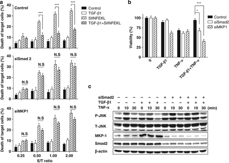
To verify participation of MKP-1 in crosstalk between TGF-β1 and TNF-α, we used siRNA specific for MKP-1 and Smad2, an upstream signaling component of MKP-1. MKP-1 knockdown efficiency at 48 h post-transfection of each siRNA was measured by immunocytochemistry and qRT-PCR (Supplementary Figure 2). These data show that both siRNAs worked well, and that MKP-1 expression was dependent on TGF-β and Smad2. After applying these siRNAs, the coculture experiments in Figure 1 were repeated. In the scrambled siRNA control sample, immunized target cells showed effector cell dose-dependent cell death, whereas pretreatment with TGF-β1 for 1 h blocked cell death. However, transfection of either siSmad2 or siMKP-1 abolished the cell death inhibitory effect of TGF-β1 (Figure 4a). Cell death was also measured in Huh-7 cells by WST-1 assay with TNF-α treatment. In control samples, TNF-α caused death in more than 30% of cells, and TGF-β1 pretreatment for 1 h protected most cells (similar to Figure 1). However, in the siSmad2 sample, rates of TNF-α-induced cell death were unaffected by TGF-β1 pretreatment. This was the same for the MKP-1 knockdown sample, indicating that knockdown of Smad2 or MKP-1 abolished the protective effect of TGF-β1 pretreatment against TNF-α-induced cell death. Instead, treatment with TGF-β1 alone caused a significant level of cell death, and TGF-β1 pretreatment exacerbated TNF-α-induced cell death in MKP-1 knockdown cells, implying that TGF-β1 cannot exert its protective functions in the absence of MKP-1 expression (Figure 4b). Intracellular signal transduction was also evaluated in Huh-7 cells after introduction of siRNA specific for Smad2. TGF-β1 did not induce MKP-1 expression in the presence of siSmad2, and phosphorylation of JNK was not attenuated by TGF-β1 pretreatment for 1 h (Figure 4c). These data collectively suggests that the link between MKP-1 and JNK is a novel point of crosstalk between the TGF-β1 and TNF-α signaling pathway.
Figure 4.
Knockdown of Smad2 or MKP-1 abolishes crosstalk between TGF-β1 and TNF-α. Knockdown of Smad2 and MKP-1 was achieved by siRNA transfection as described in the Materials and methods. Each cytokine was applied 48 h after siRNA transfection. (a) 24 h post-transfection, MC38 cells were immunized with SIINFEKL peptide. Six hours after coculture of target (MC38 cells) and effector (OT-1 mouse-derived CD8+ T cells) cells, cytotoxicity was evaluated by LDH release assay as in Figure 1a (mean±S.E.M., n=4; Bonferroni's post hoc test was applied for multiple comparisons in two-way ANOVA, ***P<0.001, N.S, not significant). (b) 24 h after treatment with each cytokine in Huh-7 cells, viability was evaluated by WST-1 assay under conditions of siRNA knockdown (mean±S.E.M., n=4; *P<0.05, ***P<0.001 by Student's t-test). (c) Huh-7 cells were incubated in the presence or absence of TGF-β1 (10 ng/ml) for 1 h, and were then treated with TNF-α (10 ng/ml) for up to 30 min. Cell lysates were subjected to immunoblot analysis as in Figure 2
Tumor-specific expression of MKP-1
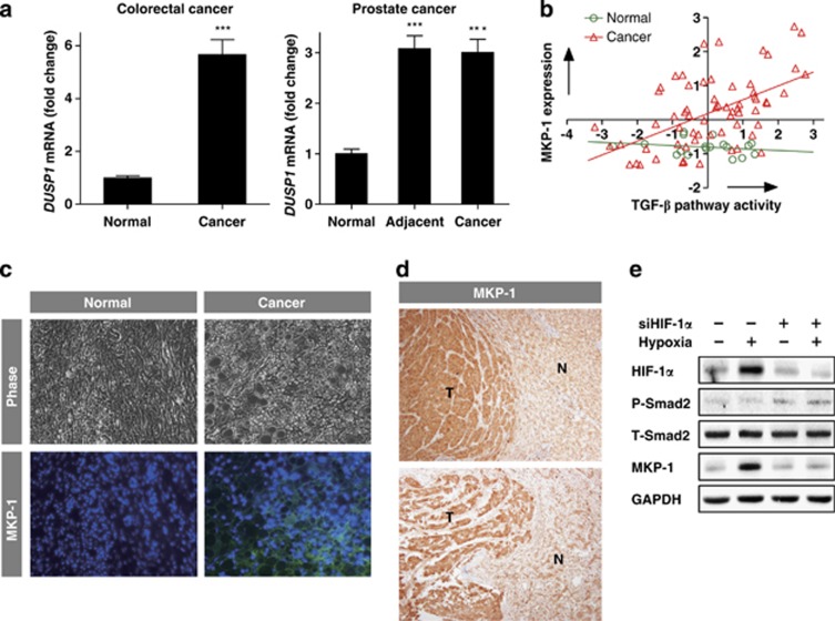
To understand the function of TGF-β1-dependent suppression of immune-mediated cell death in the tumor microenvironment, we analyzed complementary DNA microarray data provided by gene expression omnibus (GEO) databases. In human colorectal and prostate samples, MKP-1 expression was significantly higher in cancers and cancer-adjacent regions than normal regions (Figure 5a). On the basis of these data, the correlation between TGF-β pathway activity and expression of MKP-1 was evaluated in human prostate cancer tissue. The expression of MKP-1 increased according to TGF-β pathway activity, whereas normal prostate tissue showed no such correlation (Figure 5b). Correlation analysis of colorectal tissue was not included due to insufficiency of the number of samples. These data imply that TGF-β1- and MKP-1-mediated suppression of immune-mediated cell death is tumor-specific.
Figure 5.
Tumor-specific expression of MKP-1. (a) MKP-1 expression was measured in colorectal and prostate cancer patients from the GEO database using a complementary DNA microarray (mean±S.E.M.; Tukey's post hoc test was applied to significant group effects in ANOVA, P<0.0001; ***P<0.001, compared with normal tissues). (b) The correlation between TGF-β pathway activity and MKP-1 expression in prostate tissue was evaluated as described in the Materials and methods. (c) Immunofluorescence staining with FITC (green) and Hoechst (blue) of a human liver tissue array. Of the 50 HCC samples, 39 were MKP-1 positive (78%); nine normal samples did not show MKP-1 expression (0%). (d) Immunoperoxidase staining of tissue samples from HCC patients using DAB and hematoxilin. T, tumor region; N, adjacent normal region. Of 23 HCC patients, 16 showed MKP-1 expression (69.6%) (e) Cell lysates were subjected to immunoblot analysis of HIF-1α, Smad2, and MKP-1 in the presence or absence of siHIF-1α and hypoxia (oxygen concentration: 1%). GAPDH was used as the loading control
In addition to colorectal and prostate cancer, TGF-β1 is known to be abundant in many cancer tissues.28 Because MKP-1 is downstream of TGF-β1, we expected the expression of MKP-1 in cancer to also be high. We measured MKP-1 expression by immunohistochemistry. Immunofluorescence staining in human liver tissue arrays and immunoperoxidase staining in tissue samples from hepatocellular carcinoma (HCC) patients showed markedly elevated MKP-1 levels in cancer (Figure 5c and d). We also cultured Huh-7 cells in a hypoxic chamber for 24 h to mimic the in vivo tumor microenvironment, which often has an insufficient oxygen supply. Immunoblot analysis showed that MKP-1 expression was augmented under hypoxia conditions in HIF-1α-dependent manner, even though Smad2 was not phosphorylated (Figure 5e). These data confirm higher expression of MKP-1 in cancer than in normal tissues.
Chemotherapy of tumor cells with MKP-1 knockdown
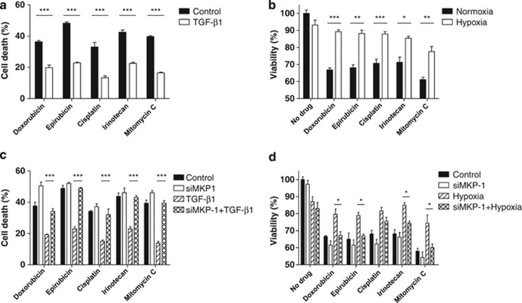
Tumors, including HCC is known to be resistant to diverse cytotoxic chemotherapies, and many clinical trials with anticancer drugs have yielded disappointing results.29, 30 To determine whether MKP-1, whose expression is only augmented in the tumor cells, can increase resistance to death receptor-mediated cell death, we conducted cytotoxicity assays with several anticancer drugs in Huh-7 cells. First, effective cytotoxic treatment concentrations of doxorubicin, epirubicin, cisplatin, irinotecan, and mitomycin C were determined by lactate dehydrogenase (LDH) release assay (data not shown). With each concentration inducing death of ∼30–40% of cells, we examined the role of TGF-β1 and hypoxia. Compared with control cells, TGF-β1 (Figure 6a) and hypoxia (Figure 6b) significantly inhibited cell death triggered by each drug. To confirm that these results were due to MKP-1 expression, we repeated the cell death assays after siRNA knockdown of MKP-1. In the absence of MKP-1, TGF-β1 (Figure 6c) and hypoxia (Figure 6d) did not exert protective roles against chemo-toxicity, indicating that MKP-1 reduced the cell death induced by the five anticancer drugs. These data suggest that resistance to anti-cancer drugs can be circumvented by MKP-1 knockdown and that systemic chemotherapy with MKP-1 knockdown can make tumor cells more susceptible to cytotoxic drugs.
Figure 6.
Knockdown of MKP-1 increases the efficacy of cytotoxic anticancer drugs. (a–b) Doxorubicin (10 ug/ml), epirubicin (10 μg/ml), cisplatin (30 μg/ml), irinotecan (500 μg/ml), and mitomycin C (50 μg/ml) were applied in Huh-7 cells with or without pretreatment with TGF-β1 (10 ng/ml) (a) or hypoxia (b). Cell death rate was evaluated by LDH release assay (mean±S.E.M., n=4; *P<0.05, **P<0.01, ***P<0.001 by Student's t-test). (c–d) 48 h post-transfection of siRNA specific for MKP-1, each drug was applied in Huh-7 cells at the same concentrations as in (a–b) with or without pretreatment with TGF-β1 (10 ng/ml) (c) or hypoxia (d). Cell death rate was evaluated by LDH release assay (mean±S.E.M., n=4; Bonferroni's post hoc test was applied for multiple comparisons in two-way ANOVA, *P<0.05, ***P<0.001)
Thus, we conclude that TGF-β1 and hypoxia ensure tumor cell survival and growth in MKP-1-dependent manner. Figure 7 demonstrates a schematic diagram of our findings. Under normal conditions, death receptor-mediated cell death signaling maintains a beneficial balance. In cancer, however, both hypoxic conditions and augmented secretion of TGF-β1 induce MKP-1 expression. MKP-1 blocks phosphorylation of JNK, which is a key mediator of death receptor-mediated cell death, and this in turn suppresses tumor cell death triggered by the immune system and anticancer drugs. This represents a novel immune escape mechanism of tumors.
Figure 7.
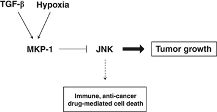
Schematic diagram of this study. We propose a novel crosstalk model involving MKP-1 expression in cancer. The tumor microenvironment is frequently hypoxic, with an overabundance of TGF-β. This triggers tumor-specific expression of MKP-1 and subsequent suppression of JNK phosphorylation. Under these conditions, the balance between cell survival and death inclines toward cell survival. Tumor cells escape from immune- and anticancer drug-mediated cell death, eventually ensuring tumor cell survival and growth
Discussion
Cells in multicellular organisms continuously face diverse molecules and integrate the signals to respond appropriately. Control of programmed cell death (whether to live or die) is one of the crucial decision-making processes. The regulation of death receptor-mediated cell death, particularly by TNF superfamily ligands, is the major determinant of apoptosis.31, 32 In this research, we elucidated a novel regulatory intersection in the death receptor-mediated cell death pathway, involving TGF-β signaling cascades and hypoxia. Our results clearly show that JNK and MKP-1 are involved in this crosstalk, with TGF-β1- and hypoxia-induced MKP-1 specifically blocking JNK phosphorylation and subsequent death receptor-mediated cell death. We also suggest that this occurs only in tumors, due to the tumor-specific constant expression of MKP-1. Even though many tumor-infiltrating immune cells secrete diverse TNF superfamily ligands,33 their effects on death receptor-mediated cell death can be blocked by hypoxic conditions and pre-existing TGF-β around tumor cells. This is an effective immune-evasion mechanism of tumor cells, and explains why hypoxia and overabundant secretion of TGF-β provide a beneficial environment for the development of cancer.34
Previous studies investigated crosstalk between the TGF-β and TNF signaling pathways. Kim, et al.35, 36 reported Smad7-involved crosstalk in several types of immune cells. In their system, Smad7 inactivates NF-κB signaling through TAB2 and TAK1, such that TGF-β shifts the TNF-α signaling balance toward cell death. In our system, however, human hepatoma and mouse colon cancer cell lines showed an opposite functional output of TGF-β and TNF-α crosstalk. Our data suggests that TGF-β1-induced MKP-1 deactivates TNF-α-induced JNK phosphorylation, whereas the NF-κB branch is not altered by the presence of TGF-β1. Interestingly, the association of the same cytokines yields different crosstalk and cellular responses according to the cell type. Even the final outputs of the two crosstalk pathways are in the counterpart, we speculate that both can contribute to immune escape of tumor cells, because excessive TGF-β simultaneously induces the death of immune cells via NF-κB inhibition and ensures tumor cell survival via JNK inactivation.
Surplus TGF-β production and hypoxic conditions are strongly correlated with various diseases, such as cancer and hepatitis.37, 38 Therefore, our experimental design is relevant to clinical issues. On the basis of TGF-β1- and hypoxia-induced expression of MKP-1, we evaluated MKP-1 as a novel drug target to overcome chemo-resistance to anticancer drugs. Because tumor cells exploit MKP-1 as a shield against immune-mediated cell death, our results provide a plausible mechanism, by which tumor cells efficiently escape from immune-mediated cell death, and might provide a rational background for development of therapeutic interventions.
Materials and Methods
Cell culture and reagents
Human HCC cell lines (Huh-7 and HepG2) and murine colorectal adenocarcinoma cell line (MC38) were maintained at 37 °C in an atmosphere containing 5% CO2 in Dulbecco's modified Eagle's medium (Welgene, Seoul, Korea) supplemented with 10% fetal bovine serum (Gibco, Gaithersburg, MD, USA), 100 U/ml penicillin and 100 μg/ml streptomycin (Invitrogen, Carlsbad, CA, USA). OT-1 mouse-derived T cells were maintained at 37 °C in an atmosphere containing 5% CO2 in RPMI 1640 medium (Welgene, Seoul, Korea) supplemented with 10% fetal bovine serum and 50 U/ml penicillin/streptomycin (PAA, Pasching, Austria).
Antibodies specific for IKK-β, phospho-IKK, phospho-IκB-α, JNK, phospho-JNK, p38, phospho-p38, p42/44, phospho-p42/44, Smad2, phospho-Smad2, MKK3, phospho-MKK3/6, MKK4, phospho-MKK4, MKK7, phospho-MKK7, TAK1, and phospho-TAK1 were purchased from Cell Signaling Technology (Beverly, MA, USA). Antibodies specific for MKP-1, MKP-2, IκB, β-actin, GAPDH, and horseradish peroxidase-conjugated secondary antibodies were obtained from Santa Cruz Biotechnology (Santa Cruz, CA, USA). An antibody specific for HIF1-α was purchased from Abcam (Cambridge, MA, USA). Human recombinant TGF-β1 and TNF-α were obtained from R&D Systems (Minneapolis, MN, USA) and TRAIL was generously provided by Dr. Kunhong Kim (Yonsei university, Seoul, Korea). An agonistic IgM type anti-Fas antibody (CH-11) was obtained from Upstate Biotechnology (Lake Placid, NY, USA). The JNK inhibitor SP600125 was purchased from Calbiochem (La Jolla, CA, USA). Five anticancer drugs, doxorubicin, epirubicin, cisplatin, irinotecan, and mitomycin C, were obtained from Sigma-Aldrich (St.Louis, MO, USA).
OT-1 mice, in vitro activation of T cells, purification, and SIINFEKL peptide loading
Eight-week-old OT-1 transgenic mice were used. Lymph nodes and spleen cells were isolated from OT-1 mice by gentle crushing of the organs and filtering through a 100-μℳ pore nylon mesh. Pooled lymph nodes and spleen cells were further treated with RBC lysis buffer (BioLegend, San Diego, CA, USA) and maintained at a density of 3–5 × 10⁶ cells/ml in 75-cm2 culture flasks. Phenotypes were determined by flow cytometric analysis using antibodies specific for CD8α and the TCR chains Vα2 and Vβ5. For in vitro OT-1T cell activation, OVA peptide (SIINFEKL) (PeproTech, Rocky Hill, NJ, USA) was added at the start of culture at a concentration of 10 μg/ml, and cells were maintained at 37 °C. After 5 days of incubation, the lymph node and spleen cells were enriched for CD8+ T cells by positive selection using MACS (Miltenyi Biotec, Auburn, CA, USA). Greater than 99% CD8+ T cell enrichment was confirmed by analysis of the surface markers CD3 and CD8α by flow cytometry. Murine colon adenocarcinoma MC38 cells were used as target cells in coculture with OT-1 mouse-derived T cells. The target cells were loaded with 10 μg/ml SIINFEKL 1 day before coculture experiments. In vitro activated OT-1 mouse CD8+ T cells were cocultured with SIINFEKL-loaded MC38 cells at given effector (E):target (T) ratios (0.25 : 1, 0.5 : 1, 1 : 1, and 2 : 1) for 6 h, and then subjected to a cell-mediated cytotoxicity assay.
Cell death and viability assays
WST-1 assays were performed to determine cell viability/death. Huh-7 cells were seeded in 24-well plates at a density of 4 × 10⁴/well in quadruplicate and WST-1 reagent (Nalgene, Rochester, NY, USA) was added to each well at a volume up to 5% that of the medium. After incubation for 2 h at 37 °C in a 5% CO2 incubator, the absorbance at 450 nm was measured using a microplate reader (Bio-Rad, Richmond, CA, USA). Cell-mediated cytotoxicity was assessed by LDH release assay (Promega, Madison, WI, USA) according to the manufacturer's protocol.
Immunoblot analysis
Immunoblot analysis was carried out to evaluate intracellular signal transduction as previously described.39 The proteins were transferred to nitrocellulose membranes and probed with antibodies specific for MKP-1, MKP-2, JNK, p38, p42/44, Smad2, IKK, IκB, MKK3/6, MKK4, MKK7, TAK1, β-actin, and GAPDH.
Immunocytochemistry
Immunocytochemistry was performed using a polyclonal antibody specific for MKP-1 (1 : 100, diluted in 1.5% BSA) as previously described.39 The slides were incubated with FITC-conjugated goat anti-rabbit IgG (Santa Cruz Biotechnology) as the secondary antibody. Slides were visualized by fluorescence microscopy (Carl Zeiss, Oberkochen, Germany).
Patients and tumor specimens
Patients admitted to Ewha Womans University Mokdong Hospital (Seoul, Korea) were considered for inclusion in the present study. Tumor tissues were dissected from 23 primary HCC patients, formalin-fixed, and paraffin-embedded for immunohistochemistry. In addition, tissue arrays containing 40 non-metastatic HCC samples, 10 metastatic HCC samples, and nine normal liver tissue samples was purchased from SuperBioChips (Seoul, Korea).
Immunohistochemistry
Paraffin-embedded tissues were deparaffinized in xylene, rehydrated in graded alcohols, and washed in water. Endogenous peroxidase activity was blocked by incubation with 3% H2O2. After treatment with 3% BSA for 30 min to block non-specific protein binding, a primary antibody specific for MKP-1 was applied (dilution: 1 : 100), and the specimens were incubated overnight. For immunoperoxidase staining, biotin-labeled secondary antibodies were applied for 30 min, followed by avidin-biotin-peroxidase complex (ABC kit; Vector Laboratories, Burlingame, CA, USA). After brief rinsing with PBS, sections were incubated with diaminobenzidine (DAB) solution for 2 min. For immunofluorescence staining, a FITC-conjugated rabbit-specific secondary antibody was applied for 30 min. Cell nuclei were counter-stained with hematoxylin for immunoperoxidase staining and with Hoechst (Sigma-Aldrich) for immunofluorescence staining.
siRNA transfection
A siRNA duplex targeted to Smad2 and MKP-1, and an siRNA with a random sequence (negative control) were synthesized by Bioneer (Daejeon, Korea). Transient transfection was carried out using Lipofectamine 2000 (Invitrogen, Carlsbad, CA, USA) according to the manufacturer's protocol. Transfection efficiency was assessed by immunoblotting and immunocytochemistry.
Microarray data sets and processing
For MKP-1 expression profile, we used two microarray data sets from the GEO database: GDS2609 (colorectal cancer) and GDS2545 (prostate cancer). In these data sets, the expression levels of mRNAs involved in TGF-β pathway from the NetPath database were averaged and defined as the TGF-β pathway activity, where expression levels were normalized by z-transformation to have same range of expression levels for all genes.
Total RNA isolation and qRT-PCR analysis
Total RNAs were isolated from Huh-7 cells using an RNeasy Mini Kit (Qiagen, Hilden, Germany). cDNA was synthesized from 1 μg of total RNA with the High-Capacity RNA-to-cDNA Kit (Applied Biosystems, Foster City, CA, USA) according to the manufacturer's protocol. RT-PCR was then carried out in triplicate to determine mRNA levels of MKP-1 using the StepOne TM RT-PCR system with SYBR Green PCR Master Mix (Applied Biosystems). The MKP-1 primer (Bioneer) sequences were 5′-CCTGTCCACTCCACGAACAGT-3′ for the forward primer and 5′-GCTGGGAGAGGTCGTAATGG-3′ for the reverse primer. Target mRNA levels were normalized to an endogenous reference GAPDH.
Statistical analysis
Data are presented as the means±S.E.M. Levels of significance for comparisons between two independent samples were determined using student's t-test. Groups were compared by one-way or two-way analysis of variance (ANOVA) with Tukey's post hoc test or Bonferroni's post hoc test applied to significant main effects.
Acknowledgments
We authors acknowledge Dr. Dong Eun Song (Ewha Womans University, Seoul, Korea) for providing human HCC samples for immunohistostaining. This work was supported by the National Research Foundation of Korea (NRF) grant funded by the Korea government (MEST; No. 2012R1A2A4A01007108).
Glossary
- TGF
transforming growth factor
- MKP
mitogen-activated protein kinase phosphatase
- JNK
c-Jun N-terminal kinase
- HCC
hepatocellular carcinoma
- DUSP
dual specificity phosphatase
- TNF
tumor necrosis factor
- TRAIL
TNF-related apoptosis-inducing ligand
- FasL
Fas ligand
- NF-κB
nuclear factor-kappaB
- MAPK
mitogen-activated protein kinase
- OVA
ovalbumin
- IKK
IκB kinase
- MKK
mitogen-activated protein kinase kinase
- TAK
transforming growth factor-beta-activated kinase
- \GEO
Gene expression omnibus
- LDH
lactate dehydrogenase
- DAB
diaminobenzidine
The authors declare no conflict of interest.
Footnotes
Supplementary Information accompanies this paper on Cell Death and Disease website (http://www.nature.com/cddis)
Edited by A Stephanou
Supplementary Material
References
- de Visser KE, Eichten A, Coussens LM. Paradoxical roles of the immune system during cancer development. Nat Rev Cancer. 2006;6:24–37. doi: 10.1038/nrc1782. [DOI] [PubMed] [Google Scholar]
- Rabinovich GA, Gabrilovich D, Sotomayor EM. Immunosuppressive strategies that are mediated by tumor cells. Annu Rev Immunol. 2007;25:267–296. doi: 10.1146/annurev.immunol.25.022106.141609. [DOI] [PMC free article] [PubMed] [Google Scholar]
- Smyth MJ, Godfrey DI, Trapani JA. A fresh look at tumor immunosurveillance and immunotherapy. Nat Immunol. 2001;2:293–299. doi: 10.1038/86297. [DOI] [PubMed] [Google Scholar]
- Drake CG, Jaffee E, Pardoll DM. Mechanisms of immune evasion by tumors. Adv Immunol. 2006;90:51–81. doi: 10.1016/S0065-2776(06)90002-9. [DOI] [PubMed] [Google Scholar]
- Dunn GP, Koebel CM, Schreiber RD. Interferons, immunity and cancer immunoediting. Nat Rev Immunol. 2006;6:836–848. doi: 10.1038/nri1961. [DOI] [PubMed] [Google Scholar]
- Shi Y, Massague J. Mechanisms of TGF-beta signaling from cell membrane to the nucleus. Cell. 2003;113:685–700. doi: 10.1016/s0092-8674(03)00432-x. [DOI] [PubMed] [Google Scholar]
- Bodmer S, Strommer K, Frei K, Siepl C, de Tribolet N, Heid I, et al. Immunosuppression and transforming growth factor-beta in glioblastoma. Preferential production of transforming growth factor-beta 2. J Immunol. 1989;143:3222–3229. [PubMed] [Google Scholar]
- von Bernstorff W, Voss M, Freichel S, Schmid A, Vogel I, Johnk C, et al. Systemic and local immunosuppression in pancreatic cancer patients. Clin Cancer Res. 2001;7 (3 Suppl:925s–932s. [PubMed] [Google Scholar]
- Hersey P. Impediments to successful immunotherapy. Pharmacol Ther. 1999;81:111–119. doi: 10.1016/s0163-7258(98)00038-2. [DOI] [PubMed] [Google Scholar]
- Yang L, Pang Y, Moses HL. TGF-beta and immune cells: an important regulatory axis in the tumor microenvironment and progression. Trends Immunol. 2010;31:220–227. doi: 10.1016/j.it.2010.04.002. [DOI] [PMC free article] [PubMed] [Google Scholar]
- Massague J. TGFbeta in Cancer. Cell. 2008;134:215–230. doi: 10.1016/j.cell.2008.07.001. [DOI] [PMC free article] [PubMed] [Google Scholar]
- Tong XK, Hamel E. Transforming growth factor-beta 1 impairs endothelin-1-mediated contraction of brain vessels by inducing mitogen-activated protein (MAP) kinase phosphatase-1 and inhibiting p38 MAP kinase. Mol Pharmacol. 2007;72:1476–1483. doi: 10.1124/mol.107.039602. [DOI] [PubMed] [Google Scholar]
- Owens DM, Keyse SM. Differential regulation of MAP kinase signalling by dual-specificity protein phosphatases. Oncogene. 2007;26:3203–3213. doi: 10.1038/sj.onc.1210412. [DOI] [PubMed] [Google Scholar]
- Mikami F, Lim JH, Ishinaga H, Ha UH, Gu H, Koga T, et al. The transforming growth factor-beta-Smad3/4 signaling pathway acts as a positive regulator for TLR2 induction by bacteria via a dual mechanism involving functional cooperation with NF-kappaB and MAPK phosphatase 1-dependent negative cross-talk with p38 MAPK. J Biol Chem. 2006;281:22397–22408. doi: 10.1074/jbc.M602124200. [DOI] [PubMed] [Google Scholar]
- Patterson KI, Brummer T, O'Brien PM, Daly RJ. Dual-specificity phosphatases: critical regulators with diverse cellular targets. Biochem J. 2009;418:475–489. doi: 10.1042/bj20082234. [DOI] [PubMed] [Google Scholar]
- Deng Y, Ren X, Yang L, Lin Y, Wu XA. JNK-dependent pathway is required for TNFalpha-induced apoptosis. Cell. 2003;115:61–70. doi: 10.1016/s0092-8674(03)00757-8. [DOI] [PubMed] [Google Scholar]
- Krueger A, Baumann S, Krammer PH, Kirchhoff S. FLICE-inhibitory proteins: regulators of death receptor-mediated apoptosis. Mol Cell Biol. 2001;21:8247–8254. doi: 10.1128/MCB.21.24.8247-8254.2001. [DOI] [PMC free article] [PubMed] [Google Scholar]
- Schmitz I, Kirchhoff S, Krammer PH. Regulation of death receptor-mediated apoptosis pathways. Int J Biochem Cell Biol. 2000;32:1123–1136. doi: 10.1016/s1357-2725(00)00048-0. [DOI] [PubMed] [Google Scholar]
- Kischkel FC, Lawrence DA, Chuntharapai A, Schow P, Kim KJ, Ashkenazi A. Apo2L/TRAIL-dependent recruitment of endogenous FADD and caspase-8 to death receptors 4 and 5. Immunity. 2000;12:611–620. doi: 10.1016/s1074-7613(00)80212-5. [DOI] [PubMed] [Google Scholar]
- Chen G, Goeddel DV. TNF-R1 signaling: a beautiful pathway. Science. 2002;296:1634–1635. doi: 10.1126/science.1071924. [DOI] [PubMed] [Google Scholar]
- Wajant H, Pfizenmaier K, Scheurich P. Tumor necrosis factor signaling. Cell Death Differ. 2003;10:45–65. doi: 10.1038/sj.cdd.4401189. [DOI] [PubMed] [Google Scholar]
- Shen HM, Pervaiz S. TNF receptor superfamily-induced cell death: redox-dependent execution. Faseb J. 2006;20:1589–1598. doi: 10.1096/fj.05-5603rev. [DOI] [PubMed] [Google Scholar]
- Davis RJ. Signal transduction by the JNK group of MAP kinases. Cell. 2000;103:239–252. doi: 10.1016/s0092-8674(00)00116-1. [DOI] [PubMed] [Google Scholar]
- Muppidi JR, Tschopp J, Siegel RM. Life and death decisions: secondary complexes and lipid rafts in TNF receptor family signal transduction. Immunity. 2004;21:461–465. doi: 10.1016/j.immuni.2004.10.001. [DOI] [PubMed] [Google Scholar]
- Chauhan D, Li G, Hideshima T, Podar K, Mitsiades C, Mitsiades N, et al. JNK-dependent release of mitochondrial protein, Smac, during apoptosis in multiple myeloma (MM) cells. J Biol Chem. 2003;278:17593–17596. doi: 10.1074/jbc.C300076200. [DOI] [PubMed] [Google Scholar]
- Nakano H. Signaling crosstalk between NF-kappaB and JNK. Trends Immunol. 2004;25:402–405. doi: 10.1016/j.it.2004.05.007. [DOI] [PubMed] [Google Scholar]
- Lin A. Activation of the JNK signaling pathway: breaking the brake on apoptosis. Bioessays. 2003;25:17–24. doi: 10.1002/bies.10204. [DOI] [PubMed] [Google Scholar]
- Elliott RL, Blobe GC. Role of transforming growth factor Beta in human cancer. J Clin Oncol. 2005;23:2078–2093. doi: 10.1200/JCO.2005.02.047. [DOI] [PubMed] [Google Scholar]
- Yau T, Chan P, Epstein R, Poon RT. Management of advanced hepatocellular carcinoma in the era of targeted therapy. Liver Int. 2009;29:10–17. doi: 10.1111/j.1478-3231.2008.01916.x. [DOI] [PubMed] [Google Scholar]
- Yu H, Park J, Lee J, Choi K, Choi C. Constitutive expression of MAP kinase phosphatase-1 confers multi-drug resistance in human glioblastoma cells. Cancer Res Treat. 2012;44:195–201. doi: 10.4143/crt.2012.44.3.195. [DOI] [PMC free article] [PubMed] [Google Scholar]
- Yousefi S, Conus S, Simon HU. Cross-talk between death and survival pathways. Cell Death Differ. 2003;10:861–863. doi: 10.1038/sj.cdd.4401262. [DOI] [PubMed] [Google Scholar]
- Heyninck K, Beyaert R. Crosstalk between NF-kappaB-activating and apoptosis-inducing proteins of the TNF-receptor complex. Mol Cell Biol Res Commun. 2001;4:259–265. doi: 10.1006/mcbr.2001.0295. [DOI] [PubMed] [Google Scholar]
- Liotta LA, Kohn EC. The microenvironment of the tumour-host interface. Nature. 2001;411:375–379. doi: 10.1038/35077241. [DOI] [PubMed] [Google Scholar]
- Moutsopoulos NM, Wen J, Wahl SM. TGF-beta and tumors--an ill-fated alliance. Current Opin Immunol. 2008;20:234–240. doi: 10.1016/j.coi.2008.04.003. [DOI] [PMC free article] [PubMed] [Google Scholar]
- Hong S, Lim S, Li AG, Lee C, Lee YS, Lee EK, et al. Smad7 binds to the adaptors TAB2 and TAB3 to block recruitment of the kinase TAK1 to the adaptor TRAF2. Nat Immunol. 2007;8:504–513. doi: 10.1038/ni1451. [DOI] [PubMed] [Google Scholar]
- Hong S, Lee C, Kim SJ. Smad7 sensitizes tumor necrosis factor induced apoptosis through the inhibition of antiapoptotic gene expression by suppressing activation of the nuclear factor-kappaB pathway. Cancer Res. 2007;67:9577–9583. doi: 10.1158/0008-5472.CAN-07-1179. [DOI] [PubMed] [Google Scholar]
- Blobe GC, Schiemann WP, Lodish HF. Role of transforming growth factor beta in human disease. N Engl J Med. 2000;342:1350–1358. doi: 10.1056/NEJM200005043421807. [DOI] [PubMed] [Google Scholar]
- Semenza GL. Oxygen sensing, homeostasis, and disease. N Engl J Med. 2011;365:537–547. doi: 10.1056/NEJMra1011165. [DOI] [PubMed] [Google Scholar]
- Park J, Kang W, Ryu SW, Kim WI, Chang DY, Lee DH, et al. Hepatitis C virus infection enhances TNFalpha-induced cell death via suppression of NF-kappaB. Hepatology. 2012;56:831–840. doi: 10.1002/hep.25726. [DOI] [PubMed] [Google Scholar]
Associated Data
This section collects any data citations, data availability statements, or supplementary materials included in this article.


