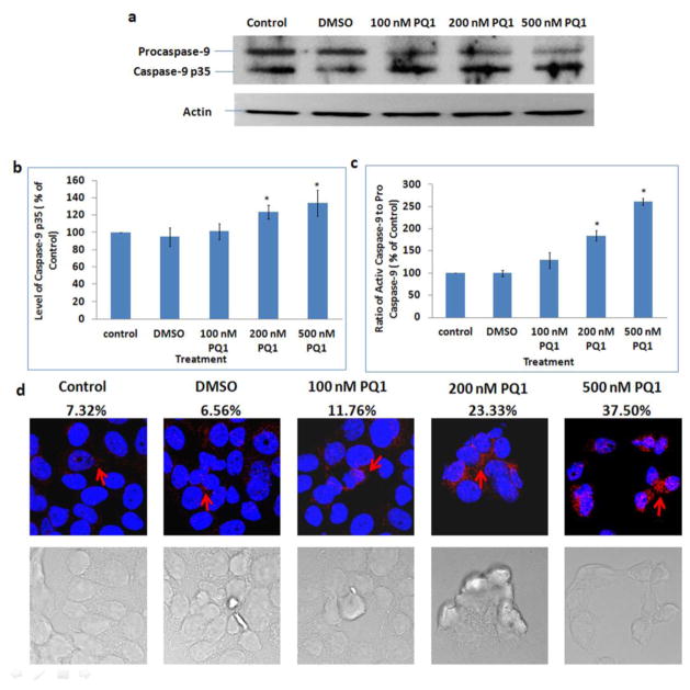
Fig. 7. PQ1 activates caspase-9 in T47D cells.
T47D cells were treated with DMSO and various concentrations of PQ1 for 48 hours. (a) Levels of procaspase-9 and caspase-9 p35 were examined by Western blot analysis using anti-caspase-9 p35 (H-170) antibody which detects the p35 subunit and precursor of caspase-9. Actin was used as a loading control. (b) Graphical presentation of three independent experiments shows the pixel intensities of caspase-9 p35 normalized to the controls. * P-value is <0.05 compared to control. (c) Graphical presentation of three independent experiments shows the ratio of active caspase-9 (caspase-9 p35) to pro-caspase-9. Results are normalized to the control. * P-value is <0.05 compared to control. (d) Immunofluorescence was performed using anti-cleaved caspase-9 (Asp315) antibody, a rabbit polyclonal antibody specific to the 35 kDa large fragment of caspase-9 following cleavage at aspartic acid 315. Red indicates caspase-9 p35 and blue indicates the nuclei. Percentages of cells with positive staining were labeled on top of relative images.

