Abstract
Vesicular stomatitis virus G glycoprotein (VSV-G) is the most widely used envelope protein for retroviral and lentiviral vector pseudotyping; however, serum inactivation of VSV-G pseudotyped vectors is a significant challenge for in vivo gene delivery. To address this problem, we conducted directed evolution of VSV-G to increase its resistance to human serum neutralization. After six selection cycles, numerous common mutations were present. Based on their location within VSV-G, we analyzed whether substitutions in several surface exposed residues could endow viral vectors with higher resistance to serum. S162T, T230N, and T368A mutations enhanced serum resistance, and additionally K66T, T368A, and E380K substitutions increased the thermostability of VSV-G pseudotyped retroviral vectors, an advantageous byproduct of the selection strategy. Analysis of a number of combined mutants revealed that VSV-G harboring T230N + T368A or K66T + S162T + T230N + T368A mutations exhibited both higher in vitro resistance to human serum and higher thermostability, as well as enhanced resistance to rabbit and mouse serum. Finally, lentiviral vectors pseudotyped with these variants were more resistant to human serum in a murine model. These serum-resistant and thermostable VSV-G variants may aid the application of retroviral and lentiviral vectors to gene therapy.
Keywords: serum-resistant, thermostable, VSV-G, directed evolution, pseudotyping
INTRODUCTION
Since the first human gene therapy trial in 1989,1 there has been remarkable progress in both understanding the genetic basis of human disease and developing improved gene delivery vector systems to treat them.2–4 For the latter, clinical gene delivery to date has focused primarily on viral vehicles due to their evolved ability to efficiently transport genetic information to into cells. However, viral vectors still face a number of challenges including immunogenencity, targeted delivery, safe genome integration, and vector production.7–9
Pseudotyping – the replacement of a virus’ attachment protein with that of a different virus – has enabled progress in addressing several of these concerns. For example, vesicular stomatitis virus G glycoprotein (VSV-G) is the most widely used glycoprotein for retroviral and lentiviral vector pseudotyping, as it offers several advantages including effective delivery to a broad range of cell types, enhanced vector stability, and increased infectious titer.16,17 That said, VSV-G is cytotoxic to producer cells,18 though the use of tetracycline-regulating promoters has alleviated this problem and facilitated the generation of stable cell lines expressing VSV-G.19 As an additional problem, however, serum inactivation of VSV-G pseudotyped viral vectors impedes their in vivo use.20 This inactivation is mediated by proteins of the complement cascade. In general, complement activation can occur via classical, alternative, and lectin pathways, and these systems are tightly regulated by the complement regulatory proteins (CRPs).21 However, the precise mechanism of VSV-G inactivation and the protein regions involved are not known.
Several approaches have been explored to overcome serum inactivation. Although known inhibitors of complement improve vector survival in serum in vitro,22 systemic delivery of complement inhibitors can be accompanied by toxicity.23 Incorporating CRPs directly into a virus – including human decay-accelerating factor (DAF)/CD55 and or membrane cofactor protein (MCP)/CD46 – enhanced the resistance of the virus to human serum inactivation.24–26 However, the efficiency of CRP incorporation into virions varied with the types of virus and producer cell, and the direct fusion of CRP to viral proteins resulted in low titers. As a result, only systemic use of modified adenovirus has been reported.27 As another approach, chemical “shielding” of VSV-G via bioconjugation of polyethylene glycol (PEG) or polyethylenimine enhanced the serum resistance of lentiviral vectors,28,29 though vector titer was reduced, and a chemical modification adds an additional step in vector production for clinical development. Alternatively, using different, serum-resistant glycoproteins – such as feline endogenous virus envelope protein RD114 or cocal virus envelope30,31 – for pseudotyping could pose a solution. However, RD114 pseudotyping resulted in lower titers (approximately 100-fold) compared to the VSV-G pseudotyping, and its use in vivo has thus been somewhat limited to date.32,33 Furthermore, the cocal envelope has not been tested in vivo to date, in contrast to the considerable in vivo characterization of VSV-G. Finally, utilizing packaging cell lines in which alpha-galactosyl-transferase genes are disrupted can generate vector lacking galactosyl-α(1,3)galactose epitopes and thereby reduce sensitivity to human complement, through this approach has not been broadly explored.25
Directed evolution has recently been developed and implemented to improve numerous properties of viral vectors, and this approach can be effective even in the absence of a mechanistic understanding of challenges facing a vector system. In particular, multiple studies have applied molecular evolution to improve vector stability, pseudotyping efficiency, transduction efficiency, resistance to antibodies, genomic integration selectivity, and other properties.34–39 Here, we explore whether directed evolution of the VSV-G envelope may enable pseudotyped viral vectors to resist neutralization by human serum. Through a combination of evolution and site-directed mutagenesis, we created VSV-G variants that are both resistant to a panel of human and animal sera and are thermostable. Furthermore, variants exhibited enhanced gene delivery in the presence of human serum in vivo.
RESULTS
Library construction and selection
VSV-G libraries were constructed by error-prone PCR at two different mutation rates: a 2 × 10⁶ mutant library with a low error rate of 1–4 nucleotide mutations per VSV-G sequence and a 2 × 10⁵ mutant library with 3–8 nucleotide mutations per sequence, as quantified by sequencing of randomly chosen clones. For selection, these libraries were inserted into a retroviral vector plasmid that was used to package a library of vector particles, where each harbored a vector genome encoding the VSV-G variant incorporated in the envelope of that particle. This requires that ~1 plasmid carrying a VSV-G variant be transfected into a producer cell during packaging. For initial optimization of this process, HEK 293T cells were transfected with 62.5 ng – 2 µg of the retroviral vector CLPIT-GFP followed by flow cytometry analysis (Supplementary data Fig. S1.). Upon transfection with 62.5 ng of CLPIT-GFP, approximately 15% of cells expressed GFP, suggesting that these conditions may introduce on average <1 plasmid per cell and could thus produce the desired virion library. Therefore, the two CLPIT VSV-G libraries were packaged separately using these conditions, and the resulting vectors were combined for selection.
To develop a strategy for selecting VSV-G variants resistant to serum neutralization, retroviral vector pseudotyped with wild type VSV-G was diluted fivefold in a mixture of human serum from 18 donors and incubated at 37°C (Supplementary data Fig. S2.). Vector infectivity progressively decreased with longer incubations, such that only ~5% of the vector remained infective after 6 hours. The VSV-G library was thus selected to evade human serum inactivation by incubating the virions with serum at 37°C for 6 hours. 293T cells were then infected with the treated viral vector library (at a MOI < 0.1 to prevent multiple infections of a single cell). Following selection with 1 µg/ml of puromycin, the selected viral pool was rescued via transfection of the pCMV gag/pol helper plasmid.
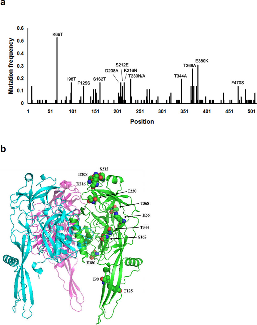
This process was repeated for six selection steps, and VSV-G variants were then recovered from 293T cell genomic DNA via PCR and inserted into the plasmid pcDNA IVS to generate helper plasmids for vector production. DNA sequencing revealed that while there were no duplicates among 36 randomly chosen VSV-G clones, common mutations were present (Figure 1A; Supplementary data Fig. S3.). The positions of these ‘hot spot’ mutations were analyzed within the structure of the prefusion form of VSV-G (Figure 1B),40 which indicated that Lys66, Ser162, Asp208, Ser212, Lys216, Thr230, Thr368, and Glu380 are located on the protein surface. In addition, Thr344 is located in the core region, Ile98 and Phe125 are located on the leg region that points toward the viral membrane, and Phe470 is located on the transmembrane domain of VSV-G. The latter, non-solvent exposed residues (Thr344, Ile98, Phe125, and Phe470) would appear unable to interact with components of human serum, and the former set of mutations was instead investigated.
Figure 1. Common mutations following directed evolution of VSV-G.
(a) The frequency of mutation at each amino acid residue of VSV-G among 36 randomly chosen VSV-G clones after 5 or 6 selection steps. (b) The location of each apparent ‘hot spot mutation’ in the crystal structure of the prefusion form of VSV-G (PDB ID: 2J6J). Figure was made using PyMol (http://www.pymol.org). Each monomer of VSV-G was colored in green, purple, and sky-blue, respectively. Green, blue, and red balls represent carbon, nitrogen, and oxygen atoms, respectively.
Functional analysis of individual VSV-G mutations
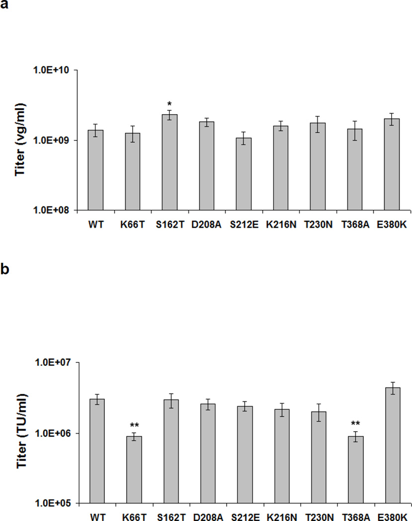
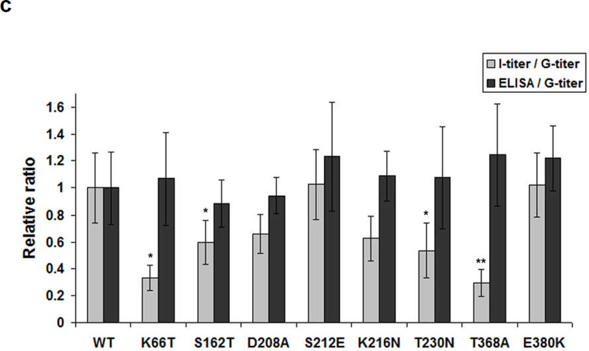
As there were no dominant individual clones following selection, we analyzed the roles of the common mutations in potential VSV-G serum resistance. Site-directed mutagenesis was used to introduce the individual, identified point mutations into the surface-exposed positions of wild type VSV-G (Figure 1), and GFP-expressing retroviral vectors were packaged. We found that, with the exception of S162T, the genomic titers (G-titers) of mutant vectors were equivalent to wild type vector (Figure 2A) suggesting that VSV-G can tolerate mutations that emerged from this library selection without loss of assembly. The mutant infectious titers (I-titers) were similar to wild type, with the exceptions that VSV-G E380K exhibited a 40% increase, and K66T and T368A showed a 70% decrease, respectively, in infectivity (Figure 2B). We investigated the relative ratio of infectious titer to genomic titer of all mutant variants (Figure 2C & Supplementary data Fig. S4C). Vector containing K66T, S162T, T230N, or T368A VSV-G showed somewhat lower I-titer/G-titer ratios compared to the wild type, suggesting that these mutations slightly decreased the infectivities of the viral vectors. We also investigated the relative ratio of VSV-G expression level, as obtained with a VSV-G ELISA assay, to G-titer (Figure 2C), and did not observe any significant differences compared to wild type VSV-G.
Figure 2. Genomic and infectious titers of VSV-G chimeric retroviral vectors.
Murine retroviral vector was packaged with the pCLPIT GFP vector plasmid, pCMV gag-pol, and pcDNA IVS VSV-G helper plasmid containing individual VSV-G variants. (a) Vector genomic titers were measured by real-time qPCR. (b) Transduction efficiencies on 293T cells were determined by flow cytometry analysis of retroviral vector mediated GFP expression. (c) Relative ratios of infectious titers to genomic titers (grey bars) titers and VSV-G ELISA data to genomic titers (black bars) of the retroviral vectors were calculated with the ratio of wild type VSV-G pseudotyped retroviral vector as 1, respectively. Error bars denote SD (n = 3). * and ** indicate statistical differences of P < 0.05 and P < 0.01, respectively, compared to infectivity of wild type VSV-G, as determined using an one-way ANOVA.
Human serum inactivation of variant VSV-G retroviral vectors
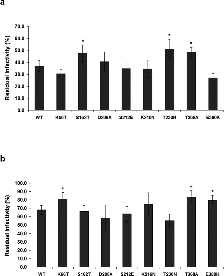
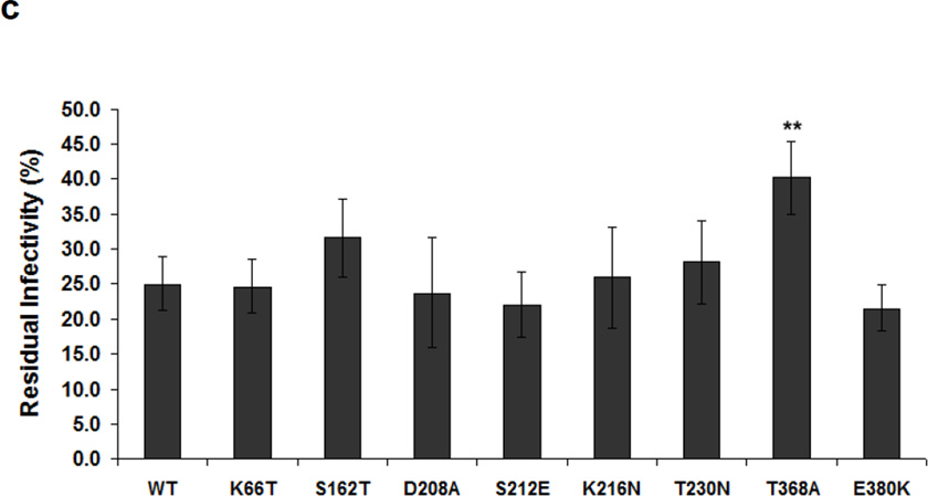
The human serum inactivation of retroviral vectors bearing wild type and single mutant VSV-G variants was examined. Since non-infectious of empty particles could conceivably affect a neutralization assay, we used the results of a VSV-G ELISA assay (Supplementary data Fig. S4C.) to equalize the amount of VSV-G added to each sample. The resulting ELISA-normalized viral vectors levels were incubated with the mixture of human serum from 18 donors. Following serum incubation at 37°C for 1 hr, titers were quantitated and reported as the percentage of remaining titer compared to that of control samples incubated at 37°C for 1 hr with phosphate-buffered saline (PBS) (pH 7.4) (Figure 3A) or heat inactivated (56°C, 30 min) human sera (HIHS) (Supplementary data Fig. S5A). Viral vectors were not inactivated in HIHS, suggesting that the inactivation may be caused by complement (Supplementary data Fig. S5B), and the results of these two normalization were thus very comparable. In particular, individual VSV-G mutants S162T (p < 0.05), T230N (p < 0.05), or T368A (p < 0.05) showed significantly increased human serum resistance compared to wild type VSV-G (Figure 3A).
Figure 3. Human serum resistance and thermostability of retroviral vectors pseudotyped with VSV-G mutants.
The amounts of viral vectors were normalized based on VSV-G ELISA assay. (a) Human serum neutralization was quantified by measuring vector titers after incubation with human serum at 37°C for 1 hr, relative to those after incubation with PBS at 37°C for 1 hr. (b) Thermal effects were determined by quantifying relative titers after incubation with PBS at 37°C for 1 hr compared to those without incubation at 37°C. (c) Human serum neutralization and thermal effects were determined by calculation of relative titers after incubation with human serum at 37°C for 1 hr compared to those without incubation at 37°C. Error bars denote SD (n = 4). * and ** indicate statistical differences of P < 0.05 and P < 0.01, respectively, compared to infectivity of wild type VSV-G, as determined using an one-way ANOVA.
Incubation in serum at 37°C may select not only for serum resistance but serendipitously also for thermostability, so we further examined the results for the vectors before and after incubation for 1 hr in PBS (Figure 3B). Mutants carrying K66T, T368A, or E380K interestingly showed higher thermostability compared to wild type VSV-G (p < 0.05). Due to the combined effects of serum resistance and thermal stability, a mutant carrying T368A showed > 1.6-fold higher infectivity (p < 0.01) compared to wild type VSV-G upon incubation in human serum at 37°C for 1 hr (Figure 3C). Based on these data, the S162T, T230N, and T368A mutations were deemed beneficial for serum resistance.
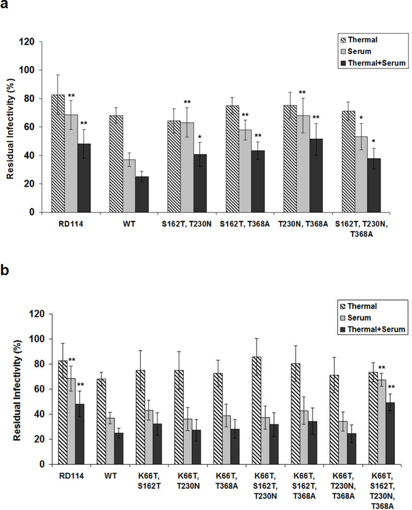
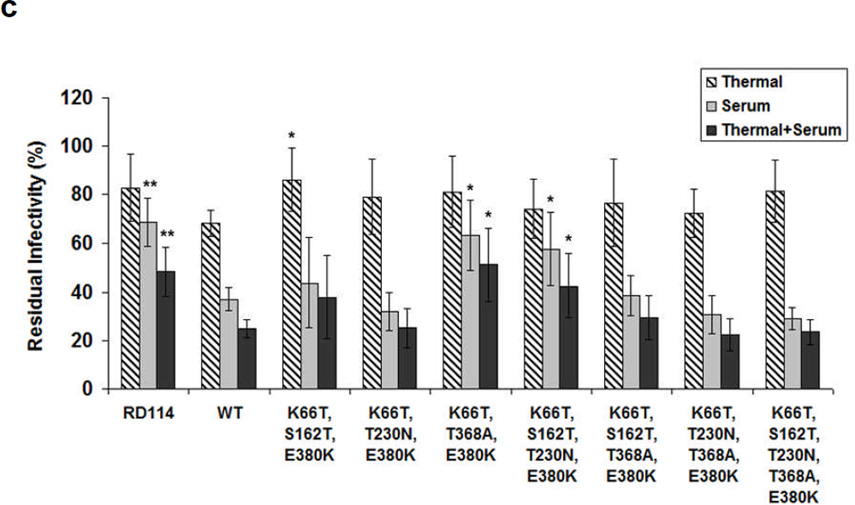
Combining mutations that individually contribute to a given property can further enhance that property.41,42 We therefore generated a number of double and triple VSV-G mutants from individual mutations shown to confer serum resistance. All VSV-G mutants analyzed showed significantly higher resistance (p < 0.01 or 0.05) to human serum (grey bars in Figure 4A). Furthermore, all such mutants showed similar thermal stability compared to wild type VSV-G (striped bars in Figure 4A). For comparison, we also prepared retroviral vector pseudotyped with an RD114 envelope, which has previously been reported to have higher serum resistance than wild type VSV-G,33,43 as another control. As reported RD114 showed increased resistance to human serum compared to wild type VSV-G, though again this envelope cannot package vector to the high titers needed for in vivo use.33 Collectively, the T230N + T368A mutant showed the highest residual infectivity (~68%) compared to the residual infectivity of parental VSV-G (~37%) and similar residual infectivity (~ 68%) of RD114 after incubation in human serum at 37°C for 1 hr.
Figure 4. Human serum neutralization and thermostability of retroviral vectors pseudotyped with VSV-G variants that combine several ‘hot spot mutations’.
VSV-G mutants with combined beneficial mutations were generated by site-directed mutagenesis. The amounts of viral vectors were normalized based on VSV-G ELISA assay. Thermal effects, human serum neutralization, and combined serum neutralization and thermal effects were determined by quantifying relative titers after incubation with PBS at 37°C for 1 hr compared to those without incubation at 37°C, after incubation with human serum at 37°C for 1 hr compared to those after incubation with PBS at 37°C for 1 hr, and after incubation with human serum at 37°C for 1 hr compared to those without incubation at 37°C, respectively. (a) Variants contained ‘hot spot mutations’ for human serum resistance. (b) K66T and (c) E380K were added to enhance the thermal stability of retroviral vectors. Error bars denote SD (n = 4). * and ** indicate statistical differences of P < 0.05 and P < 0.01, respectively, compared to the wild type VSV-G, as determined using an one-way ANOVA.
K66T, T368A, and E380K mutations increased VSV-G thermal stability (Figure 3), and we thus investigated the effects of adding these mutations to the promising serum resistant variants. Several variants containing K66T or K66T + E380K substitutions showed similar or slightly higher thermal stability compared to wild type VSV-G, and several mutants (K66T + S162T + T230N + T368A, K66T + T368A + E380K, and K66T + S162T + T230N + E380K) also showed statistically higher serum resistance (p < 0.05) (Figure 4B&C).
Human serum inactivation of variant VSV-G lentiviral vectors
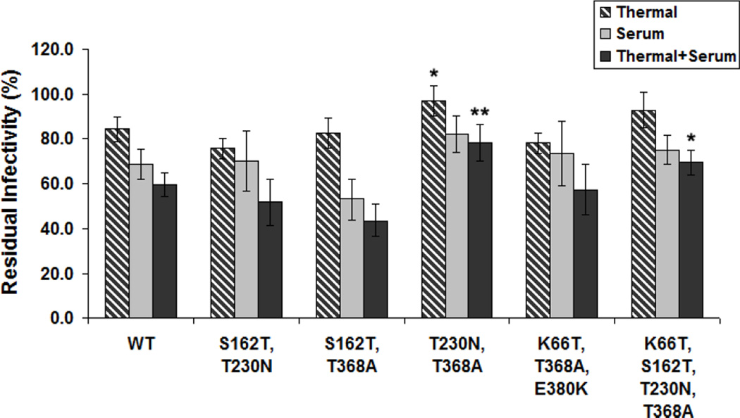
While the envelope compositions of retroviral and lentiviral vectors are likely similar, we also analyzed the ability of VSV-G variants to confer serum resistance to lentivirus. Based on results with retroviral vectors, we analyzed the top five VSV-G variants (S162T + T230N, S162T + T368A, T230N + T368A, K66T + T368A + E380K, and K66T + S162T + T230N + T368A) showing higher serum resistance for retrovirus. Equal amounts of packaged lentivirus (7 × 10⁴ GFP TU) were diluted fivefold in human serum, or PBS (pH 7.4) as a control, and incubated at 37°C for 1 hr. Vector pseudotyped with several mutant VSV-G showed a greater resistance to human serum compared to wild type VSV-G pseudotyped lentiviral vector (Figure 5). Importantly, mutants T230N + T368A or K66T + S162T + T230N + T368A had higher combined thermostability and serum-resistance than wild type VSV-G.
Figure 5. Human serum neutralization and thermostability of lentiviral vectors pseudotyped with VSV-G variants.
A standard, GFP encoding lentiviral vector was packaged with five VSV-G mutants that appeared promising in the retroviral results. Five VSV-G variants (S162T + T230N, S162T + T368A, T230N + T368A, K66T + T368A + E380K, and K66T + S162T + T230N + T368A) showing higher resistance and thermal stability for retroviral vector packaging were selected. Thermal effects, human serum neutralization, and combined serum neutralization and thermal effects were determined by quantifying relative titers after incubation with PBS at 37°C for 1 hr compared to those without incubation at 37°C, after incubation with human serum at 37°C for 1 hr compared to those after incubation with PBS at 37°C for 1 hr, and after incubation with human serum at 37°C for 1 hr compared to those without incubation at 37°C, respectively. Error bars denote SD (n = 4). * and ** indicate statistical differences of P < 0.05 and P < 0.01, respectively, compared to the wild type VSV-G, as determined using an one-way ANOVA.
Vector inactivation by animal serum
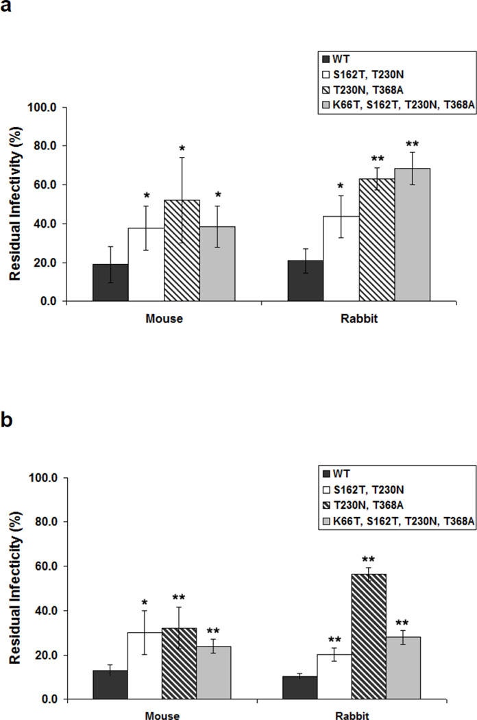
VSV-G pseudotyped viral vectors can be inactivated by sera from mouse, rat, and guinea pig, which can impact the interpretation of animal studies.25,44 The relative sensitivity of VSV-G pseudotyped retroviral or lentiviral vectors to several animal sera was thus tested (Figure 6A and 6B, respectively). S162T + T230N, T230N + T368A, or K66T + S162T + T230N + T368A VSV-G showed statistically higher resistance to mouse and rabbit sera for both retroviral and lentiviral vectors.
Figure 6. Aminal serum neutralization of retroviral and lentiviral vectors pseudotyped with VSV-G variants.
For three variants (S162T+T230N, T230N +T368A and K66T + S162T + T230N + T368A) and wild type VSV-G, neutralization by animal sera was examined. (a) Retroviral vectors and (b) lentiviral vectors were diluted fivefold in animal sera and incubated at 37°C for 1 hr. Serum inactivation was determined by quantifying relative titers after incubation with human serum at 37°C for 1 hr compared to those after incubation with PBS at 37°C for 1 hr. Error bars denote SD (n = 4). * and ** indicate statistical differences of P < 0.05 and P < 0.01, respectively, compared to the wild type VSV-G, as determined using an one-way ANOVA.
In vitro transduction of several cell lines
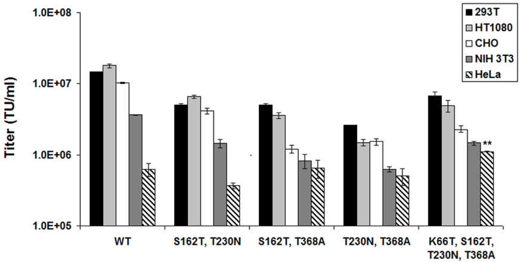
While the infectivities of 293T cells were comparable to wild type VSV-G in the absence of human serum (Figure 2), we characterized transduction of several other cell lines to determine whether the mutations affected infectivity (Figure 7). While the infectivities of the variants were somewhat lower for several cell types, the trends in infectivity for all lines was similar between wild type and mutant VSV-G. Interestingly, the mutant carrying K66T + S162T + T230N +T368A showed statistically higher transduction efficiency to HeLa cells compared to wild type (p < 0.01).
Figure 7. In vitro transduction of multiple cell lines with retroviral vectors pseudotyped with VSV-G variants.
Retroviral vectors expressing GFP were used to transduce a panel of cell lines: HEK 293T, HT1080 (human fibrosarcoma cell line), CHO K1, NIH 3T3 (mouse embryonic fibroblast cell line), and HeLa cells to assess the transduction profile of the novel VSV-G variants. Error bars denote SD (n = 3). ** indicates statistical differences of P < 0.01, as determined using an one-way ANOVA.
In vivo analysis of human serum resistance
The ability of two promising variants – which we now term mutant 1 (T230N + T368A) and mutant 2 (K66T + S162T + T230N + T368A) – to resist human serum neutralization in a murine model was analyzed. For the in vivo neutralization assays, vector could be added to 100% human serum; however, human serum administration in vivo results in a ~ tenfold dilution of the serum into circulation. To tune the level of vector that should be administered to observe neutralization, a preliminary study with wild type VSV-G was conducted, and the interval between serum and lentiviral vector administration was also varied. BALB/c mice were injected with 200 µl of human serum, or PBS as a control, into the tail vein. One, 5, or 24 hours later, lentiviral vector encoding firefly luciferase was administered. For an 8 × 10¹⁰ vector genome administration, after two weeks expression was observed in the liver, the primary site of lentiviral vector transduction.45 Also, expression levels were lower for vector administered 1 or 5 hours, but not 24 hours, after infection of human serum, compared to the PBS control (data not shown).
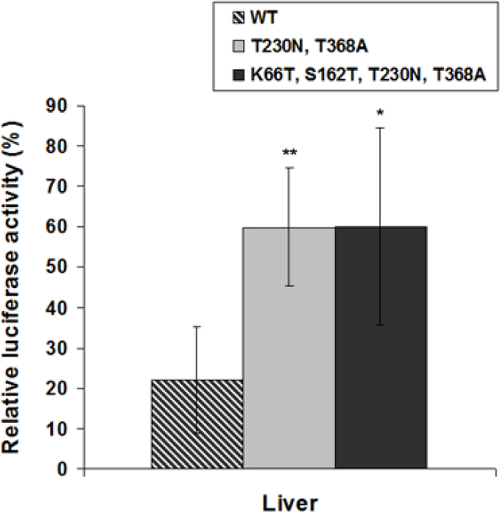
To assess neutralization of the VSV-G mutants, equal levels of lentiviral vector (8 × 10¹⁰ vector genomes) were injected via the tail vein 1 hour after the administration of 200 µl of human serum or PBS. After two weeks, the mice were sacrificed, and luciferase expression levels were analyzed in liver. The presence of human serum reduced luciferase expression mediated by wild type VSV-G to only 22.1% of PBS-injected controls in liver. The human serum reduced mutants 1 and 2 to only 60% of PBS controls, levels that were nearly 3-fold higher than WT VSV-G under the same conditions (Figure 8).
Figure 8. Human serum neutralization in vivo.
For two variants (T230N +T368A and K66T + S162T + T230N + T368A) and wild type VSV-G, lentiviral vectors encoding luciferase were administered via tail vein injection to female BALB/c mice one hour after human serum or PBS introduction. After two weeks, levels of luciferase activity were determined and normalized to total protein for each sample analyzed. Relative luciferase expression in liver was determined by quantifying relative enzyme activity from human serum-primed mice relative to activity from naïve mice. Error bars denote SD (n = 4). * and ** indicate statistical differences of P < 0.05 and P < 0.01, respectively, compared with wild type VSV-G, as determined using an one-way ANOVA.
DISCUSSION
Due to their broad tropism and high stability, VSV-G pseudotyped retroviral and lentiviral vectors are promising vehicles for gene transfer gene in vitro and in vivo. However, while VSV-G pseudotyped vectors have been increasingly explored for ex vivo gene delivery in clinical trials,46,47 serum neutralization of the VSV-G pseudotyped viral vectors is a significant obstacle for direct, in vivo administration. In this study, we conducted directed evolution of VSV-G to create serum-resistant VSV-G mutants. After 6 selection steps, followed by functional analysis of common mutations, we identified several mutations (S162T, T230N, and T368A) that enhanced VSV-G serum resistance. Furthermore, since incubation at 37°C was inherent to the selection, we also identified several adventitious mutations (K66T, T368A, and E380K) that apparently increased VSV-G thermostability. Upon combining these two classes of mutations, retroviral and lentiviral vectors pseudotyped with two resulting VSV-G variants (T230N + T368A and K66T + S162T + T230N +T368A) showed higher resistance to human and animal sera as well as increased thermostability compared to wild type VSV-G. While adeno-associated virus (AAV) has been evolved to resist antibody neutralization,38,48,49 to our knowledge this is the first effort to harness iterative random mutagenesis and selection to endow retroviral or lentiviral vectors with the capacity to resist serum inactivation.
VSV-G pseudotyped viral vectors can be inactivated in serum by complement,20,30 a major element of the innate immune response that in general functions via classical, alternative, and lectin pathways.21 All pathways can mediate virus opsonization, virolysis, and anaphylatoxin and chemotaxin production. However, the particular mechanisms by which complement inactivates VSV-G presenting virions are unknown. The mutation sites (Ser162, Thr230, and Thr368) we identified do not lie within several known antibody binding epitopes50, indicating that these residues may lie in previously uncharacterized epitopes or that VSV-G may interact with complement via mechanisms independent of antibody neutralization. Despite the lack of mechanistic information underlying a given gene delivery problem, directed evolution can still be employed to create enhanced variants, which in this case could aid future efforts to investigate mechanisms of retroviral or lentiviral vector inactivation by complement.
As a byproduct of the evolution, mutations that enhanced the thermal stability of VSV-G were also identified, which can potentially enhance vector production and reduce dosage of administration. The extracellular half-life of retroviral vectors lies between 3.5 hrs and 8 hrs at 37°C,35,51,52 and our measurement (t1/2 = ~ 3 hrs) is close to this range. Therefore, our incubation of the library for 6 hours in human serum at 37°C also selected for enhanced thermostability. An increased protein thermal stability can be caused by several factors such as disulfide bond formation, hydrophobic interactions, or change of electrostatic interactions of the surface.53 The E380K mutation in VSV-G may eliminate a like-charge repulsion with Asp381 and create opposite-charge attraction on the protein surface to confer thermostability to the protein. By comparison, potential mechanisms for K66T and T368A thermostabilization are not as readily apparent. At any rate, combining these mutations with others that conferred serum-resistance resulted in vectors with both properties. In particular, mutants 1 and 2 showed a two-fold increase in half-life at 37°C (t1/2 = ~ 6 hrs). These improvements of thermal stability may have also contributed slightly to a greater stability to vector concentration by ultracentrifugation (data not shown).
Cocal-pseudotyped lentiviral vectors may have potential for in vivo gene therapy, due to their high titers, broad tropism, stability, and reported increased resistance to human serum compared to VSV-G-pseudotyped vectors.31 However, unlike VSV-G, their in vivo use has not yet been reported. RD114-pseudotped vectors also exhibit resistance to human serum. Green et al. conducted immunostaining for RD114 receptors,54 which were present in colon, testis, bone marrow, skeletal muscle, and skin epithelia. However, the receptors are absent in lung, thyroid, and artery, suggesting that the application of RD114-pseudotyped vectors to treat some diseases may be limited. By comparison, VSV-G pseudotyped lentiviral vectors have potential for broad tropism.55
In this study, we evolved VSV-G and successfully created variants with higher resistance to human, rabbit, and mouse sera in vitro and human serum in vivo. This work therefore further establishes the power of directed evolution to improve viral vectors, and these results may enhance the utility of retroviral and lentiviral vectors to treat human disease. In addition, vesicular stomatitis virus (VSV) itself has emerged as a promising candidate in the field of oncolytic virus therapy. Therefore, incorporating a VSV variant encoding a human serum-resistant and thermostable VSV-G may enhance the therapeutic potential of VSV for treating human cancer.
MATERIALS AND METHODS
Library construction and selection
Random mutagenesis libraries were generated by standard error-prone PCR of a VSV-G template using VSV-G fwd and VSV-G rev as primers (see Supplementary Table S1 for all primer sequences). The amplified PCR fragments were digested with Sfi I and Xho I, and the resulting fragments were ligated into the corresponding sites of the retroviral vector pCLPIT, which contains a puromycin resistance gene and places VSV-G under a tetracycline responsive promoter.
HEK 293T cells were cultured in Iscove’s modified Dulbecco’s medium (IMDM) supplemented with 10% FBS (Invitrogen, Calrsbad, CA) and 1% penicillin/streptomycin (Invitrogen) at 37°C and 5% CO2. The VSV-G library was packaged into retroviral vectors via calcium phosphate transfection of 62.5 ng of pCLPIT VSV-G Lib, 6 µg of pCMV gag-pol, and 14 µg of pBluescript II SK (Stratagene, Santa Clara, CA) in a ~ 80% confluent 10-cm dish of HEK 293T cells. The library vector supernatant was twice harvested, 2 and 3 days post-transfection, and then concentrated by ultracentrifugation. For selection, 18 individual human serum samples were obtained from Innovative Research, Inc. (Southfield, MI). The viral vector library was diluted fivefold in the pooled human serum and incubated for 6 hours at 37°C. These viral vectors were used to infect 293T cells at a MOI of < 0.1. The infected cells were selected and enriched using 1 µg/ml of puromycin for two or three weeks. Packaging-competent virus variants were rescued through transfection of pCMV gag/pol into the expanded 293T cells and concentrated by ultracentrifugation for the next round of screening.
Sequence analysis and site-directed mutagenesis
After 5 and 6 rounds of the selection process, genomic DNA of the infected 293T cells was extracted using the QIAamp® DNA Mini kit (Qiagen, Valencia, CA). For sequencing of individual VSV-G clones, the VSV-G genes were amplified from this genomic DNA by PCR (using VSV-G IVS fwd and VSV-G IVS rev primers). The resulting PCR fragments were digested with Eco RI, and the fragments were ligated into the corresponding site of pcDNA IVS.56 Randomly chosen clones were sequenced (with IVS seq fwd, IVS seq rev, and VSV-G seq in primers).
Site-directed mutagenesis was conducted on the plasmid pcDNA IVS VSV-G using the QuickChange PCR-based technique, with Pfu polymerase and primers described in Supplementary Table S1. The amplified PCR products were treated with Dpn I and propagated into E. coli DH10B. Mutations were verified by DNA sequencing.
Viral production
The individual VSV-G mutant retroviral vectors were packaged with 10 µg of pCLPIT GFP, 6 µg of pCMV gag-pol, and 4 µg of pcDNA IVS VSV-G (or pcDNA IVS VSV-G mut) in a ~ 80% confluent 10-cm dish of HEK 293T cells. RD114 envelope pseudotyped retroviral vector was packaged with pcDNA IVS RD114 which was obtained by substitution of VSV-G gene by RD114 gene from pLTR-RD114A (Addgene, Cambridge, MA). The culture medium was changed 12 hours post-transfection, and the viral supernatant was collected both 24 h and 48 h later and concentrated by ultracentrifugation. To determine the titers of GFP-expressing vectors, 4 × 10⁵ 293T cells were infected with at least three different volumes of vector supernatant or concentrate. The cells were assayed for GFP expression by flow cytometry 3 days after infection. Vector genomic titers were measured by real-time qPCR using the iCycler iQ Real Time Detection System (Bio-Rad) and SYBR Green I (Invitrogen) with primers 5′-ATTGACTGAGTCGCCCGG-3′ (forward) and 5′-AGCGAGACCACAAGTCGGAT-3′ (reverse). Individual VSV-G mutant lentiviral vectors were packaged with 10 µg of lentiviral transfer vector HIV-CSCG, 5 µg of pMDLg/pRRE, 1.5 µg of pRSV Rev, and 3.5 µg of pcDNA IVS VSV-G (or pcDNA IVS VSV-G mut) and a process similar to retroviral vector production. Lentiviral vectors harboring firefly luciferase were packaged with 10 µg of Fluc, 5 µg of pMDLg/pRRE, 1.5 µg of pRSV Rev, and 3.5 µg of pcDNA IVS VSV-G (or pcDNA IVS VSV-G mut).57
Serum inactivation assay
In addition to the 18 human serum samples were obtained from Innovative Research, Inc. (Southfield, MI), mouse and rabbit serum were purchased from Sigma (St. Louis, MO). Serum inactivation experiments were carried out as described previously with some modifications.58 Briefly, 20 µl of viral vector normalized based on VSV-G ELISA data was diluted fivefold in human sera, heat-inactivated human sera (56°C, 30 min), or PBS (pH 7.4) as a control. Each sample was incubated for 1 hour at 37°C. All samples were then serially diluted and titered on 293T cells. Gene transfer was evaluated by flow cytometer for GFP expression 3 days after infection. To determine the change in titer after exposure to serum, the percentage of GFP-expressing cells for vector incubation in the serum was divided by the percentage of GFP-expressing cells for vector in PBS and reported as the percentage of titer recovery. To investigate thermal stability of the viral vectors, samples in PBS without incubation at 37°C was used as a control.
ELISA assay
To evaluate the expression level of VSV-G of purified viral vectors the binding of viral vector populations to a VSV-G polyclonal antibody was monitored by ELISA.59 MaxiSorp (Nunc) 96 well plates were coated at 4°C overnight with 50 µl of viral vectors diluted in carbonate coating buffer (100 mM, pH 9.6) and washed three times with PBS (pH 7.4). The wells were blocked by adding 200 µl of PBS with 5% non-fat dry milk at room temperature (RT) for 2 hours and washed twice with PBS. 100 µl of biotin conjugated anti-VSV-G tag antibody (1:3,000) (Abcam, Cambridge, MA) in carbonate coating buffer was added. After incubation overnight at 4°C, the plates were washed four times with PBS and 100 µl of streptavidin-alkaline phosphatase (1: 1,000) (Invitrogen) was added and incubated for 2 hours at RT. After four times washing with PBS, p-nitrophenly-phosphate (pNPP) substrate solution (Invitrogen) was added. Plates were read to determine the OD405 after 30 min incubation at RT.
Transduction analysis in vitro
To determine the relative transduction efficiencies the selected mutants compared to parental wild-type VSV-G pseudotyped retroviral vectors, HEK293T, CHO K1, NIH 3T3 (mouse embryonic fibroblast cell line), HeLa, and HT1080 cells were plated at a density of 4.0 × 10⁵ cells per well 24 hours prior to infection. Cells were infected with wild type or VSV-G variants pseudotyped retroviral vectors. The fraction of GFP positive cells was assessed 72 hours post infection using a Beckman-Coulter Cytomics FC-500 flow cytometer.
In vivo human serum inactivation
Eight-week-old female BALB/c mice (Jackson Laboratories, Bar Harbor, ME; n = 4) were used for all experiments. 200 µl of pooled human serum, or PBS as a control, was administered into the tail vein with a 30-gauge needle. One hour after human serum or PBS introduction, recombinant lentiviral vectors carrying cDNA encoding firefly luciferase under the control of human ubiquitin promoter was injected into the tail vein. Two weeks after virus injection, animals were transcardially perfused with PBS, and liver was harvested and frozen. Frozen tissue samples were homogenized in 1× reporter lysis buffer (Promega, Madison, WI) and clarified by centrifugation for 10 min at 10,000 g. Luciferase reporter activities were determined as previously described,60 and the luciferase signal was normalized to total protein content as determined by a bicinchoninic acid assay (Pierce, Rockford, IL). Animal protocols were approved by the UCB Animal Care and Use Committee and conducted in accordance with National Institutes of Health guidelines.
Supplementary Material
ACKNOWLEDGMENTS
We acknowledge a research grant from Virxsys Corporation as well as NIH R01 GM07305.
Footnotes
CONFLICT OF INTEREST
The authors declare no conflict of interest.
Supplementary information is available at Gene Therapy's website (http://www.nature.com/gt).
REFERENCES
- 1.Rosenberg SA, Aebersold P, Cornetta K, Kasid A, Morgan RA, Moen R, et al. Gene transfer into humans--immunotherapy of patients with advanced melanoma, using tumor-infiltrating lymphocytes modified by retroviral gene transduction. N Engl J Med. 1990;323:570–578. doi: 10.1056/NEJM199008303230904. [DOI] [PubMed] [Google Scholar]
- 2.Edelstein ML, Abedi MR, Wixon J. Gene therapy clinical trials worldwide to 2007--an update. J Gene Med. 2007;9:833–842. doi: 10.1002/jgm.1100. [DOI] [PubMed] [Google Scholar]
- 3.Schaffer DV, Koerber JT, Lim KI. Molecular engineering of viral gene delivery vehicles. Annu Rev Biomed Eng. 2008;10:169–194. doi: 10.1146/annurev.bioeng.10.061807.160514. [DOI] [PMC free article] [PubMed] [Google Scholar]
- 4.Sheridan C. Gene therapy finds its niche. Nat Biotechnol. 2011;29:121–128. doi: 10.1038/nbt.1769. [DOI] [PubMed] [Google Scholar]
- 5.Braun S. Muscular gene transfer using nonviral vectors. Curr Gene Ther. 2008;8:391–405. doi: 10.2174/156652308786070998. [DOI] [PubMed] [Google Scholar]
- 6.Nishikawa M, Takakura Y, Hashida M. Pharmacokinetic considerations regarding non-viral cancer gene therapy. Cancer Sci. 2008;99:856–862. doi: 10.1111/j.1349-7006.2008.00774.x. [DOI] [PMC free article] [PubMed] [Google Scholar]
- 7.Schmidt-Wolf GD, Schmidt-Wolf IG. Non-viral and hybrid vectors in human gene therapy: an update. Trends Mol Med. 2003;9:67–72. doi: 10.1016/s1471-4914(03)00005-4. [DOI] [PubMed] [Google Scholar]
- 8.Follenzi A, Santambrogio L, Annoni A. Immune responses to lentiviral vectors. Curr Gene Ther. 2007;7:306–315. doi: 10.2174/156652307782151515. [DOI] [PubMed] [Google Scholar]
- 9.Nair V. Retrovirus-induced oncogenesis and safety of retroviral vectors. Curr Opin Mol Ther. 2008;10:431–438. [PubMed] [Google Scholar]
- 10.Cronin J, Zhang XY, Reiser J. Altering the tropism of lentiviral vectors through pseudotyping. Curr Gene Ther. 2005;5:387–398. doi: 10.2174/1566523054546224. [DOI] [PMC free article] [PubMed] [Google Scholar]
- 11.Morizono K, Ku A, Xie Y, Harui A, Kung SK, Roth MD, et al. Redirecting lentiviral vectors pseudotyped with Sindbis virus-derived envelope proteins to DC-SIGN by modification of N-linked glycans of envelope proteins. J Virol. 2010;84:6923–6934. doi: 10.1128/JVI.00435-10. [DOI] [PMC free article] [PubMed] [Google Scholar]
- 12.Miletic H, Bruns M, Tsiakas K, Vogt B, Rezai R, Baum C, et al. Retroviral vectors pseudotyped with lymphocytic choriomeningitis virus. J Virol. 1999;73:6114–6116. doi: 10.1128/jvi.73.7.6114-6116.1999. [DOI] [PMC free article] [PubMed] [Google Scholar]
- 13.Sinn PL, Burnight ER, Shen H, Fan H, McCray PB., Jr Inclusion of Jaagsiekte sheep retrovirus proviral elements markedly increases lentivirus vector pseudotyping efficiency. Mol Ther. 2005;11:460–469. doi: 10.1016/j.ymthe.2004.10.022. [DOI] [PubMed] [Google Scholar]
- 14.Funke S, Schneider IC, Glaser S, Mühlebach MD, Moritz T, Cattaneo R, et al. Pseudotyping lentiviral vectors with the wild-type measles virus glycoproteins improves titer and selectivity. Gene Ther. 2009;16:700–705. doi: 10.1038/gt.2009.11. [DOI] [PubMed] [Google Scholar]
- 15.Sakuma T, De Ravin SS, Tonne JM, Thatava T, Ohmine S, Takeuchi Y, et al. Characterization of retroviral and lentiviral vectors pseudotyped with xenotropic murine leukemia virus-related virus envelope glycoprotein. Hum Gene Ther. 2010;21:1665–1673. doi: 10.1089/hum.2010.063. [DOI] [PMC free article] [PubMed] [Google Scholar]
- 16.Burns JC, Friedmann T, Driever W, Burrascano M, Yee JK. Vesicular stomatitis virus G glycoprotein pseudotyped retroviral vectors: concentration to very high titer and efficient gene transfer into mammalian and nonmammalian cells. Proc Natl Acad Sci USA. 1993;90:8033–8037. doi: 10.1073/pnas.90.17.8033. [DOI] [PMC free article] [PubMed] [Google Scholar]
- 17.Yee JK, Friedmann T, Burns JC. Generation of high-titer pseudotyped retroviral vectors with very broad host range. Methods Cell Biol. 1994;43:99–112. doi: 10.1016/s0091-679x(08)60600-7. [DOI] [PubMed] [Google Scholar]
- 18.Yang YP, Vanin EF, Whitt MA, Fornerod M, Zwart R, Schneiderman RD, et al. Inducible, high-level production of infectious murine leukemia retroviral vector particles pseudotyped with vesicular stomatitis virus G envelope protein. Hum Gene Ther. 1995;6:1203–1213. doi: 10.1089/hum.1995.6.9-1203. [DOI] [PubMed] [Google Scholar]
- 19.Ory DS, Neugeboren BA, Mulligan RC. A stable human-derived packaging cell line for production of high titer retrovirus/vesicular stomatitis virus G pseudotypes. Proc Natl Acad Sci USA. 1996;93:11400–11406. doi: 10.1073/pnas.93.21.11400. [DOI] [PMC free article] [PubMed] [Google Scholar]
- 20.DePolo NJ, Reed JD, Sheridan PL, Townsend K, Sauter SL, Jolly DJ, et al. VSV-G pseudotyped lentiviral vector particles produced in human cells are inactivated by human serum. Mol Ther. 2000;2:218–222. doi: 10.1006/mthe.2000.0116. [DOI] [PubMed] [Google Scholar]
- 21.Gasque P. Complement: a unique innate immune sensor for danger signals. Mol Immunol. 2004;41:1089–1098. doi: 10.1016/j.molimm.2004.06.011. [DOI] [PubMed] [Google Scholar]
- 22.Hofmann C, Strauss M. Baculovirus-mediated gene transfer in the presence of human serum or blood facilitated by inhibition of the complement system. Gene Ther. 1998;5:531–536. doi: 10.1038/sj.gt.3300607. [DOI] [PubMed] [Google Scholar]
- 23.Hoare J, Waddington S, Thomas HC, Coutelle C, McGarvey MJ. Complement inhibition rescued mice allowing observation of transgene expression following intraportal delivery of baculovirus in mice. J Gene Med. 2005;7:325–333. doi: 10.1002/jgm.671. [DOI] [PubMed] [Google Scholar]
- 24.Schauber-Plewa C, Simmons A, Tuerk MJ, Pacheco CD, Veres G. Complement regulatory proteins are incorporated into lentiviral vectors and protect particles against complement inactivation. Gene Ther. 2005;12:238–245. doi: 10.1038/sj.gt.3302399. [DOI] [PubMed] [Google Scholar]
- 25.Magre S, Takeuchi Y, Langford G, Richards A, Patience C, Weiss R. Reduced sensitivity to human serum inactivation of enveloped viruses produced by pig cells transgenic for human CD55 or deficient for the galactosyl-alpha(1–3) galactosyl epitope. J Virol. 2004;78:5812–5819. doi: 10.1128/JVI.78.11.5812-5819.2004. [DOI] [PMC free article] [PubMed] [Google Scholar]
- 26.Tani H, Limn CK, Yap CC, Onishi M, Nozaki M, Nishimune Y, et al. In vitro and in vivo gene delivery by recombinant baculoviruses. J Virol. 2003;77:9799–9808. doi: 10.1128/JVI.77.18.9799-9808.2003. [DOI] [PMC free article] [PubMed] [Google Scholar]
- 27.Spitzer D, Hauser H, Wirth D. Complement-protected amphotropic retroviruses from murine packaging cells. Hum Gene Ther. 1999;10:1893–1902. doi: 10.1089/10430349950017572. [DOI] [PubMed] [Google Scholar]
- 28.Croyle MA, Callahan SM, Auricchio A, Schumer G, Linse KD, Wilson JM, et al. PEGylation of a vesicular stomatitis virus G pseudotyped lentivirus vector prevents inactivation in serum. J Virol. 2004;78:912–921. doi: 10.1128/JVI.78.2.912-921.2004. [DOI] [PMC free article] [PubMed] [Google Scholar]
- 29.Kim YK, Choi JY, Jiang HL, Arote R, Jere D, Cho MH, et al. Hybrid of baculovirus and galactosylated PEI for efficient gene carrier. Virology. 2009;387:89–97. doi: 10.1016/j.virol.2009.02.001. [DOI] [PubMed] [Google Scholar]
- 30.Sandrin V, Boson B, Salmon P, Gay W, Nègre D, Le Grand R, et al. Lentiviral vectors pseudotyped with a modified RD114 envelope glycoprotein show increased stability in sera and augmented transduction of primary lymphocytes and CD34+ cells derived from human and nonhuman primates. Blood. 2002;100:823–832. doi: 10.1182/blood-2001-11-0042. [DOI] [PubMed] [Google Scholar]
- 31.Trobridge GD, Wu RA, Hansen M, Ironside C, Watts KL, Olsen P, et al. Cocal-pseudotyped lentiviral vectors resist inactivation by human serum and efficiently transduce primate hematopoietic repopulating cells. Mol Ther. 2010;18:725–733. doi: 10.1038/mt.2009.282. [DOI] [PMC free article] [PubMed] [Google Scholar]
- 32.Gatlin J, Melkus MW, Padgett A, Petroll WM, Cavanagh HD, Garcia JV, et al. In vivo fluorescent labeling of corneal wound healing fibroblasts. Exp Eye Res. 2003;76:361–371. doi: 10.1016/s0014-4835(02)00302-0. [DOI] [PubMed] [Google Scholar]
- 33.Ting-De Ravin SS, Kennedy DR, Naumann N, Kennedy JS, Choi U, Hartnett BJ, et al. Correction of canine X-linked severe combined immunodeficiency by in vivo retroviral gene therapy. Blood. 2006;107:3091–3097. doi: 10.1182/blood-2005-10-4057. [DOI] [PMC free article] [PubMed] [Google Scholar]
- 34.Powell SK, Kaloss MA, Pinkstaff A, McKee R, Burimski I, Pensiero M, et al. Breeding of retroviruses by DNA shuffling for improved stability and processing yields. Nat Biotechnol. 2000;18:1279–1282. doi: 10.1038/82391. [DOI] [PubMed] [Google Scholar]
- 35.Vu HN, Ramsey JD, Pack DW. Engineering of a stable retroviral gene delivery vector by directed evolution. Mol Ther. 2008;16:308–314. doi: 10.1038/sj.mt.6300350. [DOI] [PubMed] [Google Scholar]
- 36.Merten CA, Stitz J, Braun G, Poeschla EM, Cichutek K, Buchholz CJ. Directed evolution of retrovirus envelope protein cytoplasmic tails guided by functional incorporation into lentivirus particles. J Virol. 2005;79:834–840. doi: 10.1128/JVI.79.2.834-840.2005. [DOI] [PMC free article] [PubMed] [Google Scholar]
- 37.Excoffon KJ, Koerber JT, Dickey DD, Murtha M, Keshavjee S, Kaspar BK, et al. Directed evolution of adeno-associated virus to an infectious respiratory virus. Proc Natl Acad Sci USA. 2009;106:3865–3870. doi: 10.1073/pnas.0813365106. [DOI] [PMC free article] [PubMed] [Google Scholar]
- 38.Maheshri N, Koerber JT, Kaspar BK, Schaffer DV. Directed evolution of adeno-associated virus yields enhanced gene delivery vectors. Nat Biotechnol. 2006;24:198–204. doi: 10.1038/nbt1182. [DOI] [PubMed] [Google Scholar]
- 39.Lim KI, Klimczak R, Yu JH, Schaffer DV. Specific insertions of zinc finger domains into Gag-Pol yield engineered retroviral vectors with selective integration properties. Proc Natl Acad Sci USA. 2011;107:12475–12480. doi: 10.1073/pnas.1001402107. [DOI] [PMC free article] [PubMed] [Google Scholar]
- 40.Roche S, Rey FA, Gaudin Y, Bressanelli S. Structure of the prefusion form of the vesicular stomatitis virus glycoprotein G. Science. 2007;315:843–848. doi: 10.1126/science.1135710. [DOI] [PubMed] [Google Scholar]
- 41.Kuchner O, Arnold FH. Directed evolution of enzyme catalysts. Trends Biotechnol. 1997;15:523–530. doi: 10.1016/S0167-7799(97)01138-4. [DOI] [PubMed] [Google Scholar]
- 42.Hamamatsu N, Aita T, Nomiya Y, Uchiyama H, Nakajima M, Husimi Y, et al. Biased mutation-assembling: an efficient method for rapid directed evolution through simultaneous mutation accumulation. Protein Eng Des Sel. 2005;18:265–271. doi: 10.1093/protein/gzi028. [DOI] [PubMed] [Google Scholar]
- 43.Pensiero MN, Wysocki CA, Nader K, Kikuchi GE. Development of amphotropic murine retrovirus vectors resistant to inactivation by human serum. Hum Gene Ther. 1996;7:1095–1101. doi: 10.1089/hum.1996.7.9-1095. [DOI] [PubMed] [Google Scholar]
- 44.Kaname Y, Tani H, Kataoka C, Shiokawa M, Taguwa S, Abe T, et al. Acquisition of complement resistance through incorporation of CD55/decay-accelerating factor into viral particles bearing baculovirus GP64. J Virol. 2010;84:3210–3219. doi: 10.1128/JVI.02519-09. [DOI] [PMC free article] [PubMed] [Google Scholar]
- 45.Pan D, Gunther R, Duan W, Wendell S, Kaemmerer W, Kafri T, et al. Biodistribution and toxicity studies of VSVG-pseudotyped lentiviral vector after intravenous administration in mice with the observation of in vivo transduction of bone marrow. Mol Ther. 2002;6:19–29. doi: 10.1006/mthe.2002.0630. [DOI] [PubMed] [Google Scholar]
- 46.Cartier N, Hacein-Bey-Abina S, Bartholomae CC, Veres G, Schmidt M, Kutschera I, et al. Hematopoietic stem cell gene therapy with a lentiviral vector in X-linked adrenoleukodystrophy. Science. 2009;326:818–823. doi: 10.1126/science.1171242. [DOI] [PubMed] [Google Scholar]
- 47.DiGiusto DL, Krishnan A, Li L, Li H, Li S, Rao A, et al. RNA-based gene therapy for HIV with lentiviral vector-modified CD34(+) cells in patients undergoing transplantation for AIDS-related lymphoma. Sci Transl Med. 2010;2:36–43. doi: 10.1126/scitranslmed.3000931. [DOI] [PMC free article] [PubMed] [Google Scholar]
- 48.Maersch S, Huber A, Buning H, Hallek M, Perabo L. Optimization of stealth adeno-associated virus vectors by randomization of immunogenic epitopes. Virology. 2010;397:167–175. doi: 10.1016/j.virol.2009.10.021. [DOI] [PubMed] [Google Scholar]
- 49.Grimm D, Lee JS, Wang L, Desai T, Akache B, Storm TA, et al. In vitro and in vivo gene therapy vector evolution via multispecies interbreeding and retargeting of adeno-associated viruses. J Virol. 2008;82:5887–5911. doi: 10.1128/JVI.00254-08. [DOI] [PMC free article] [PubMed] [Google Scholar]
- 50.Vandepop SB, Lefrancois L, Holland JJ. Sequences of the major antibody epitopes of the Indiana serotype Vesicular stomatitis virus. Virology. 1986;148:312–325. doi: 10.1016/0042-6822(86)90328-4. [DOI] [PubMed] [Google Scholar]
- 51.Paul RW, Morris D, Hess BW, Dunn J, Overell RW. Increased viral titer through concentration of viral harvests from retroviral packaging lines. Hum Gene Ther. 1993;4:609–615. doi: 10.1089/hum.1993.4.5-609. [DOI] [PubMed] [Google Scholar]
- 52.Andreadis ST, Brott D, Fuller AO, Palsson BO. Moloney murine leukemia virus-derived retroviral vectors decay intracellularly with a half-life in the range of 5.5 to 7.5 hours. J Virol. 1997;71:7541–7548. doi: 10.1128/jvi.71.10.7541-7548.1997. [DOI] [PMC free article] [PubMed] [Google Scholar]
- 53.Yano JK, Poulos TL. New understandings of thermostable and peizostable enzymes. Curr Opin Biotechnol. 2003;14:360–365. doi: 10.1016/s0958-1669(03)00075-2. [DOI] [PubMed] [Google Scholar]
- 54.Green BJ, Lee CS, Rasko JEJ. Biodistribution of the RD114/mammalian type D retrovirus receptor, RDR. J Gene Med. 2004;6:249–259. doi: 10.1002/jgm.517. [DOI] [PubMed] [Google Scholar]
- 55.Jimenez DF, Lee CI, O’Shea CE, Kohn DB, Tarantal AF. HIV-1-derived lentiviral vectors and fetal route of administration on transgene biodistribution and expression in rhesus monkeys. Gene Ther. 2005;12:821–830. doi: 10.1038/sj.gt.3302464. [DOI] [PubMed] [Google Scholar]
- 56.Yu JH, Schaffer DV. Selection of novel vesicular stomatitis virus glycoprotein variants from a peptide insertion library for enhanced purification of retroviral and lentiviral vectors. J Virol. 2006;80:3285–3292. doi: 10.1128/JVI.80.7.3285-3292.2006. [DOI] [PMC free article] [PubMed] [Google Scholar]
- 57.Yang L, Yang H, Rideout K, Cho T, Joo KI, Ziegler L, et al. Engineered lentivector targeting of dendritic cells for in vivo immunization. Nat Biotechnol. 2008;26:326–334. doi: 10.1038/nbt1390. [DOI] [PMC free article] [PubMed] [Google Scholar]
- 58.Friedman HM, Wang L, Fishman NO, Lambris JD, Eisenberg RJ, Cohen GH, et al. Immune evasion properties of herpes simplex virus type 1 glycoprotein gC. J Virol. 1996;70:4253–4260. doi: 10.1128/jvi.70.7.4253-4260.1996. [DOI] [PMC free article] [PubMed] [Google Scholar]
- 59.Kweon CH, Kwon BJ, Kim IJ, Lee SY, Ko YJ. Development of monoclonal antibody-linked ELISA for sero-diagnosis of vesicular stomatitis virus (VSV-IN) using baculovirus expressed glycoprotein. J Virol Methods. 2005;130:7–14. doi: 10.1016/j.jviromet.2005.05.023. [DOI] [PubMed] [Google Scholar]
- 60.Koerber JT, Jang JH, Schaffer DV. DNA shuffling of adeno-associated virus yields functionally diverse viral progeny. Mol Ther. 2008;16:1703–1709. doi: 10.1038/mt.2008.167. [DOI] [PMC free article] [PubMed] [Google Scholar]
Associated Data
This section collects any data citations, data availability statements, or supplementary materials included in this article.


