Abstract
Lipomas are benign tumours that consist of mature adipocytes. They are the commonest soft tissue tumours, most frequently seen in the trunk and proximal extremities. Lesions in the hand are uncommon, and giant lipomas of the hand, defined as greater than 5 cm in size, are particularly rare. We present a case of an exceptionally large giant lipoma of the hand, presenting as an extremely large inconvenient swelling of the palm in a 67-year-old woman. The diagnosis of lipoma was suggested via ultrasonography, and confirmed via MRI and histology. The lesion was successfully excised with no postoperative neurovascular deficit. The excised lesion measured 8×6×3 cm, one of the largest giant lipomas of the hand reported to date. When patients present with large lesions such as these a malignant cause must always be considered, and appropriate early imaging is essential when assessing these patients.
Background
This case gives an example of the diagnostic approach to an unusually large soft tissue swelling of the hand. We emphasise the importance of appropriate radiology in assessing these lesions, and discuss the relevant sonographic and MRI features of lipomas.
Case presentation
A retired 67-year-old right hand dominant woman presented with a 2-year history of arthritic pain at the base of the left thumb. The patient also reported extensive swelling of the left hand, slowly increasing in size over the past 2 years, causing significant inconvenience. No neurological symptoms were reported. On examination a 10×8 cm soft, fluctuant, non-tender and trans-illuminable swelling was noted at the left thenar eminence, with two further swellings in the line of the left index finger ray, which were contiguous with the main swelling. There was no tendon dysfunction or neurovascular deficit. The patient was referred for ultrasound examination with the provisional diagnosis of a ganglion.
Investigations
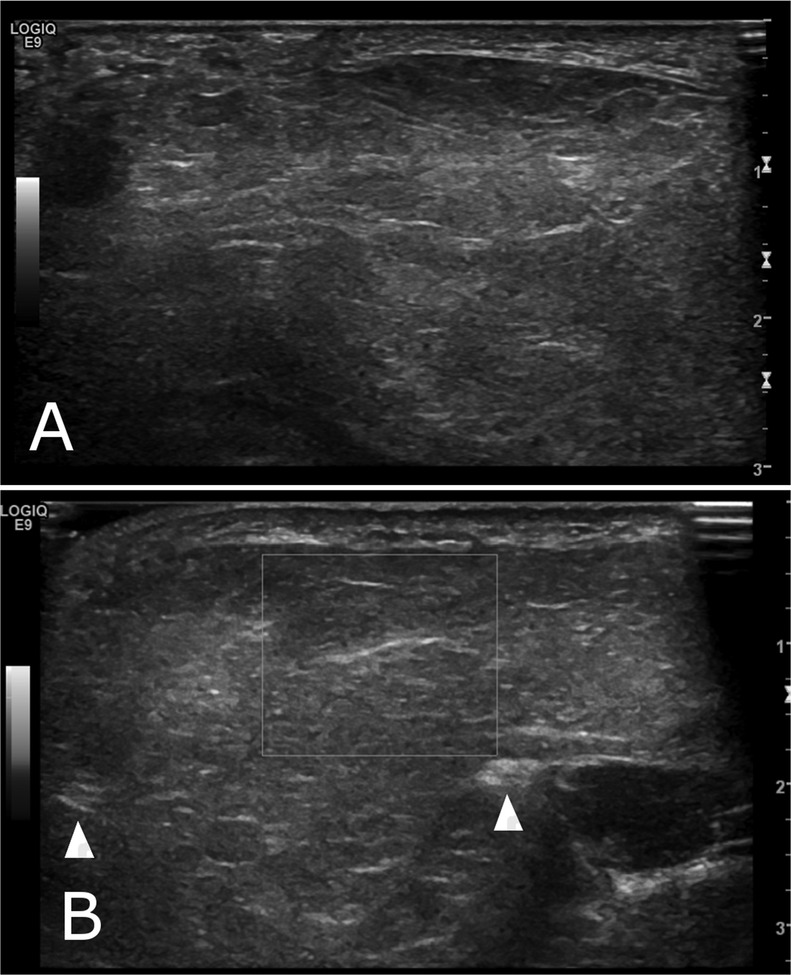
Ultrasonography revealed a bilobed solid lesion spreading along the course of the index finger flexor tendon and on either side in between the tendon sheaths of the thumb and middle finger. It was isoechogenic to subcutaneous fat with homogenous echotexture, and no vascularity was seen on Doppler interrogation. The median nerve and radial artery were clear of the mass. These appearances were strongly suggestive of a fat containing soft tissue mass (figure 1).
Figure 1.

Longitudinal (A) and axial (B) ultrasound images of the left hand showing a large homogenous soft tissue mass which is isoechoic to subcutaeneous fat and lacks vascularity on colour Doppler (B). The mass is noted on either side of second and third common flexor tendon sheaths (arrows, B).
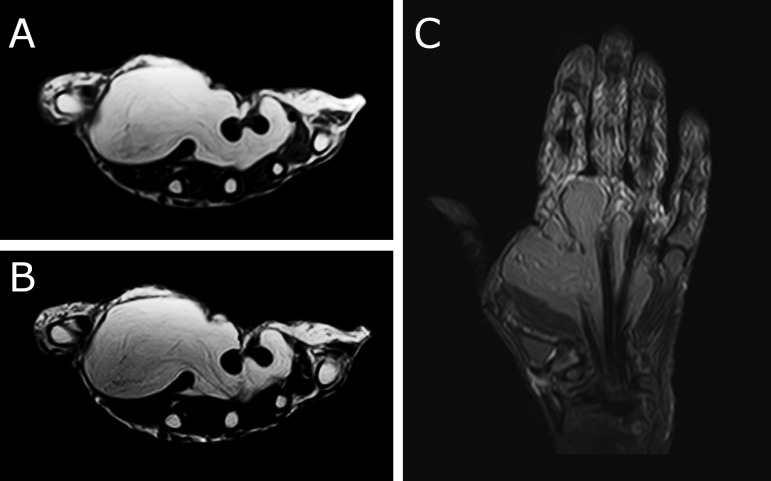
To assess the full extent of the lesion and to aid in preoperative planning, MRI was performed. The lesion appeared bright on T1-weighted and T2-weighted (T1W, T2W) images, with complete suppression of signal on short tau inversion recovery (STIR) sequences. Minimal peripheral enhancement was seen after gadolinium. There was no non-adipose component within the lesion, in keeping with a fatty mass (figure 2).
Figure 2.
Axial T1-weighted (A) and T2-weighted (B) images of the left hand showing a large hyperintense soft tissue mass occupying most of the hand, extending from the first to fourth metacarpals (B). On Coronal STIR image (C) it extends from distal carpal row to the metacarpophalangeal joint and shows uniform suppression of signal, indicating its’ fatty nature.
Differential diagnosis
The clinical and radiological findings in this instance point towards a benign fat containing mass, most likely a giant lipoma. However, the size of this lesion means that malignancy must be considered. The differential here therefore includes liposarcoma, of which well-differentiated liposarcomas are the most common type.1 This distinction is important as a different management approach and referral to a specialist sarcoma unit would be required in malignant disease.2
Treatment
The patient underwent elective excision of the lesion. The carpal tunnel was decompressed and the surgical wound was extended distally using a modified Brunner's incision. An 8×6×3 cm lesion was dissected out from the surrounding tissues, preserving the local neurovascular structures.
Outcome and follow-up
Histology confirmed a lipoma composed of mature fatty cells, with no evidence of malignancy. At postoperative assessment the patient had a well-healed scar with no signs of infection, and importantly no neurovascular deficit was noted. The patient went on to have a trapeziectomy for first left carpometacarpal joint arthritis.
Discussion
Lipomas are benign tumours of mature adipocytes that can occur anywhere in the body where adipose tissue is present. They are the commonest soft tissue tumour, usually presenting between the ages of 40 and 60 years.3 They commonly occur in the trunk, neck, shoulder and upper arm and are rarely found in the hands or feet.4
Giant lipomas of the hand, greater than 5 cm in size, are extremely rare, with the extant literature limited to case reports and small case series.5 They present as slowly growing painless masses, although functional deficit and symptoms of nerve compression may be present depending on the size and location of the lesion.5 The presented case is one of the largest giant lipomas of the hand reported to date.1 5 6 The treatment of these giant lipomas is surgical, via marginal excision. The risk of malignant transformation in benign lesions is low, and therefore the indication for surgery is usually cosmesis and functional impairment due to compression of local structures.5
Any soft tissue mass greater than 5 cm must be considered malignant until proven otherwise.2 The important distinction in cases such as this is between a benign lipoma and liposarcoma, which is the commonest form of soft tissue sarcoma.7 Of these, well-differentiated liposarcomas are the most common type, accounting for 40%.6 The identification of malignant disease is important, as more radical treatment may be required.5 The appropriate choice of investigation is therefore imperative when presented with a large soft tissue swelling.
We believe that sonography has a valuable role to play in assessing large swellings of the hand. Ultrasound is a non-ionising, quick, cost-effective and readily available test which can be used for rapid assessment of a lesion and to provide patient reassurance. On ultrasonography lipomas are well-defined, isoechoic to neighbouring fat, and show no vascularity on colour Doppler. Features including poorly defined margins, non-homogenous echotexture (eg,focal nodularity and necrotic areas), and vascularity on Doppler interrogation should raise suspicion of sarcomatous change in such masses.8
MRI can also be used to assess these swellings, and has been reported to provide the correct diagnosis in 94% of cases of masses of the hand and wrist.9 A benign lipoma will be well demarcated, shows the same signal intensity as subcutaeneous fat in all sequences, suppressed signal on fat suppression sequences and small numbers of thin (<2 mm) septae on T1W and T2W sequences. Enhancement is generally not seen with contrast, apart from at the fibrous capsule.10 11 In contrast, liposarcomas have traditionally been described as showing increased lesion size, irregular and large septae and increased non-adipose content.10
In this case the ultrasound features provided the provisional diagnosis of benign giant lipoma, which was subsequently supported by the MRI findings and histological analysis. If suspicious features are seen on imaging, or if the findings are inconclusive, then further investigation with biopsy and referral to a sarcoma centre is warranted.2
Learning points.
Giant lipomas of the hand are rare, but should be considered in the differential of any large swelling of the hand.
Ultrasonography can be used as a quick screening tool to initially assess large swellings of the hand and guide further investigation.
Larger lesions should also be further investigated by MRI as malignancy must be ruled out in any soft tissue tumour greater than 5 cm in size.
If suspicious features are seen on ultrasound/MRI, then further investigation and referral to a sarcoma centre is required.
Footnotes
Contributors: All the authors have been actively involved in the generation of this manuscript, and have seen and agreed to the final version prior to submission.
Competing interests: None.
Patient consent: Obtained.
Provenance and peer review: Not commissioned; externally peer reviewed.
References
- 1.Grivas TB, Psarakis SA, Kaspiris A, et al. Giant lipoma of the thenar—case study and contemporary approach to its aetiopathogenicity. Hand (New York) 2009;2013:173–6 [DOI] [PMC free article] [PubMed] [Google Scholar]
- 2.Grimer RJ, Briggs TW. Earlier diagnosis of bone and soft-tissue tumours. J Bone Joint Surg Br 2010;2013:1489–92 [DOI] [PubMed] [Google Scholar]
- 3.Salam GA. Lipoma excision. Am Fam Physician 2002;2013:901–4 [PubMed] [Google Scholar]
- 4.Bancroft LW, Kransdorf MJ, Peterson JJ, et al. Benign fatty tumors: classification, clinical course, imaging appearance and treatment. Skeletal Radiol 2006;2013:719–33 [DOI] [PubMed] [Google Scholar]
- 5.Cribb GL, Cool WP, Ford DJ, et al. Giant lipomatous tumours of the hand and forearm. J Hand Surg Br 2005;2013:509–12 [DOI] [PubMed] [Google Scholar]
- 6.Pagonis T, Givissis P, Christodoulou A. Complications arising from a misdiagnosed giant lipoma of the hand and palm: a case report. J Med Case Rep 2011;2013:552. [DOI] [PMC free article] [PubMed] [Google Scholar]
- 7.Dei Tos AP. Liposarcoma: new entities and evolving concepts. Ann Diagn Pathol 2000;2013:252–66 [DOI] [PubMed] [Google Scholar]
- 8.Cheng JW, Tang SF, Yu TY, et al. Sonographic features of soft tissue tumors in the hand and forearm. Chang Gung Med J 2007;2013:547–54 [PubMed] [Google Scholar]
- 9.Capelastegui A, Astigarraga E, Fernandez-Canton G, et al. Masses and pseudomasses of the hand and wrist: MR findings in 134 cases. Skeletal Radiol 1999;2013:498–507 [DOI] [PubMed] [Google Scholar]
- 10.Brisson M, Kashima T, Delaney D, et al. MRI characteristics of lipoma and atypical lipomatous tumor/well-differentiated liposarcoma: retrospective comparison with histology and MDM2 gene amplification. Skeletal Radiol 2013;2013:635–47 [DOI] [PubMed] [Google Scholar]
- 11.Ergun T, Lakadamyali H, Derincek A, et al. Magnetic resonance imaging in the visualization of benign tumors and tumor-like lesions of hand and wrist. Curr Probl Diagn Radiol 2010;2013:1–16 [DOI] [PubMed] [Google Scholar]


