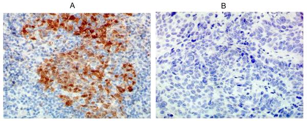
Figure 1. p16 by IHC for two patient samples.

(A) Case 1 shows brown nuclear and cytoplasmic staining indicating p16 positive status; (B) Case 2 shows no staining indicating p16 negative status. 40X magnification. (IHC - immunohistochemistry)

(A) Case 1 shows brown nuclear and cytoplasmic staining indicating p16 positive status; (B) Case 2 shows no staining indicating p16 negative status. 40X magnification. (IHC - immunohistochemistry)