Summary
Mast cell-derived group III phospholipase A2 (PLA2G3) and fibroblast prostaglandin synthase contribute to a PGD2 and mast-cell-DP-1-dependent mast cell-fibroblast paracrine axis that can enhance mast cell maturation and mediator secretion.
Mast cells are derived from hematopoietic precursors that complete their differentiation and maturation in the microenvironments of virtually all vascularized tissues1. Mast cells are best known as key effector cells in acute IgE-associated disorders, including anaphylaxis, an immediate and potentially catastrophic systemic response initiated when antigen crosslinks antigen-specific IgE antibodies bound to high-affinity FcεRI receptors on the mast cell plasma membrane, thereby initiating mast cell activation2. Mast cells activated by IgE and antigen degranulate (i.e., rapidly release pre-formed, granule-stored bioactive compounds) and also secrete newly formed lipid mediators and a large variety of growth factors, cytokines and chemokines that participate in hypersensitivity reactions2.
The types and/or amounts of mediators released by mast cells activated by particular signals (including IgE and specific antigen) can be influenced, or “tuned”, by many factors3. Among them, the stage of mast cell maturation influences the types and quantities of mediators stored in mast cell cytoplasmic granules and other features of mast cell phenotype1,3. The local development and maturation of mast cells is thought to be regulated by signals provided by structural cells in local tissue microenvironments, such as the c-Kit ligand, SCF (stem cell factor)1,3. Indeed, mice with loss-of-function mutations in both copies of the genes encoding SCF or c-Kit (CD117) are profoundly mast cell-deficient, reflecting the importance of SCF and c-Kit in mast cell development and survival in vivo1,3. However, our understanding of the regulation of mast cell maturation is by no means complete. In the current issue of Nature Immunology, Taketomi et al. describe a novel lipid-based mast cell-fibroblast interaction pathway which contributes to normal maturation of mouse peritoneal, skin and small intestinal mast cells in vivo4. The authors report that a mast cell-secreted group III phospholipase A2 (PLA2G3) plays a central role in this maturation mechanism and consequently also increases the intensity of biological responses involving IgE-dependent mast cell activation4.
Phospholipase A2s (PLA2s) were originally discovered as toxic components of snake venoms5. PLA2s have a high proportion of cysteine (more than 10% of all amino acids) and a high prevalence of disulfide bridges5. The PLA2 superfamily is divided into subgroups based on the number and location of disulfide bridges, with groups I, II and III being originally characterized by PLA2s isolated from cobra, rattlesnake and honeybee venoms, respectively (with honeybee PLA2 being significantly different structurally from the two snake venom PLA2s)5. However, all PLA2s share the ability to cleave fatty acids at the sn-2 position of glycerophospholipids, thereby generating free fatty acids and lysophospholipids5. Major biological functions of secreted mammalian PLA2s (sPLA2s; including groups I, II and III enzymes) include, but are not limited to, the metabolism of dietary phospholipids and contributions to skin homeostasis and host defense against bacteria and viruses5,6 (Fig. 1, step 3).
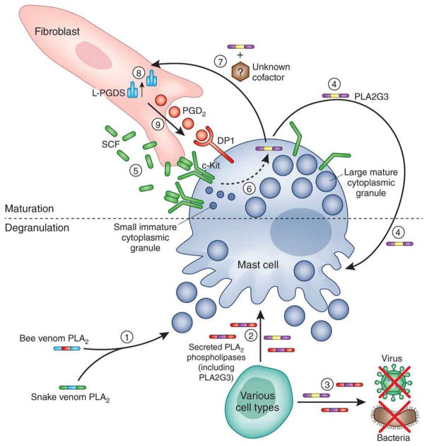
Figure 1. Endogenous PLA2G3 can promote mast cell maturation and induce and enhance mast cell activation.
Exogenous phospholipase A2 (PLA2) contained in venoms of different species (1), and mammalian sPLA2s (secreted by various cell types) (2), can induce mast cell degranulation and mediator release. Mammalian sPLA2s are also involved in many other biological processes, including defense against bacteria and viruses (3). Taketomi et al. show that PLA2G3 (which can be produced by mast cells) can induce mast cell degranulation directly (4) and also can enhance antigen- and IgE-dependent mast cell degranulation (not shown). These findings suggest the following model of how PLA2G3 can promote mast cell maturation in peripheral tissues. Stem cell factor (SCF) produced by fibroblasts binds to and induces dimerization of its receptor c-Kit on mast cells (5). c-Kit signaling enhances mast cell survival and promotes mast cell maturation and, under certain circumstances, also can induce mast cells to secrete PLA2G3 (6). Mast cell-secreted PLA2G3, in the presence of an additional, presumably mast cell-derived, factor(s) yet to be identified, interacts with fibroblasts, in a way that remains to be determined (7), and increases expression of the prostaglandin D2 (PGD2) synthesizing enzyme L-PGDS (8). Fibroblast L-PGDS supplies a local pool of PGD2, which, by binding to DP1 expressed by mast cells, locally contributes to terminal mast cell maturation (9), inducing increasing the mast cell’s content of cytoplasmic granule-associated mediators, e.g., histamine and proteases, and also increasing surface levels of FcεRI (not shown). Such “maturation effects” prime tissue mast cells to respond more strongly to stimulation with IgE and specific antigen and thus contribute to the intensity of IgE-dependent biological responses (not shown).
PLA2s isolated from certain snake venoms and, with less potency, from honeybee venom, are known to induce antigen-independent degranulation of mouse and human mast cells in vitro7 (Fig. 1, step 1). Earlier work by Murakami et al. showed that rat or mouse mast cells also can be activated in vitro by purified rat group II PLA28. Such group II sPLA2s can be released by mast cells, platelets and other cell types8,9, raising the possibility of autocrine or paracrine mechanisms of sPLA2-dependent mast cell activation in vivo (Fig. 1, steps 2 and 4). The ability of sPLA2s to induce mast cell degranulation was reported to depend on their enzymatic activity7–9. However, the systemic toxicity of venom sPLA2s (e.g., neurotoxicity) is thought also to be related to complex formation with other toxic venom compounds and their interaction with target molecules expressed in different tissues and organs10.
Mammalian PLA2G3 is a multidomain protein with a central 150 amino acid (AA) PLA2 domain flanked by N-terminal (130 AA) and C-terminal (219 AA) extensions with unknown functions11. Compared to venom PLA2s, the PLA2 domain of PLA2G3 has moderate overall amino acid sequence homology to PLA2s from the venoms of the Gila monster lizard and the honeybee (46% and 31%, respectively) and relatively high homology in the conserved calcium binding loop (73% and 64%, respectively) and active site regions (54%), and has therefore been assigned to group III11. It was shown recently that PLA2G3 potentially contributes to sperm maturation and to the development of atherosclerosis in mice5,6.
In the current study, Taketomi et al. first show that high amounts of purified honeybee venom PLA2 and recombinant human PLA2G3 can induce tissue oedema in mouse ear pinnae by inducing mast cell degranulation, and identify PLA2G3 as a cytoplasmic (and perhaps granule-associated) sPLA2 in mouse mast cells4 (Fig. 1). Using mice systemically deficient for Pla2g3 or other sPLA2s, mice overexpressing human Pla2g3 and mast cell-deficient mice engrafted with bone marrow-derived cultured mast cells from different transgenic mouse strains, the authors show that PLA2G3 (but none of the other tested sPLA2s) can contribute to the intensity of IgE- and mast cell-dependent passive systemic anaphylaxis in vivo. This effect of PLA2G3 seems to be cell-intrinsic and related, at least in part, to the ability of PLA2G3 to promote normal maturation of mast cell granules (e.g., PLA2G3-deficient mast cells have substantially reduced amounts of histamine and granule-stored proteases) and in part to its ability to enhance levels of surface FcεRI. These data convincingly identify an important role for mast cell-derived PLA2G3 in mast cell maturation and function, but do not rule out potential contributions of PLA2G3 derived from other cell types. Indeed, the authors showed that Pla2g3 is also expressed in bone marrow-derived mouse basophils, but they did not comment on possible production of PLA2G3 by other hematopoietic cells, such as neutrophils and eosinophils. In this regard, the decreased cutaneous anaphylaxis observed in Pla2g3-deficient mice actively sensitized with ovalbumin and alum might also be related to PLA2G3 deficiency in basophils or neutrophils, since the dependency of this model on mast cells alone was not directly tested.
The authors also used a mast cell-fibroblast co-culture system to provide an additional line of evidence that PLA2G3 is important for normal mast cell maturation. By analyzing different transgenic mouse models targeting components of lipid pathways with potential effects on mast cell maturation or function (e.g., via actions of eicosanoid mediators)12, Taketomi et al. investigated the hypothesis that the observed effects of PLA2G3 on mast cells are associated with the role of PLA2G3 in lipid metabolism. Like Pla2g3−/− mice, Ptgdr−/− mice, which are systemically deficient in DP-1 (prostaglandin D receptor-1, which is expressed in several different cell types), or Ptgds−/− mice, deficient in L-PGDS, an enzyme involved in PGD2 synthesis that is expressed by fibroblasts, developed abnormal mast cell phenotypes and attenuated passive cutaneous anaphylaxis. Upon co-culture with wild type fibroblasts, mast cells derived from Ptgdr−/− mice exhibited markedly reduced enhancement of expression of histidine decarboxylase (HDC), an enzyme required for mast cell histamine synthesis and for normal formation of cytoplasmic granules. Furthermore, wild type mast cells co-cultured with L-PGDS-deficient fibroblasts also exhibited impaired granule formation and decreased expression of HDC, implying that maturational effects of fibroblast L-PGDS require mast cell expression of DP-1. Finally, the authors provided evidence that this fibroblast-associated, lipid-based mast cell maturation pathway also exists in humans. Specifically, the robust HDC expression observed in human lung mast cells after co-culture with lung fibroblasts was significantly decreased by neutralization of PLA2G3, L-PGDS or DP-1. These novel findings, based on an impressive amount of work, support a model in which mast cell-secreted PLA2G3 (Fig. 1, step 7) induces the expression of fibroblast-derived PGD2 synthase (L-PGDS) (Fig. 1, step 8), leading to increased local production of PGD2 which in turn promotes mast cell maturation via DP-1 receptors (Fig. 1, step 9).
A notable conclusion of this study is that PLA2G3 not only can activate mast cells (as has been shown previously for other mammalian sPLA2s7,8) but can also contribute to mast cell maturation in the tissues. In this case, the molecule of interest was shown first to be a mast cell activator and then a contributor to mast cell maturation. By contrast, SCF was originally described as a critical mast cell survival and maturation factor, but subsequent studies showed that SCF also can activate mast cells directly and can enhance IgE- and antigen-dependent mast cell activation1,3. It is tempting to speculate that such a dual role in enhancing mast cell maturation, as well as in certain circumstances directly inducing and/or enhancing mast cell secretory function, might emerge as a feature shared by many mast cell “maturation factors”.
An obvious goal for future studies is to understand in detail the mechanisms leading to the enhanced production of fibroblast L-PGDS upon PLA2G3 release by mast cells, and subsequently to local PGD2 generation contributing to mast cell maturation. While one can speculate that the enzymatic activity of PLA2G3 plays an important role in this paracrine interaction pathway, an additional mechanism might involve the direct interaction of PLA2G3’s unique N- or C-terminal domains with molecules expressed by fibroblasts or other structural cells5. It also will be interesting to determine whether other mast cell-secreted factors can importantly contribute to this process (Fig. 1, step 7), and to assess in detail how features of this axis are influenced by SCF, which can augment the IgE-dependent generation of PGD2 by mouse mast cells at least in part by up-regulating the mast cell expression of the three enzymes in the pathway which generates PGD2 from membrane phospholipids: cPLA2, prostaglandin H synthase, and hematopoietic PGDS12.
The work of Taketomi et al. thus makes important contributions to our understanding of the potential roles of secreted phospholipases and lipid mediators in paracrine and autocrine mechanisms contributing to the regulation of mast cell development and function. Their fascinating observations also suggest that additional findings of interest remain to be uncovered by extending such investigations to other types of hematopoietic cells.
References
- 1.Gurish MF, Austen KF. Developmental origin and functional specialization of mast cell subsets. Immunity. 2012;37:25–33. doi: 10.1016/j.immuni.2012.07.003. [DOI] [PubMed] [Google Scholar]
- 2.Galli SJ, Tsai M. IgE and mast cells in allergic disease. Nat Med. 2012;18:693–704. doi: 10.1038/nm.2755. [DOI] [PMC free article] [PubMed] [Google Scholar]
- 3.Galli SJ, et al. Mast cells as “tunable” effector and immunoregulatory cells: recent advances. Annu Rev Immunol. 2005;23:749–786. doi: 10.1146/annurev.immunol.21.120601.141025. [DOI] [PubMed] [Google Scholar]
- 4.Taketomi Y, et al. Mast cell maturation is driven via a novel group III phospholipase A2-prostaglandin D2-DP1 receptor paracrine axis. Nat Imm. 2013 doi: 10.1038/ni.2586. [DOI] [PMC free article] [PubMed] [Google Scholar]
- 5.Dennis EA, Cao J, Hsu YH, Magrioti V, Kokotos G. Phospholipase A2 enzymes: physical structure, biological function, disease implication, chemical inhibition, and therapeutic intervention. Chem Rev. 2011;111:6130–6185. doi: 10.1021/cr200085w. [DOI] [PMC free article] [PubMed] [Google Scholar]
- 6.Murakami M, Lambeau G. Emerging roles of secreted phospholipase A2 enzymes: an update. Biochimie. 2013;95:43–50. doi: 10.1016/j.biochi.2012.09.007. [DOI] [PubMed] [Google Scholar]
- 7.Varsani M, Pearce FL. Role of phospholipase A2 in mast cell activation. Inflamm Res. 1997;46 (Suppl 1):S9–10. [PubMed] [Google Scholar]
- 8.Murakami M, Hara N, Kudo I, Inoue K. Triggering of degranulation in mast cells by exogenous type II phospholipase A2. J Immunol. 1993;151:5675–5684. [PubMed] [Google Scholar]
- 9.Murakami M, Kudo I, Suwa Y, Inoue K. Release of 14-kDa group-II phospholipase A2 from activated mast cells and its possible involvement in the regulation of the degranulation process. Eur J Biochem. 1992;209:257–265. doi: 10.1111/j.1432-1033.1992.tb17284.x. [DOI] [PubMed] [Google Scholar]
- 10.Pungercar J, Krizaj I. Understanding the molecular mechanism underlying the presynaptic toxicity of secreted phospholipases A2. Toxicon. 2007;50:871–892. doi: 10.1016/j.toxicon.2007.07.025. [DOI] [PubMed] [Google Scholar]
- 11.Valentin E, Ghomashchi F, Gelb MH, Lazdunski M, Lambeau G. Novel human secreted phospholipase A2 with homology to the group III bee venom enzyme. J Biol Chem. 2000;275:7492–7496. doi: 10.1074/jbc.275.11.7492. [DOI] [PubMed] [Google Scholar]
- 12.Boyce JA. Mast cells and eicosanoid mediators: a system of reciprocal paracrine and autocrine regulation. Immunol Rev. 2007;217:168–185. doi: 10.1111/j.1600-065X.2007.00512.x. [DOI] [PubMed] [Google Scholar]


