

Abstract
Lipomas of the colon are relatively rare benign tumors of mesenchymatic origin. They are usually asymptomatic but as they become larger they can cause symptoms including abdominal pain, diarrhea, nausea, constipation, haematochezia, loss of body weight, anemia or even intussusception and colonic obstruction. We present a 52 year old male patient who visited the emergency room complaining of constipation, rectal bleeding, mucus in stools and a palpable rectal mass. Colonoscopy revealed a polypoid mass of the sigmoid colon lying about 30 cm from the anal verge. Sigmoidectomy was performed. The postoperative recovery was uneventful and he was discharged five days later. At follow up a month after surgery the patient was asymptomatic. The pathological examination revealed a transmural tumor of the sigmoid colon measuring a 9x5x2.5cm and histologically compatible with a lipoma.
Keywords: colonic, lipoma, benign, tumor, intussusception
Lipomas of the colon are fatty benign tumors with a reported incidence between 0.2% and 4.4%1. Though rare, colonic lipomas are the most common benign nonepithelial tumors found in the gastrointestinal tract and are the third commonest tumors after hyperplastic and adenomatous polyps2. They may attain substantial size of up to several centimeters in diameter. The principal site of their occurrence is the right colon and females are more commonly affected. Giant lipomas of the colon are even less common, they may cause symptoms and therefore surgical removal should be considered. Intermittent subacute obstruction of the colon has been described in the literature by very large colonic lipomas which may require surgical resection. Intussusception may also occur as a result of a giant lipoma of the colon, as well as bleeding from the ulcerated tip of the lesion3. Malignant transformation and recurrence is very rare2.
Case report
A 52 year old male patient presented to our institution’s emergency department with symptoms of constipation, rectal bleeding, and an intermittent rectum mass prolapse. On admission blood count and chest x-ray were normal. Digital examination revealed a palpable mobile soft mass in the rectum. Colonoscopy was performed the next day which revealed a polypoid mass of the sigmoid colon about 30 cm from the anal verge (Figure 1). The biopsy of the lesion revealed changes compatible with a lipoma. There was no preoperative CT examination performed. Due to the size of the mass partial sigmoidectomy and an end to end anastomosis was performed (Figure 2). The postoperative period was without complications and the patient was discharged 5 days later. At follow up a month later the patient was without symptoms and the physical examination normal. The pathological examination of the tumor revealed a 9x5x2.5 cm transmural lipoma of the sigmoid colon that presented a few cell aberrations (Figure 3, Figure 4).
Figure 1. Endoscopy revealed a pendicular tumor at 30 cm.

Figure 2. Surgical specimen: the size of the tumor is 9 x 5 x 2,5cm.
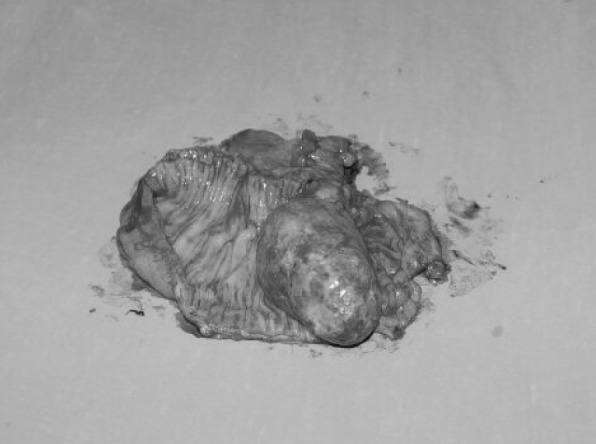
Figure 3. The tumor is histologically compatible with a lipoma (hematoxylin and eosin, original magnification x 200).

Figure 4. Transmural expansion of the lipoma (hematoxylin and eosin, original magnification x 200).

Discussion
Colonic lipomas are relatively uncommon tumors of mesenchymal origin, composed of well-differentiated adipose tissue supported by fibrous tissue, that rarely cause symptoms and are usually detected incidentally. They usually arise from the submucosa, but occasionally extend into the muscularis propria; up to 10% are subserosal4 but transmural location is extremely rare. Considering that the commonest location for solitary colonic lipoma is the ascending colon and the cecum5, we present a rare case of a giant transmural lipoma of the left colon. Lipomas of the colon should be resected when they are symptomatic or have a diameter of over 2cm. When the lipomas of the colon are small in size, usually less than 5 cm in diameter they can be resected via endoscopy6. Nevertheless the risk of perforation or hemorrhage is notably increased when the lesion is sessile or broadly-based7. Surgical resection seems to be the ideal choice of treatment for large lipomas, especially when malignancy cannot be completely excluded. Colotomy excision or segmental colon resection is recommended for complete removal of the lipoma. If the preoperative diagnosis of colon lipoma can be made correctly, extent of surgery may be appropriately limited6. One has to remember that their accurate preoperative diagnosis is difficult and they can be mistaken for malignancy, especially when the lesion is large in size, and with ulceration. A surgical approach remains the treatment of choice for large and complicated cases or in cases that malignancy cannot be completely excluded8.
References
- 1.Paškauskas S, Latkauskas T, Valeikaitė G, Paršeliūnas A, Svagždys S, Saladžinskas Z, et al. Colonic intussusception caused by colonic lipoma: a case report. Medicina (Kaunas) 2010;46:477–481. [PubMed] [Google Scholar]
- 2.Arora R, Kumar A, Bansal V. Giant rectal lipoma. Abdom Imaging. 2011;36:545–547. doi: 10.1007/s00261-010-9668-7. [DOI] [PubMed] [Google Scholar]
- 3.Bahadursingh AM, Robbins PL, Longo WE. Giant submucosal sigmoid colon lipoma. Am J Surg. 2003;18:81–82. doi: 10.1016/s0002-9610(03)00111-9. [DOI] [PubMed] [Google Scholar]
- 4.Katsinelos G, Chatzimavroudis G, Zavos GC, Pilpilidis I, Lazaraki G, Papaziogas B, et al. Cecal lipoma with pseudomalignant features: A case report and review of the literature. World J Gastroenterol. 2007;13:2510–2513. doi: 10.3748/wjg.v13.i17.2510. [DOI] [PMC free article] [PubMed] [Google Scholar]
- 5.Gidirim G, Mishin I, Gutsu E, Gagauz I, Danch A, Russu S. Giant submucosal lipoma of the cecum: A report of a case and review of the literature. Rom J Gastroenterol. 2005;14:393–396. [PubMed] [Google Scholar]
- 6.Ladurner R, Mussack T, Hohenbleicher F, Folwaczny C, Siebeck M, Hallfeldt K. Laparoscopic- assisted resection of giant sigmoid lipoma under colonoscopic guidance. Surg Endosc. 2003;17:160. doi: 10.1007/s00464-002-4232-3. [DOI] [PubMed] [Google Scholar]
- 7.Zhang H, Cong C, Chen CS, Qiao L, Liu EQ. Submucous colon lipoma: A case report and review of the literature. World J Gastroenterol. 2005;11:3167–3169. doi: 10.3748/wjg.v11.i20.3167. [DOI] [PMC free article] [PubMed] [Google Scholar]
- 8.Zhang X, Ouyang J, Kim D. Large ulcerated cecal lipoma mimicking malignancy. World J Gastrointest Oncol. 2010;2:304–306. doi: 10.4251/wjgo.v2.i7.304. [DOI] [PMC free article] [PubMed] [Google Scholar]