Abstract
To evaluate the utility of three-dimensional (3D) reconstruction when planning the surgical treatment of nasal bone fractures. The axial scan of high-resolution facial bone CT was reconstructed in 3D using the program V-works 4.0 (CyberMed, Seoul, Korea) with a volume and surface rendering technique. For detailed stereoscopic examination of the nasal valve area, an air-bone view with the volume rendering technique was obtained using thresholds for air, cartilage, and bone. In most nasal bone fractures, 2D and 3D CT had similar detection rates. However, to determine the fracture angle and dimpled area, and identify multiple fractures, surgeons can get better information to help with the reduction of the fractured bone from 3D reconstruction images. Additionally, with a septal deformity, this view helps in deciding on the need for septal surgery during nasal reduction. The air view of the nasal passage provides clues to obstruction of the nasal cavity. We could identify the contour and location of the fracture site accurately from 3D CT images. The detection rate of fractures was similar to that of 2D CT. However, 3D CT enabled the accurate determination of the distance and direction of the fractured bony fragment from normal bone structure. Additionally, a stereoscopic image of the fracture site facilitated an understanding of the location and range of reduction. The air-bone view gave more information about the pathological obstruction of the nasal air passage.
Keywords: Nasal bone, Fracture, Computed tomography, Three dimensional
Introduction
Computed tomography (CT) is useful in the accurate diagnosis of nasal bone fractures [1–3]. Three-dimensional (3D) CT is reliable when it is obtained simultaneously with two-dimensional (2D) CT [4]. Others have reported that 2D CT is the diagnostic technique of choice for nasal bone fractures [5]. The goals of treatment of nasal bone fractures are both esthetic and functional. From a functional perspective, the nasal valve area is critical in terms of nasal stuffiness and olfactory function. The nasal valve area can be narrowed following a nasal bone fracture. Thus, in this study, we evaluated the utility of 3D reconstruction in the diagnosis of nasal bone fractures for planning the surgical treatment and evaluated a new modality, the air-bone view of the volume rendering technique, for identifying pathological obstruction of the air passage.
Methods
Six patients with nasal bone fractures were enrolled in this study. Exclusion criteria were a history of sinus surgery, clinical sinusitis, radiological sinusitis, sinus mass, allergic rhinitis, and age < 16 years.
High-resolution CT of the facial bone was obtained in 2-mm slices using a Somatom Definition AS + 128 (Siemens, Seoul, Korea) from the vertex to the mandible (Fig. 1). From the axial scan, a 3D reconstruction was performed using the V-works 4.0 software (CyberMed, Seoul, Korea) using a volume and surface rendering technique with a threshold of 290 HU for both. For a detailed stereoscopic examination of the nasal valve area, an imaginary cut was made and a basal view was obtained orthogonal to the anteroposterior view. The thresholds for the air column ranged from –1024 to –302 HU and those for cartilage and bone were from 489 HU to 3071 HU.
Fig. 1.
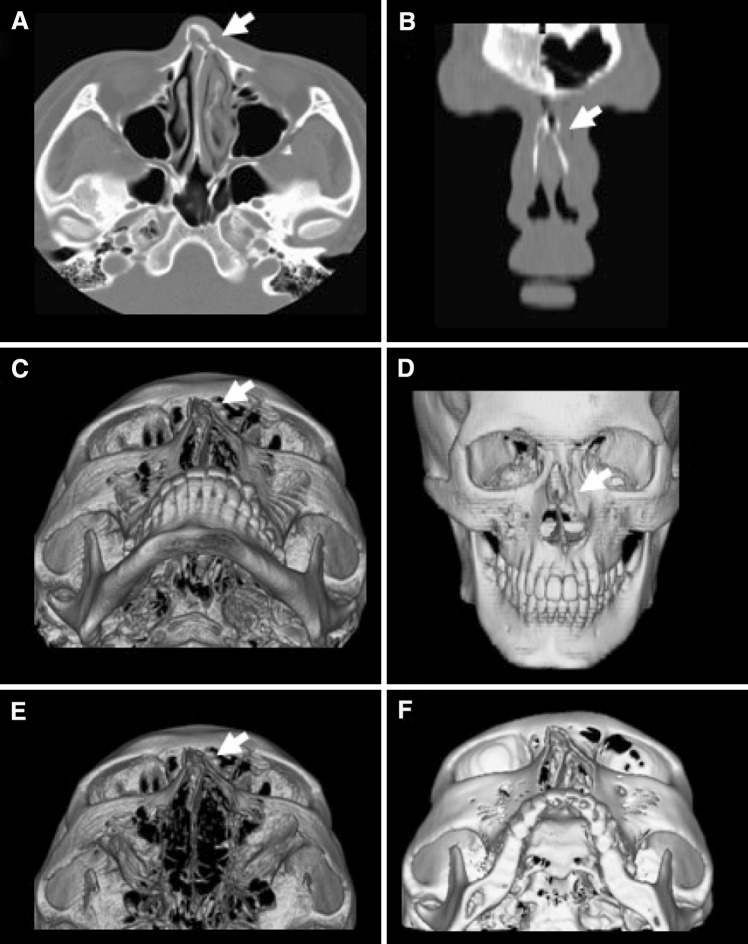
Axial and coronary CT shows a bilateral nasal bone fracture with a concurrent septal fracture (a, b). Volume rendering image shows a three-dimensional figure, which helps to understand the adjacent structures, including the deviated nasal septum (c–e). The surface rendering technique helps to estimate the angle of the fracture (f)
For a control image, the same exclusion criteria and CT technique were applied to a normal adult.
Results
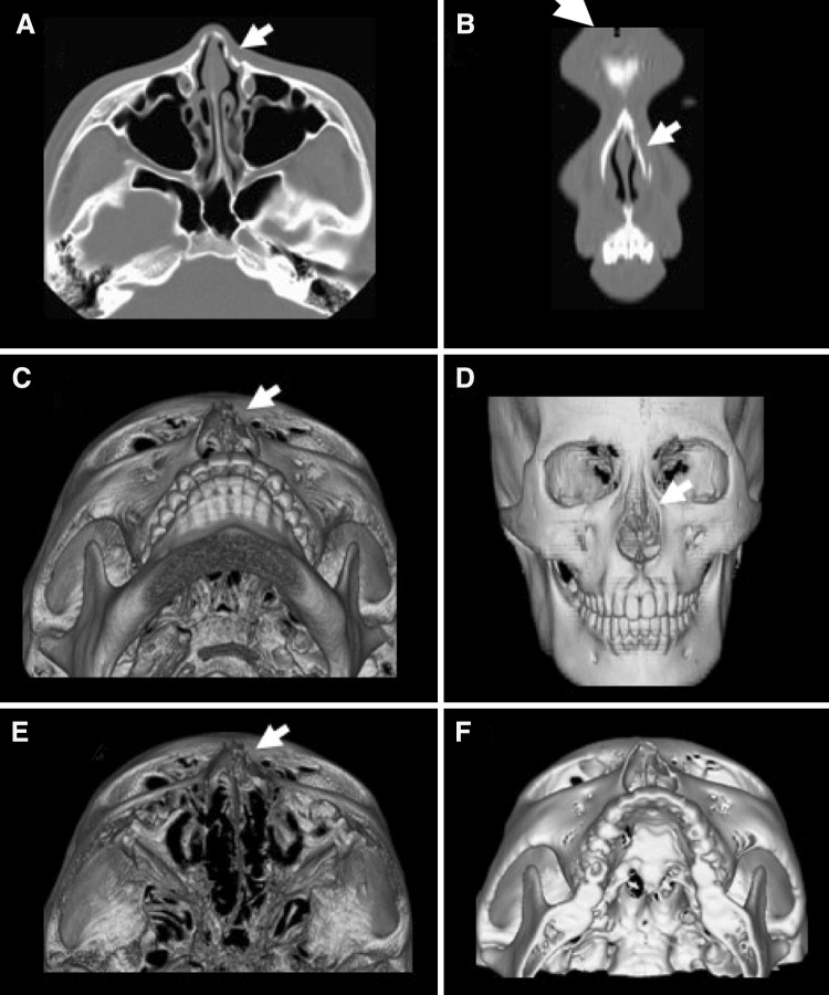
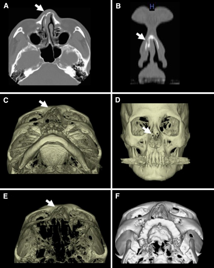
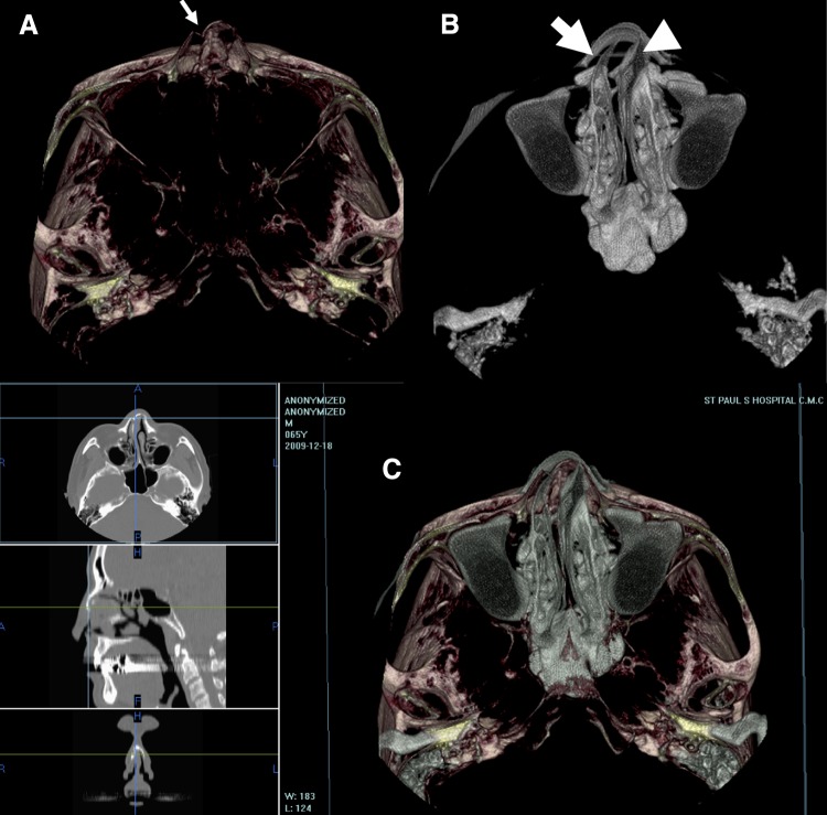
We could get a more detailed image of nasal bone fractures using 3D reconstruction images with both volume and surface rendering technique than with conventional 2D CT images. When there was a bilateral nasal bone fracture with a concurrent septal fracture (Fig. 1), the volume-rendering image produced 3D pictures, which helped in understanding adjacent structures, including any septal fracture (Fig. 1c–e). The surface rendering technique helps when estimating the angle of the fracture (Fig. 1f). Another advantage of 3D CT over 2D CT is identifying a gross dimpled area of nasal bone (Fig. 2), which is difficult to identify with 2D CT or on a physical examination when there is acute swelling of the nasal soft tissues. The third advantage of 3D CT is in identifying the angle of the fracture in a stereoscopic view, which helps in determining the direction of closed reduction of nasal bone fracture. The final advantage of 3D reconstruction of a nasal bone fracture is that it gives an image of each tissue according to its density, which is reflected in the Hounsfield units. This allows obtaining air, bone, and cartilage images of the nasal cavity using thresholds for each. In a nasal bone fracture with septal deviation, a conventional 3D image (Fig. 3) was obtained together with an air-bone view (Fig. 4) of the nasal bone fracture using the volume rendering technique. The air-bone view of the fracture site gives more detailed information on the air passage. This helps with the surgical planning, such as whether to perform a septoplasty during nasal bone reduction. Figure 5 is an air view of a normal human, which shows a standard image of the normal air passage and can be compared with an image of a pathological obstruction of the nasal air passage.
Fig. 2.
Axial and coronary CT shows a dimpling fracture of the nasal bone, left (a, b). No septal fracture was seen (c, d). The volume rendering view enables the acquisition of a cross-section of any part (e). The surface rendering technique helps to understand the direction of the fracture (f)
Fig. 3.
Unilateral fracture of the nasal bone, right. A severely deviated septum and dimpling are prominent in the 3D CT (e, f). This patient may need a septoplasty
Fig. 4.
A right nasal bone fracture (thin arrow) was observed in the bone cartilage view (a), and a decreased right air column (thick arrow), compared with the left side (arrowhead), was seen in the air (b) and air and bone cartilage (c) views in the 3D reconstruction
Fig. 5.
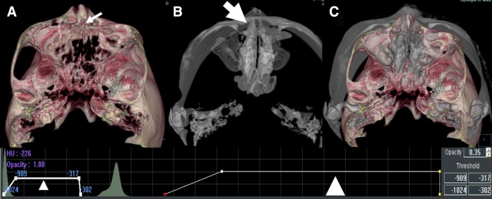
The air (a), bone cartilage (b), and air and bone cartilage (c) views in a normal adult. The thresholds (d) for the air column were from –1024 to –302 HU (thin arrowhead) and those for cartilage and bone (thick arrowhead) were from 489 to 3071 HU
Discussion
Plain films of nasal bone fractures have high rates of false-positive and -negative findings, and are inadequate for an accurate diagnosis of nasal bone fractures [6–9]. The nasal bone is a small, thin bone in the central part of the face and is the bone fractured most commonly. Treatment usually involves a simple closed reduction, but an incorrect diagnosis and inadequate reduction can cause secondary deformity [10]. Additional operation like corrective rhinoplasty or septoplasty could be needed later. An accurate history and physical examination, including clinical signs, such as crepitus, are more useful than a simple X-ray for diagnosing nasal bone fractures. The nasal bone forms from multiple ossification centers and involves multiple suture lines, which can be misidentified as fractures. Additionally, the bony groove that contains the nasociliary nerve is difficult to distinguish from a fracture line on plain films. Third, soft tissue swelling may hide a nasal bone fracture line [11]. Also, a septal bone fracture can be missed on a simple X-ray film, potentially leading to secondary deformity. The utility of 3D CT is that it enables the precise reconstruction of the extent of the nasal bone fracture and displaced fragments with a low dose of radiation. In one study, 3D CT helped to recognize not only the position and direction of fractures, but also the number of bone pieces and the amount of dislodgement in acute trauma cases [12, 13]. Another study preferred 3D CT to 2D CT for evaluating complex midfacial fractures [4].
We obtained 3D images of nasal bone fractures using the axial scan in facial bone CT. The ability to detect the fracture site is similar in both 2D and 3D CT. However, the 3D image of nasal bone fracture is superior to the 2D image for understanding the spatial relationship of adjacent structures, such as the nasal septum, and for identifying a dimpled area of nasal bone fragments and multiple fractures. The volume rendering and surface rendering techniques were equally good at identifying nasal cartilage and bone. The volume rendering technique has the advantage of real time operation of the object mass. In the surface rendering technique, we could determine the distance and angle of each structure. Using an air-bone view, we could understand the air passage in a stereoscopic view of the fracture site. Consequently, we could evaluate the air passage, providing useful information to help with the surgical planning of the restoration of the air passage. The airway around the turbinates is critical for smell conduction [14]. Therefore, reduction operation has to restore this space for preserving olfactory function. Better understanding of deformity with 3D rendering technique could lead to better results after reduction procedure.
We could identify the contour and location of the fracture from the 3D image. The detection rate was similar to that of 2D CT, but 3D CT with the air-bone view enabled us to identify narrowing of the nasal valve area caused by the nasal bone fracture, which is critical for nasal stuffiness and olfaction. The air-bone view may help the surgeon to decide on the need for a septoplasty. Additionally, the structure can be analyzed in three orthogonal planes, which can be changed at an arbitrary angle, to facilitate understanding of the structures of the septum and external nasal bone.
Conclusions
We could identify the accurate contour and location of the fracture site from 3D CT images. The 3D reconstruction technique enables one to determine the distance and direction of the fractured bony fragment from the normal nasal plane accurately. The diagnostic value of the stereoscopic 3D CT air-bone view is in assessing the depressed edge and location of the fractured bone in the nasal valve area. We expect that more precise information about the fracture site will help us to determine the direction and severity of the fracture, and to improve the therapeutic success rate, based on a good surgical plan.
Conflict of Interest
The authors declare that they have no conflict of interest.
References
- 1.Shimada K, Kamei K. Evaluation of K-wire fixation for nasal bone fracture using CT Images. J Jpn Plast Reconstr Surg. 1996;16:314. [Google Scholar]
- 2.Yabe T, Matamura H, Mitinari M. Post-operative evaluation of nasal bone fractures with CT images and its necessity. Jpn J Plast Reconstr Surg. 1999;42:303. [Google Scholar]
- 3.Hirota K, Shimizu Y, Iimuma T. Image diagnosis of nasal bone fracture. J Jpn Otorhinolaryngol. 1988;91:539. doi: 10.3950/jibiinkoka.91.539. [DOI] [PubMed] [Google Scholar]
- 4.Wang P, Yu Q, Zhi SH (2001) Zhonghua Kou Qiang Yi Xue Za 36(4):256–258 [PubMed]
- 5.Kim JH, Kim MG, Park JH, Joo JB, Kim YJ. The practical role of nasal bone CT in the diagnosis of nasal bone fractures. Korean J Otolaryngol. 2001;44:405–411. [Google Scholar]
- 6.Logan M, O’Driscoll K, Masterson J. The utility of nasal bone radiographs in nasal trauma. Clinical Radiol. 1994;49:192–194. doi: 10.1016/S0009-9260(05)81775-1. [DOI] [PubMed] [Google Scholar]
- 7.Lacey GJ, Chir B, Wignall BK, Hussain S, Reidy JR. The radiology of nasal injuries: problems of interpretation and clinical relevance. British J Radiol. 1977;50:412–414. doi: 10.1259/0007-1285-50-594-412. [DOI] [PubMed] [Google Scholar]
- 8.Bartkiw TP, Pynn BR, Brown DH. Diagnosis and management of nasal fractures. Int J Trauma Nurs. 1995;1:11–18. doi: 10.1016/S1079-2104(05)80405-6. [DOI] [PubMed] [Google Scholar]
- 9.Illum P. Legal aspects in nasal fractures. Rhinology. 1991;29:263–266. [PubMed] [Google Scholar]
- 10.Rohrich RJ, Adams WP., Jr Nasal fracture management: minimizing secondary nasal deformities. Plast Reconstr Surg. 2000;106:266. doi: 10.1097/00006534-200008000-00003. [DOI] [PubMed] [Google Scholar]
- 11.Byun US, Seok UH, Park CS, Hwang K, Suh CH, Chung WK. Nasal bone fracture, evaluation with thin-section CT. J Korean Radiol Soc. 1995;33:197–203. [Google Scholar]
- 12.Altobelli DE, Kikinis R, Mulliken JB, Cline H, Lorensen W, Jolesz F. Computer assisted three-dimensional planning in craniofacial surgery. Plast Reconstr Surg. 1993;92(4):576–585. doi: 10.1097/00006534-199309001-00003. [DOI] [PubMed] [Google Scholar]
- 13.Ohkawa M, Tanabe M, Toyama Y, Kimura N, Uematsu K, Satoh G. The role of three-dimensional computed tomography in the management of maxillofacial bone fractures. Acta Med Okayama. 1997;51(4):219–225. doi: 10.18926/AMO/30760. [DOI] [PubMed] [Google Scholar]
- 14.Jun BC, Song SW, Kim BG, Kim BY, Seo JH, Kang JM, Park YJ, Cho JH. A comparative analysis of intranasal volume and olfactory function using a three-dimensional reconstruction of paranasal sinus computed tomography, with a focus on the airway around the turbinates. Eur Arch Otorhinolaryngol. 2010;267:1389–1395. doi: 10.1007/s00405-010-1217-z. [DOI] [PubMed] [Google Scholar]


