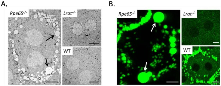
Figure 3.
(A) Horizontal EM images of RPE in 3 month old Rpe65−/−, Lrat−/− and wild-type mice. Of particular interest are the large retinosomes present around the perimeter of RPE cells in Rpe65−/− mice (black arrows), these formations are indicative of excessive ester accumulation in the retina. (B) Two photon imaging of the RPE in 3 month old Rpe65−/−, Lrat−/− and wild-type mice. Large autofluorescent spots are observed in the RPE of Rpe65−/− mice (white arrows), while such spots are absent in Lrat−/− mice, and are minimally observed in wild type mice. Scale bar 5.0 µm.

