Abstract
Lkb1 is a central regulator of cell polarity and energy metabolism through its capacity to activate the AMPK-related family of protein kinases. Germ line inactivating mutation of Lkb1 leads to Peutz-Jeghers syndrome, which is characterized by benign hamartomas and a susceptibility to malignant epithelial tumors. Mutations in Lkb1 are also found in sporadic carcinomas— most frequently in lung cancers associated with tobacco carcinogen exposure. The basis for Lkb1-dependent tumor suppression is not defined. Here, we uncover a marked sensitivity of Lkb1 mutant mice to the chemical carcinogen, DMBA. Lkb1+/- mice are highly prone to DMBA-induced squamous cell carcinoma (SCC) of the skin and lung. Confirming a cell autonomous tumor suppressor role of Lkb1, mice with epidermal-specific Lkb1 deletion are also susceptible to DMBA-induced SCC, and develop spontaneous SCC with long latency. Restoration of wild type Lkb1 causes senescence in tumor-derived cell lines, a process that can be partially bypassed by inactivation of the Rb pathway, but not by inactivation of p53 or AMPK. Our data indicate that Lkb1 is a potent suppressor of carcinogen-induced skin and lung cancers and that downstream targets beyond the AMPK-mTOR pathway are likely mediators of Lkb1-dependent tumor suppression.
Keywords: Lkbl, squamous cell carcinoma, mouse, carcinogen, tumor suppressor
Introduction
The Lkb1 tumor suppressor encodes a serine-threonine kinase that is mutated in individuals with the Peutz-Jeghers polyposis and cancer syndrome (PJS) (1, 2). PJS patients develop benign polyps—hamartomas—during adolescence and have a high incidence of carcinomas of the gastrointestinal tract and lung as adult (3, 4). Carcinomas in PJS patients arise independently of the hamartomas, hence the mechanisms by which Lkb1 controls benign polyposis and malignancy may be distinct (5). Lkb1 is also mutated in sporadic cancers whose spectrum of tumor types suggests cooperation with exposure to environmental carcinogens. Lkb1 alterations are most prevalent in lung cancers—with mutations detected in ∼30% of specimens (6-8). Significantly, Lkb1 mutations in lung cancer are highly correlated with a history of tobacco smoking and show a preferential occurrence of GC:TA substitutions, suggesting a mutational effect of polycyclic aromatic hydrocarbon adducts from tobacco carcinogens (8). Other carcinomas exhibiting Lkb1 mutations include head and neck squamous cell carcinoma and pancreatic cancer (9), which are also associated with tobacco smoking.
Lkb1 regulates cellular energy metabolism and cell polarity through its capacity to phosphorylate and activate AMP-activated protein kinase (AMPK) as well as other members of the AMPK subfamily (10-14). Activated AMPK restores ATP levels by promoting ATP-producing catabolic processes (e.g. glycolysis) and blocking ATP-consuming biosynthetic processes (including mTOR-directed protein synthesis) (15). Lkb1-mediated regulation of AMPK has tissue specific effects on glucose metabolism. In skeletal muscle, Lkb1 deletion results in loss of AMPK function and improved glucose uptake (16). Lkb1 knockout in the liver causes inactivation of both AMPK and of the AMPK family member, SIK2, and produces metabolic defects—including deregulated gluconeogenesis and lipogenesis (17). LKB1 is also an essential modulator of cellular structure and polarity through activation of AMPK as well as the microtubule affinity-regulating kinases (MARK1, 2, 3 and 4) and SAD/Brsk kinases (SAD-A and SAD-B) (18-24). Collectively, these data indicate that energy sensing and cell polarity may be broadly integrated under the control of LKB1-AMPK family signaling.
The specific signaling pathways by which Lkb1 suppresses both malignant and benign tumorigenesis, and the relative contributions of the AMPK-related family members to these processes are unknown. In this regard, it is provocative that PTEN, TSC and LKB1—tumor suppressor genes associated with hamartoma and cancer syndromes—are all negative regulators of mTOR activity (25). Hence, the AMPK-mTOR pathway is a plausible mediator of Lkb1 in restraining tumor development. In support of this notion, both Lkb1 mutant hamartomas and lung cancer cell lines show deregulation of mTOR signaling (11, 26), although the biological significance of mTOR activation has yet to be defined in these tumors. At the cellular level, Lkb1 has been implicated in the direct control of cell growth in vitro through several mechanisms. Lkb1 overexpression in melanoma and lung cancer cell lines blocks colony formation associated with the induction of the p53-p21 and PTEN pathways, respectively (27, 28). Mouse embryonic fibroblasts (MEFs) lacking Lkb1 escape culture-induced senescence, suggesting a potential role in cellular response to oxidative stress (29). Despite their immortal growth, Lkb1-/- MEFs are resistant to transformation by activated H-ras, a property that distinguishes Lkb1 deficient MEFs from those with classical immortalizing lesions in the Rb and p53 pathways.
Genetic models in which Lkb1 inactivation promotes carcinoma formation enable the study of Lkb1 tumor suppressor pathways in vivo. Given the association of Lkb1 mutation with carcinogen-associated malignancies and the need for Lkb1 mutant cancer models we assessed the impact of 7,12-dimethylbenzanthracene (DMBA) exposure on Lkb1 deficient mice.
Materials and Methods
Genetically engineered mouse strains
All mice were housed and treated in accordance with protocols approved by the institutional care and use committees for animal research at the Massachusetts General Hospital. Lkb1+/- and Lkb1L/L strains have been previously described (29). To generate K14-Cre Lkb1L/L and littermate control K14-Cre/Lkb1L/+ males were crossed with Lkb1L/L females. The Lkb1+/- mice and littermates were on an inbred FVB/n genetic background. The K14-Cre Lkb1 L/L mice and control animals were ∼87.5% FVB/n.
DMBA treatment
Pups (5-7 days old) were treated with a single dose of 50 microliters of DMBA (Sigma) in acetone (0.5% w/v) applied directly onto mouse's back.
Antibodies
Keratin 14, Involucrin, Keratin 1 (Covance), p63 (Sigma), phospho- and total-AMPK, p-ACC, phospho- and total-p70 S6Kinase, phospho- and total-S6, phospho- and total-Erk, phospho- and total-Akt, phospho-Jnk, phospho- MEK, phospho-p38, cyclin D1, phospho- and total- Rb, (Cell Signaling), phospho-EGFR (abcam) p16Ink4a (M-156) p21 (C-19) (Santa Cruz Biotechnology) p19Arf (Ab-80, Abcam), BrdU (BD; Transduction Laboratories). Rabbit Lkb1 1K antibody was described previously (29). Secondary antibodies used were Alexa fluor 488 (Molecular Probes), anti-Rabbit- and -mouse-biotin and –HRP antibodies (Vector labs). MOM kit and ABC Vector kit (Vector Labs).
Immunostaining
Dorsal skin samples were fixed for overnight in 4% paraformaldehyde then embedded in paraffin or frozen in OCT. Prior to freezing in OCT, tissue was shaken in PBS, 10, 20 and 30% sucrose for 1hr each. 10 micron sections were used for all experiments. Following immunohistochemical staining images were taken using Leica DM100 microscope and Leica DC500 camera. Following immunofluorescent staining images were taken with a Leica confocal microscope under a 40x oil immersion objective using the same laser intensity and z-settings and analyzed with LCS Advanced software. For each marker at least three independent samples were evaluated for each genotype.
H-ras mutational analysis
H-ras codon-61 status was ascertained by allele-specific PCR analysis. Oligonucleotides HrasF-wt: 5′-GACATCTTAGACACAGCAGGTCA and HrasR: 5′-TGGTGTTGTTGATGGCAAATACT were used to detect the wild type allele. HrasF-mut: 5′-GACATCTTAGACACAGCAGGTCT and HrasR were used to detect the CAA→CTA mutation. Sequence analysis of Hras codons 12, 13 and 61 was performed following amplification of genomic DNA sequences flanking these regions.
SCC cell lines
Cell lines were derived from SCC tumor tissue as described in (30). Briefly, tumor tissue was rinsed in PBS, minced finely with sterile razor blades, incubated in 0.25% trypsin-EDTA at 37°C for 20 minutes with intermittent tituration and plated in fibronectin/vitrogen coated plates in keratinocyte media (Ca+- free EMEM, 8% chelexed serum, 50ng/ml cholera toxin, 1ug/ml hydrocortisone and 2mM glutamine, 0.1-0.3mM Ca). The AB-B9 cell line—a murine DMBA-TPA induced SCC cell line with wild type Lkb1—was used as a control (31).
Molecular and Cellular Analysis
RNA and DNA isolation was performed as described using standard procedures. SCC or papilloma tissue were homogenized in lysis buffer (50mM HEPES, 250mM NaCl, 2mM EDTA, 25% Glycerol, 1% NP-40, 0.1% SDS, protease inhibitors (Roche) and phosphatase inhibitors (Calbiochem)) on ice. SCC cell lines were infected with retroviruses expressing vector, GFP, Lkb1 or Lkb1-KD, selected with puromycin and harvested in lysis buffer at indicated time points and subjected to SDS-PAGE and immunobloted with indicated antibody. For cell proliferation assays, the cells were plated in 96 well plate 24h post-infection and assayed using the WST-1 Cell Proliferation Assay reagent (Roche).
Results
Lkb1+/- mice are highly sensitive to DMBA-induced Squamous Cell Carcinoma
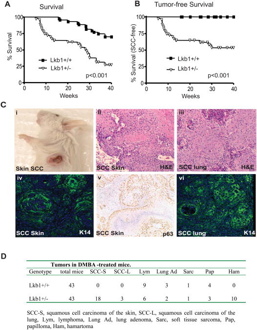
We examined the function of Lkb1 in suppression of carcinogen-induced tumors by treating 5-7 day old Lkb1+/+ and Lkb1+/- mice (29) with a single topical dose of DMBA. DMBA is acts systemically under this protocol, promoting the gradual development of lymphomas, sarcomas, and lung adenomas in wild type mice (32). The mouse cohorts were monitored for tumor incidence and spectrum up to 40 weeks of age. Lkb1+/- mice showed a significantly reduced cancer-free survival—28.6 weeks versus > 40 weeks—and altered tumor spectrum relative to Lkb1+/+ animals (Fig. 1A and table 1D; Lkb1+/- mice developing benign hamartomas were censored in the survival analysis). The increased mortality in the Lkb1+/- cohort was due to the development of invasive skin and lung cancers (present in 18/42 Lkb1+/- mice), tumors types that were not observed in wild type animals (Fig. 1B, 1Ci, and Table in 1D). Histological analysis and staining for both cytokeratin-14 and p63 revealed that these lung and skin tumors were all malignant squamous cell carcinomas (SCC) (Fig. 1Cii-vi). Lymphomas were observed in both wild type and mutant cohorts with similar incidence and latency. DMBA exposure did not affect the incidence or histopathology of hamartomas in Lkb1+/- animals (not shown). Hence, Lkb1 heterozygosity specifically sensitizes mice to DMBA-induced SCC.
Figure 1. Lkb1+/- mice are highly prone to DMBA-induced squamous cell carcinomas.
A) Survival analysis of Lkb1+/- and Lkb1+/+ mice treated with DMBA at ages 5-7 days. Lkb1+/- mice developing hamartomas were censored from the survival analysis. B) Survival analysis of the DMBA-treated cohorts documenting mortality due to SCC. C) i) Gross image of SCC in an Lkb1+/- mouse 15 wks post-DMBA application. ii) Histology of cutaneous SCC and iii) lung SCC. Immunostaining of cutaneous SCC for iv) keratin 14 and v) p63. vi) Immunofluorescence staining for keratin 14 in lung SCC.
Lkb1 mutant SCC do not evolve from the classical DMBA-induced papilloma-to-SCC sequence
The molecular progression of DMBA-induced SCC in wild type mice is well described (33). Specifically, A—T transversions at H-ras codon 61 (CAA→CTA) are a hallmark of DMBA-induced skin carcinogenesis, serving to initiate the development of benign papillomas that undergo gradual multistage progression to malignant SCC (33, 34). Mutations of various tumor suppressor genes can increase papilloma number or accelerate papilloma-to-SCC progression. Notably, papillomas were not observed prior to SCC development in serially monitored DMBA-treated Lkb1+/- mice. Furthermore, we did not detect papillomatous changes adjacent to carcinoma in our histological analysis. Finally, the incidence of papillomas was comparable in the wildtype and mutant cohorts (4/42 Lkb1+/+ mice and 3/42 Lkb1+/- mice developed papillomas).
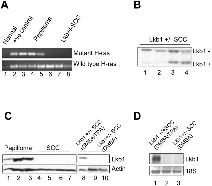
We used allele-specific PCR and direct sequencing to test the mutational status of H-ras in the papillomas and SCC arising in our study. All papillomas had activating H-ras mutations, regardless of Lkb1 genotype (4/4 papillomas from Lkb1+/+ mice and 3/3 from Lkb1+/- mice; Fig 2C and data not shown). On the other hand, none of the SCC arising in the Lkb1+/- mice (0 of 17 SCC tested) exhibited H-ras mutations (Fig. 2A and data not shown), a finding consistent with the lack of an observed papilloma-SCC sequence in these mice. Together, these results suggest that SCC associated with Lkb1 loss may involve pathways distinct from those in H-ras induced SCC. Alternatively, Lkb1 loss could lead to activation of H-ras or its effectors obviating the need for concurrent H-ras mutations (see below).
Figure 2. Genetic analysis of SCC in Lkb1+/- mice.
A) Allele-specific PCR to detect H-ras codon-61 wild type (lower panel) and CAA→CTA mutant alleles (upper panel). Lkb1 deficient SCC have only wild type H-ras (lanes 6-9) whereas papillomas (lanes 3-5) and SCC from mice with wild type Lkb1 (lane 2) harbor the mutant H-ras allele. B) Southern blot analysis showing loss of wild type Lkb1 in a subset of SCC from Lkb1+/- mice (note loss of the Lkb1+ band in lanes 1 and 2). C) Western blot showing absence of Lkb1 expression in SCC from Lkb1+/- mice (lanes 4-7) and cell lines derived from SCC from Lkb1+/- (lanes 9 and 10). Lkb1 expression is readily detected in papillomas (lanes 1-3) and in an SCC cell line derived from a DMBA/TPA-treated wild type mouse (lane 8). D) Northern blot analysis showing Lkb1 mRNA expression in an SCC cell line from a wild type mouse (lane 1) and absence of Lkb1 expression in SCC from Lkb1+/- mice (lanes 2 and 3; the tumor in lane 3 showed retention of the wild type Lkb1 allele at DNA level). 18S rRNA is shown as a loading control.
The Wild type Lkb1 allele is inactivated in SCC from Lkb1+/- mice
The benign hamartomas arising in Lkb1+/- mice and PJS patients appear to be driven primarily by Lkb1 haploinsufficiency since loss of wild type Lkb1 expression is not an obligate event in these tumors (29, 35-37). We sought to assess the status of the wild type Lkb1 allele in the malignant SCC arising in Lkb1+/- mice. Southern blot analysis of tumor DNA revealed loss of wild type Lkb1 in 8 of 20 specimens (Fig. 2B and data not shown). Furthermore, western and northern blot analyses showed that all SCC and SCC-derived cell lines lacked Lkb1 expression regardless of the status of the wild type allele (Fig. 2C and D), suggesting that the Lkb1 wild type allele is inactivated by multiple mechanisms in SCC, including deletion and possibly point mutation or promoter hypermethylation. In comparison, robust Lkb1 expression was detected in papillomas from both Lkb1+/- and Lkb1+/+ mice, in SCC cell lines generated from DMBA-12-O-tetradecanoyl-phorbol-13-acetate (TPA)-treated wild type mice (31), and in epidermal keratinocytes, the normal cellular counterparts to SCC (Fig. 2C and 3B). Hence, inactivation of the wild type Lkb1 allele is specifically associated with SCC pathogenesis in Lkb1+/- mice.
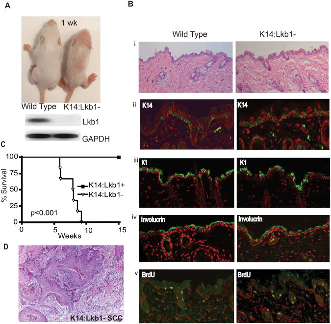
Figure 3. Analysis of mice with epidermal-specific deletion of Lkb1.
A) Gross image of K14:Lkb1- and litter mate control mice at 1 wk (upper panel) and Northern blot showing efficient deletion of Lkb1 in K14:Lkb1- keratinocytes compared to keratinocytes from littermate controls (lower panel); B i) Histological analysis of K14:Lkb1- and litter mate control dorsal skin. Epidermis from K14:Lkb1- and wild type mice show comparable staining for the differentiation markers, ii) Keratin 14, iii) Keratin 1 and iv) involucrin and comparable rates of BrdU staining (v). The immunofluorescence staining is green. DAPI stained nuclei are pseudo-colored red. C) Survival analysis of the DMBA-treated cohorts documenting mortality due to SCC in K14:Lkb1-1 mice and littermate control mice; D) Histology of SCC from a DMBA-treated K14:Lkb1-1 mouse.
Selective inactivation of Lkb1 in the epidermis sensitizes mice to carcinogen-induced and spontaneous SCC
Cutaneous SCC arises from the transformation of epidermal progenitors (38). We sought to examine Lkb1 function in the epidermis and to determine whether homozygous inactivation in this compartment is sufficient to promote SCC development. To this end, we generated mice with selective epidermal deletion of Lkb1 by crossing the Lkb1L/L and Keratin-14-Cre strains (29, 39). K14-Cre Lkb1L/L mice (hereafter, designated K14:Lkb1-) were born at the expected frequency, but were smaller than Lkb1lox/lox and K14-Cre Lkb1+/+ controls. The K14:Lkb1- animals exhibited delays in hair growth, and had wavy and less dense hair as adults (Fig. 3A). Northern blot analysis of primary keratinocytes confirmed Lkb1 was specifically inactivated in the epidermis of these mice (Fig. 3A lower panel). Cutaneous histology revealed a diminution in hair shaft diameter, increased erythema of the skin (reddening associated with congestion of the capillaries), and mild follicular plugging (filling of follicular openings with keratinous debris) (Fig. 3Bi). In addition, these animals had corneal opacity associated with hyperkeratinization of the corneal epithelium (data not shown). Immunofluorescence analysis of K14, K1, and involucrin revealed comparable staining in the epidermis of K14:Lkb1- and control mice, indicating that epidermal differentiation is not compromised in the absence of Lkb1 (Fig. 3B ii-iv). BrdU analysis of the epidermal compartment showed similar rates of proliferation between 3-week old control and K14:Lkb1- animals (the frequency of BrdU+ nuclei/field was 8 +/- 1.7 and 7.5 +/- 1.7, respectively), indicating that Lkb1 does not influence keratinocyte proliferation in untreated mice (Fig. 3B v). Similarly, histological analysis failed to reveal significant alterations in epidermal cell death or defects in epidermal polarity in the K14:Lkb1- animals. Despite their grossly normal epidermal development and homeostasis, a subset of K14:Lkb1- mice developed spontaneous SCC by the age of 40 weeks (3/20 mice). The relatively long latency and absence of an early hyperproliferative phenotype in these mice suggest that Lkb1 functions as a tumor suppressor in the epidermis but that the development of SCC requires additional oncogenic changes.
In contrast to the long latency in spontaneous tumor development, the K14:Lkb1- mice were highly tumor-prone following DMBA administration. Invasive skin cancers were observed in 6/6 DMBA-treated K14:Lkb1- mice (average latency 8 weeks) whereas 0/10 DMBA-treated control animals developed tumors by 15 weeks (Fig. 3C). Histological analysis confirmed that these tumors were SCC resembling those observed in the DMBA-treated Lkb1+/- mice (Fig. 3D). Hence homozygous deletion of Lkb1 in the epidermis renders mice highly sensitive to SCC initiated by a chemical carcinogen.
Signaling pathways in Lkb1 mutant SCC
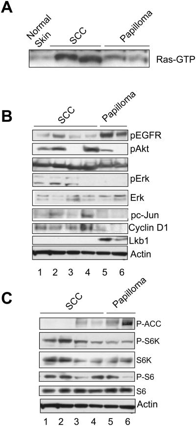
Having demonstrated a critical role for Lkb1 in suppression of DMBA-induced SCC, we wished to define molecular alterations associated with tumorigenesis in Lkb1 mutant mice. RAF pulldown assays showed elevated levels of activated Ras-GTP in Lkb1 mutant SCC relative to papillomas and normal skin despite the absence of H-ras mutations in these tumors (Fig. 4A). Previous genetic studies have shown that RAF-MEK-ERK-cyclin D1 and EGFR-PI3K-AKT pathways are critical effectors of Ras directed skin carcinogenesis and that the activity of these pathways increases gradually during papilloma-SCC progression (40-43). Western blots analysis showed that 3/4 Lkb1 mutant SCC tested expressed p-ERK, whereas p-ERK was absent in papillomas (Fig. 4B). Cyclin D1 and p-c-Jun levels were elevated in SCC relative to papillomas. Finally, p-EGFR was detectable in all SCC and robust p-AKT levels were noted in 3/4 of these tumors (Fig. 4B). Together, these results indicate that although no activating H-Ras mutations are present in the Lkb1 mutant SCC, Ras signaling pathways are deregulated in these tumors.
Figure 4. Signaling pathways in Lkb1 mutant SCC.
A) RAF pulldown assay showing levels of activated Ras-GTP in normal skin (lane 1), SCC from from Lkb1+/- mice (lanes 2 and 3), and papillomas (lanes 4 and 5). B) Western blot analysis of ERK and PI3K pathway components in papillomas (lanes 5 and 6) and Lkb1-mutant SCC (lanes 1-4). C) Western blot analysis of the AMPK/mTOR signaling pathway in papillomas (lanes 5 and 6) and SCC (lanes 1-4).
The AMPK-TSC-mTOR pathway is a candidate mediator of Lkb1-dependent tumor suppression. Western blot analysis showed that the Lkb1 mutant SCC had diminished AMPK Thr-308 phosphorylation relative to papillomas, consistent with loss AMPK activity in these tumors (Fig. 4C). Correspondingly, p-S6-kinase levels were increased in the Lkb1 mutant SCCs, whereas there was heterogeneous expression of p-S6. Hence, AMPK activity is compromised in Lkb1 mutant SCC, although, the level of mTOR signaling was not markedly increased in these tumors relative to papillomas.
Restoration of Lkb1 in SCC cell lines results in growth arrest that cannot be rescued by disruption of AMPK signaling
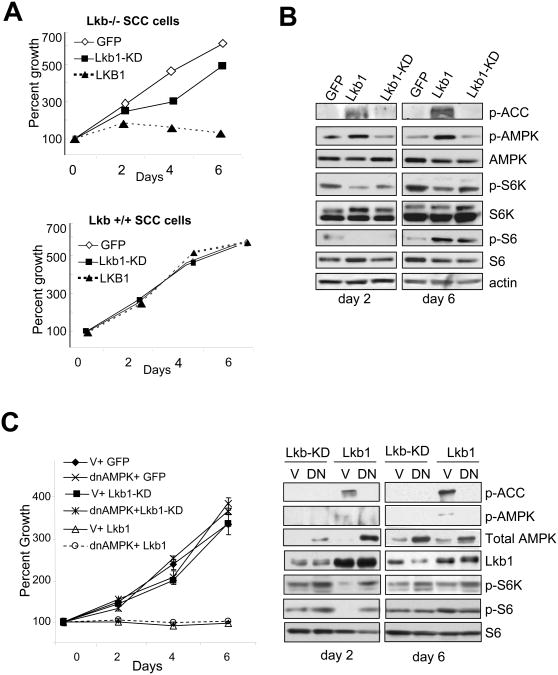
The Lkb1 mutant SCC cell lines that we established from DMBA-treated mice provided a system to address the mechanisms of Lkb1-dependent tumor suppression. SCC cell lines generated from DMBA/TPA-treated wild type mice served as controls for these studies (31). Human cancer genetics studies indicate that Lkb1 kinase activity is likely to be critical for tumor suppression since most cancer-associated Lkb1 mutations result in impaired catalytic activity (9). Correspondingly, introduction of retroviruses expressing wild type Lkb1, but not a kinase-dead mutant, resulted in rapid induction of growth arrest in these cell lines (Fig. 5A upper panel). This phenotype was specific since Lkb1 overexpression did not affect the growth characteristics of SCC cell lines harboring wild type Lkb1 (Fig. 5A lower panel).
Figure 5. Lkb1 restoration provokes AMPK-independent growth arrest in Lkb1-mutant SCC cells.
A) Growth curves of Lkb1-/- (upper panel) and Lkb1+/+ SCC cells (lower panel) following transduction with retroviruses expressing GFP, Lkb1 or a kinase-dead Lkb1 mutant. B) Western blot analysis of AMPK-mTOR pathway at 2 days (left panel) and 6 days (right panel) following retroviral transduction. Note the Lkb1 provokes sustained p-AMPK and p-ACC expression, and that p-S6K levels remain repressed whereas p-S6 reduction is only transiently observed. C) Left panel: Growth curves of SCC cells infected with adenoviruses expressing dominant-negative AMPK or GFP and subsequently transduced with retroviruses encoding wild type Lkb1 or Lkb-KD mutant. Right panel: western blot analysis showing that DN-AMPK blocks Lkb1 mediated activation of AMPK measured by pAMPK and p-ACC and the effect on mTOR pathway measured by p-S6K and pS6 at 2 days and 6 days post-retroviral transduction.
Next we assessed role of the AMPK pathway in Lkb1-directed growth arrest. Wild type Lkb1 specifically activated AMPK in SCC cells, as reflected by increased phosphorylation of AMPKα Thr-172, and the of AMPK target, Acetyl Co-A Carboxylase Ser-79 (Fig. 5B). Consistent with AMPK activation, mTOR activity was repressed by wild type Lkb1 as shown by a reduction in levels of p-S6-Kinase. P-S6 levels were also reduced 24-48 hours following Lkb1 restoration, however, this effect was transient, and elevations in p-S6 were noted at later time points (>96 hrs).
To test directly the requirement of AMPK in growth arrest, we introduced adenoviruses expressing dominant-negative AMPK into the SCC cells and assessed whether Lkb1 arrest was abrogated. While DN-AMPK expression blocked Lkb1-induced increases in p-AMPK and its target p-ACC, and a concomitant decrease in p-S6K and p-S6 at early time points, this disruption in AMPK signaling did not rescue the gross arrest phenotype (Fig. 5C left and right panels). Along these lines, the pharmacological AMPK inhibitor, compound C, was unable to rescue Lkb1-mediated growth arrest (data not shown). These results indicate that AMPK-mTOR signaling is not required for Lkb1 induced growth inhibition of SCC cell lines, and therefore may not be critical for tumor suppression downstream of Lkb1.
Lkb1-mediated growth arrest shows features of oncogene-induced senescence
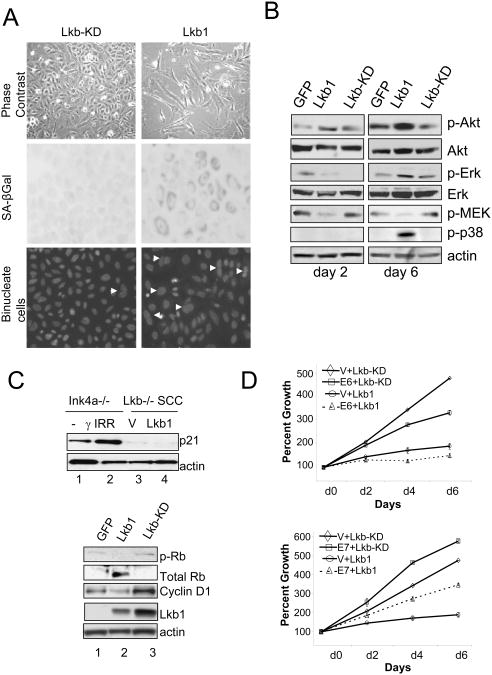
Oncogene-induced senescence refers to a specific type of growth arrest that occurs in primary cells in response to strong oncogenic signals, and therefore serves as a barrier to tumor progression (44). We noted that Lkb1-arrested SCC cells became enlarged, took on a flattened appearance, were frequently bi-nucleated, and stained for senescence-associated beta-galactosidase—indicating that wild type Lkb1 restored a senescence response in these cancer cells (Fig 6A).
Figure 6. Analysis of Lkb1-dependent growth arrest in SCC cells.
A) Expression of wild type, but not kinase-dead Lkb1 induces senescence in Lkb1 mutant SCC cells, as reflected by enlarged, flattened appearance (upper panels), senescence-associated β-gal staining (middle panels) and binucleated cells (bottom panels). B) Signaling pathways activated in SCC cells during Lkb1-mediated growth inhibition and senescence measured by western blot analysis at early (day 2) and late time points (day 6). Despite inhibition of p-MEK levels, Lkb1 expression resulted in increase in p-Akt, pErk and p-38. C) Western blot showing that Lkb1-expressing retroviruses fail to induce the p53 target gene in SCC cells (lane 4) in comparison to empty vector (lane 3) (upper panel). Untreated and γ-irradiated fibroblasts (lanes 1 and 2) were used as controls for p53 activation. C lower panel) Western blot analysis showing Rb pathway activation by Lkb1 in SCC cells. Lkb1 (lane 2) induced a loss of p-Rb-S795, a increase in hypophorylated Rb and a decrease in cyclin D1 relative to Lkb1-KD mutant (lane 3) or GFP (lane 1. D) Growth curves showing that expression of E6 had no effect on Lkb1-mediated growth inhibition (upper panel) whereas E7 partially rescued Lkb1-mediated growth inhibition (lower panel). Error bars are depicted but may be too small to see.
Oncogene-induced senescence is associated with feedback signals that inactivate the RAF-MAPK and PI3K-AKT pathways, with the induction of p38-mediated oxidative stress responses (45, 46). Western blot analysis showed that Lkb1-mediated growth arrest did not require inactivation of PI3K signaling since p-AKT levels were elevated following Lkb1 expression (Fig. 6B), a finding consistent with reduced mTOR/S6K signaling and a resulting loss of feedback inhibition IGFI/PI3K signaling (47). Lkb1 expression led to an acute and sustained decrease in p-MEK, whereas p-ERK levels were only transiently decreased, suggesting that Lkb1 represses the RAF-MAPK pathway at a level of upstream of p-ERK. Finally, we found that Lkb1 restoration resulted in the pronounced activation of p38, indicating that Lkb1 restoration provokes an acute stress response in these cells.
Role of the Rb and p53 pathways in Lkb1-induced growth arrest
Intact Rb and p53 pathway function are broadly required for senescence responses. Lkb1 restoration did not lead to increased expression of p53 or p21, a p53 target gene indicating that arrest provoked by Lkb1 was likely to be p53-independent (Fig. 6C upper panel, and not shown). In contrast, the wild type Lkb1-expressing SCC cells showed increased levels of the hypophosphorylated, activated form of Rb and decreases in hyperphosphorylated, inactivated Rb (Fig. 6C lower panel). Rb phosphorylation is controlled by cyclin/cyclin-dependent kinase (CDK) complexes. Both wild type and kinase-dead Lkb1 induced expression of the CDK4/6 inhibitor, p16Ink4a, (data not shown), whereas wild type Lkb1 expressing cells showed a specific reduction in cyclin D1 levels. These data indicate that Lkb1-induced growth arrest is associated with activation of Rb and a corresponding altered balance of negative and positive regulators of the Rb checkpoint.
We sought to directly test the requirement for the Rb and p53 pathways in Lkb1 mediated growth arrest of SCC cells by expression of viral oncogenes that inactivate these pathways. Prior to introduction of Lkb1 retroviruses, the SCC cells were transduced with retroviruses encoding the human papilloma virus proteins, E6 (to inactivate p53), E7 (to inactivate Rb), or both E6 and E7. The expression of E6 had no impact on Lkb1-induced arrest (Fig. 6E upper panel), while expression of either E7 alone, or E6 and E7, led to a partial rescue of this growth arrest phenotype (Fig. 6E lower panel, and data not shown). These results suggest that Lkb1 suppression of SCC proliferation involves the Rb function, but not p53 function, and that additional pathways are required to mediate Lkb1 activity.
Discussion
In this study, we describe the development of genetic models that demonstrate an important role of Lkb1 in suppression of carcinogen-induced tumorigenesis. Mice with germ line heterozygous mutations of Lkb1, or with selective deletion of Lkb1 in the epidermis, were highly prone to the development of DMBA-induced SCC. Restoration of wild type Lkb1 in tumor-derived SCC cell lines resulted in a senescence-like growth arrest that involved Rb function but was independent of the p53 and AMPK pathways. This genetic model and associated cell lines provide a framework to elucidate the mechanisms of Lkb1-dependent suppression of epithelial cancers and may uncover specific roles for Lkb1 in response to environmental carcinogens.
We observed that the wild type Lkb1 allele was inactivated in all DMBA-induced SCC from Lkb1+/- mice, and that absence of Lkb1 in the epidermis led to greatly accelerated SCC progression (SCC latency was 15 weeks in Lkb1+/- mice and 7 weeks in K14:Lkb1- mice). Regardless of allelic loss, all SCC lacked expression of Lkb1. Hence, in contrast to the benign gastrointestinal polyposis associated with Lkb1 deficiency, malignant SCC pathogenesis appears to require biallelic inactivation of Lkb1.
The AMPK-TSC-mTOR pathway has been a prime candidate for mediating tumor suppression downstream of the Lkb1 kinase (48). Consistent with this, deregulation of this pathway was observed in Lkb1 mutant SCCs in vivo and in derivative cell lines. However, the inactivation of AMPK using pharmacological inhibitors or expression of dominant negative AMPK mutants had no effect on Lkb1-induced growth arrest in SCC cell lines. The results indicate that in Lkb1-deficient cancers, pathways other than AMPK-TSC-mTOR are likely to be the critical downstream effectors of Lkb1. It remains possible that deregulation of this pathway may be important for the initiating stages of Lkb1 mutant tumors, and dispensable in the later stages of cancer progression.
Our demonstration that Lkb1 restoration causes senescence in SCC cell lines is notable in light of our previous observation that Lkb1 forms a barrier to passage induced senescence in primary MEFs. Senescence in primary cells arises due to the generation of reactive oxygen species (ROS) that are genotoxic and provoke Rb and p53 dependent checkpoint responses (44, 45). Genetic alterations that reduce ROS production or that bypass these checkpoints can result in escape from senescence. We noted that Lkb1 restoration in SCC cell lines was associated with induction of p38 suggesting the Lkb1 provokes an acute stress response. We speculate that tumor suppressor role of Lkb1 may involve induction of senescence in cells receiving aberrant growth signals.
Based on the requirement of Lkb1 in squamous tumor suppression it was surprising that mice with epidermal-specific deletion of Lkb1 showed largely normal skin development. The overall architecture of the skin was not impaired in these mice and the normal expression of keratin-1, keratin-14 and involucrin indicated the Lkb1 mutant keratinocytes underwent normal differentiation. The prominent increase in SCC development in the context of DMBA exposure may indicate that Lkb1 plays a particular role in restraining proliferation in response to chemical carcinogens or more broadly to stresses that result in increased cell turnover. Such a role could account for the strong association between Lkb1 mutations and smoking-associated cancers.
Acknowledgments
The authors would like to thank Ron DePinho for helpful discussions during the course of this work. We are grateful to Alice Yu and Shan Zhou for their dedication and expertise in maintenance of the mouse colonies. We thank Stephen Lessnick for the E6 and E7 plasmids. These studies were supported by grants to N.B. from the NIH (5 K01 CA104647 and 5 P01 CA117969), the Waxman Foundation, the Harvard Stem Cell Institute, and the Linda Verville Foundation.
References
- 1.Jenne DE, Reimann H, Nezu J, et al. Peutz-Jeghers syndrome is caused by mutations in a novel serine threonine kinase. Nat Genet. 1998;18(1):38–43. doi: 10.1038/ng0198-38. [DOI] [PubMed] [Google Scholar]
- 2.Hemminki A. The molecular basis and clinical aspects of Peutz-Jeghers syndrome. Cell Mol Life Sci. 1999;55(5):735–50. doi: 10.1007/s000180050329. [DOI] [PMC free article] [PubMed] [Google Scholar]
- 3.Hearle N, Schumacher V, Menko FH, et al. Frequency and spectrum of cancers in the Peutz-Jeghers syndrome. Clin Cancer Res. 2006;12(10):3209–15. doi: 10.1158/1078-0432.CCR-06-0083. [DOI] [PubMed] [Google Scholar]
- 4.Schreibman IR, Baker M, Amos C, McGarrity TJ. The hamartomatous polyposis syndromes: a clinical and molecular review. Am J Gastroenterol. 2005;100(2):476–90. doi: 10.1111/j.1572-0241.2005.40237.x. [DOI] [PubMed] [Google Scholar]
- 5.Jansen M, de Leng WW, Baas AF, et al. Mucosal prolapse in the pathogenesis of Peutz-Jeghers polyposis. Gut. 2006;55(1):1–5. doi: 10.1136/gut.2005.069062. [DOI] [PMC free article] [PubMed] [Google Scholar]
- 6.Sanchez-Cespedes M, Parrella P, Esteller M, et al. Inactivation of LKB1/STK11 is a common event in adenocarcinomas of the lung. Cancer Res. 2002;62(13):3659–62. [PubMed] [Google Scholar]
- 7.Ji H, Ramsey MR, Hayes DN, et al. LKB1 modulates lung cancer differentiation and metastasis. Nature. 2007;448(7155):807–10. doi: 10.1038/nature06030. [DOI] [PubMed] [Google Scholar]
- 8.Matsumoto S, Iwakawa R, Takahashi K, et al. Prevalence and specificity of LKB1 genetic alterations in lung cancers. Oncogene. 2007 doi: 10.1038/sj.onc.1210418. [DOI] [PMC free article] [PubMed] [Google Scholar]
- 9.Sanchez-Cespedes M. A role for LKB1 gene in human cancer beyond the Peutz-Jeghers syndrome. Oncogene. 2007 doi: 10.1038/sj.onc.1210594. [DOI] [PubMed] [Google Scholar]
- 10.Shaw RJ, Kosmatka M, Bardeesy N, et al. The tumor suppressor LKB1 kinase directly activates AMP-activated kinase and regulates apoptosis in response to energy stress. Proc Natl Acad Sci U S A. 2004 doi: 10.1073/pnas.0308061100. [DOI] [PMC free article] [PubMed] [Google Scholar]
- 11.Shaw RJ, Bardeesy N, Manning BD, et al. The LKB1 tumor suppressor negatively regulates mTOR signaling. Cancer Cell. 2004;6(1):91–9. doi: 10.1016/j.ccr.2004.06.007. [DOI] [PubMed] [Google Scholar]
- 12.Corradetti MN, Inoki K, Bardeesy N, DePinho RA, Guan KL. Regulation of the TSC pathway by LKB1: evidence of a molecular link between tuberous sclerosis complex and Peutz-Jeghers syndrome. Genes Dev. 2004;18(13):1533–8. doi: 10.1101/gad.1199104. [DOI] [PMC free article] [PubMed] [Google Scholar]
- 13.Hawley SA, Boudeau J, Reid JL, et al. Complexes between the LKB1 tumor suppressor, STRADalpha/beta and MO25alpha/beta are upstream kinases in the AMP-activated protein kinase cascade. J Biol. 2003;2(4):28. doi: 10.1186/1475-4924-2-28. [DOI] [PMC free article] [PubMed] [Google Scholar]
- 14.Woods A, Johnstone SR, Dickerson K, et al. LKB1 is the upstream kinase in the AMP-activated protein kinase cascade. Curr Biol. 2003;13(22):2004–8. doi: 10.1016/j.cub.2003.10.031. [DOI] [PubMed] [Google Scholar]
- 15.Shaw RJ. Glucose metabolism and cancer. Current opinion in cell biology. 2006;18(6):598–608. doi: 10.1016/j.ceb.2006.10.005. [DOI] [PubMed] [Google Scholar]
- 16.Koh HJ, Arnolds DE, Fujii N, et al. Skeletal muscle-selective knockout of LKB1 increases insulin sensitivity, improves glucose homeostasis, and decreases TRB3. Mol Cell Biol. 2006;26(22):8217–27. doi: 10.1128/MCB.00979-06. [DOI] [PMC free article] [PubMed] [Google Scholar]
- 17.Shaw RJ, Lamia KA, Vasquez D, et al. The kinase LKB1 mediates glucose homeostasis in liver and therapeutic effects of metformin. Science. 2005;310(5754):1642–6. doi: 10.1126/science.1120781. [DOI] [PMC free article] [PubMed] [Google Scholar]
- 18.Lee JH, Koh H, Kim M, et al. Energy-dependent regulation of cell structure by AMP-activated protein kinase. Nature. 2007;447(7147):1017–20. doi: 10.1038/nature05828. [DOI] [PubMed] [Google Scholar]
- 19.Baas AF, Kuipers J, van der Wel NN, et al. Complete polarization of single intestinal epithelial cells upon activation of LKB1 by STRAD. Cell. 2004;116(3):457–66. doi: 10.1016/s0092-8674(04)00114-x. [DOI] [PubMed] [Google Scholar]
- 20.Zheng B, Cantley LC. Regulation of epithelial tight junction assembly and disassembly by AMP-activated protein kinase. Proc Natl Acad Sci U S A. 2007;104(3):819–22. doi: 10.1073/pnas.0610157104. [DOI] [PMC free article] [PubMed] [Google Scholar]
- 21.Mirouse V, Swick LL, Kazgan N, St Johnston D, Brenman JE. LKB1 and AMPK maintain epithelial cell polarity under energetic stress. J Cell Biol. 2007;177(3):387–92. doi: 10.1083/jcb.200702053. [DOI] [PMC free article] [PubMed] [Google Scholar] [Retracted]
- 22.Lizcano JM, Goransson O, Toth R, et al. LKB1 is a master kinase that activates 13 kinases of the AMPK subfamily, including MARK/PAR-1. Embo J. 2004;23(4):833–43. doi: 10.1038/sj.emboj.7600110. [DOI] [PMC free article] [PubMed] [Google Scholar]
- 23.Shelly M, Cancedda L, Heilshorn S, Sumbre G, Poo MM. LKB1/STRAD promotes axon initiation during neuronal polarization. Cell. 2007;129(3):565–77. doi: 10.1016/j.cell.2007.04.012. [DOI] [PubMed] [Google Scholar]
- 24.Barnes AP, Lilley BN, Pan YA, et al. LKB1 and SAD kinases define a pathway required for the polarization of cortical neurons. Cell. 2007;129(3):549–63. doi: 10.1016/j.cell.2007.03.025. [DOI] [PubMed] [Google Scholar]
- 25.Tee AR, Blenis J. mTOR, translational control and human disease. Seminars in cell & developmental biology. 2005;16(1):29–37. doi: 10.1016/j.semcdb.2004.11.005. [DOI] [PubMed] [Google Scholar]
- 26.Carretero J, Medina PP, Blanco R, et al. Dysfunctional AMPK activity, signalling through mTOR and survival in response to energetic stress in LKB1-deficient lung cancer. Oncogene. 2007;26(11):1616–25. doi: 10.1038/sj.onc.1209951. [DOI] [PubMed] [Google Scholar]
- 27.Tiainen M, Vaahtomeri K, Ylikorkala A, Makela TP. Growth arrest by the LKB1 tumor suppressor: induction of p21(WAF1/CIP1) Hum Mol Genet. 2002;11(13):1497–504. doi: 10.1093/hmg/11.13.1497. [DOI] [PubMed] [Google Scholar]
- 28.Jimenez AI, Fernandez P, Dominguez O, Dopazo A, Sanchez-Cespedes M. Growth and molecular profile of lung cancer cells expressing ectopic LKB1: down-regulation of the phosphatidylinositol 3′-phosphate kinase/PTEN pathway. Cancer Res. 2003;63(6):1382–8. [PubMed] [Google Scholar]
- 29.Bardeesy N, Sinha M, Hezel AF, et al. Loss of the Lkb1 tumour suppressor provokes intestinal polyposis but resistance to transformation. Nature. 2002;419(6903):162–7. doi: 10.1038/nature01045. [DOI] [PubMed] [Google Scholar]
- 30.Pera MF, Gorman PA. In vitro analysis of multistage epidermal carcinogenesis: development of indefinite renewal capacity and reduced growth factor requirements in colony forming keratinocytes precedes malignant transformation. Carcinogenesis. 1984;5(5):671–82. doi: 10.1093/carcin/5.5.671. [DOI] [PubMed] [Google Scholar]
- 31.Oft M, Akhurst RJ, Balmain A. Metastasis is driven by sequential elevation of H-ras and Smad2 levels. Nat Cell Biol. 2002;4(7):487–94. doi: 10.1038/ncb807. [DOI] [PubMed] [Google Scholar]
- 32.Sharpless NE, Bardeesy N, Lee KH, et al. Loss of p16Ink4a with retention of p19Arf predisposes mice to tumorigenesis. Nature. 2001;413(6851):86–91. doi: 10.1038/35092592. [DOI] [PubMed] [Google Scholar]
- 33.Quintanilla M, Brown K, Ramsden M, Balmain A. Carcinogen-specific mutation and amplification of Ha-ras during mouse skin carcinogenesis. Nature. 1986;322(6074):78–80. doi: 10.1038/322078a0. [DOI] [PubMed] [Google Scholar]
- 34.Frame S, Balmain A. Integration of positive and negative growth signals during ras pathway activation in vivo. Curr Opin Genet Dev. 2000;10(1):106–13. doi: 10.1016/s0959-437x(99)00052-0. [DOI] [PubMed] [Google Scholar]
- 35.Rossi DJ, Ylikorkala A, Korsisaari N, et al. Induction of cyclooxygenase-2 in a mouse model of Peutz-Jeghers polyposis. Proc Natl Acad Sci U S A. 2002;99(19):12327–32. doi: 10.1073/pnas.192301399. [DOI] [PMC free article] [PubMed] [Google Scholar]
- 36.Jishage K, Nezu J, Kawase Y, et al. Role of Lkb1, the causative gene of Peutz-Jegher's syndrome, in embryogenesis and polyposis. Proc Natl Acad Sci U S A. 2002;99(13):8903–8. doi: 10.1073/pnas.122254599. [DOI] [PMC free article] [PubMed] [Google Scholar]
- 37.Miyoshi H, Nakau M, Ishikawa TO, Seldin MF, Oshima M, Taketo MM. Gastrointestinal hamartomatous polyposis in Lkb1 heterozygous knockout mice. Cancer Res. 2002;62(8):2261–6. [PubMed] [Google Scholar]
- 38.Perez-Losada J, Balmain A. Stem-cell hierarchy in skin cancer. Nat Rev Cancer. 2003;3(6):434–43. doi: 10.1038/nrc1095. [DOI] [PubMed] [Google Scholar]
- 39.Indra AK, Li M, Brocard J, et al. Targeted somatic mutagenesis in mouse epidermis. Hormone research. 2000;54(5-6):296–300. doi: 10.1159/000053275. [DOI] [PubMed] [Google Scholar]
- 40.Sibilia M, Fleischmann A, Behrens A, et al. The EGF receptor provides an essential survival signal for SOS- dependent skin tumor development. Cell. 2000;102(2):211–20. doi: 10.1016/s0092-8674(00)00026-x. [DOI] [PubMed] [Google Scholar]
- 41.Dlugosz AA, Hansen L, Cheng C, et al. Targeted disruption of the epidermal growth factor receptor impairs growth of squamous papillomas expressing the v-ras(Ha) oncogene but does not block in vitro keratinocyte responses to oncogenic ras. Cancer Res. 1997;57(15):3180–8. [PubMed] [Google Scholar]
- 42.Robles AI, Rodriguez-Puebla ML, Glick AB, et al. Reduced skin tumor development in cyclin D1-deficient mice highlights the oncogenic ras pathway in vivo. Genes Dev. 1998;12(16):2469–74. doi: 10.1101/gad.12.16.2469. [DOI] [PMC free article] [PubMed] [Google Scholar]
- 43.Segrelles C, Ruiz S, Perez P, et al. Functional roles of Akt signaling in mouse skin tumorigenesis. Oncogene. 2002;21(1):53–64. doi: 10.1038/sj.onc.1205032. [DOI] [PubMed] [Google Scholar]
- 44.Hemann MT, Narita M. Oncogenes and senescence: breaking down in the fast lane. Genes Dev. 2007;21(1):1–5. doi: 10.1101/gad.1514207. [DOI] [PubMed] [Google Scholar]
- 45.Yaswen P, Campisi J. Oncogene-induced senescence pathways weave an intricate tapestry. Cell. 2007;128(2):233–4. doi: 10.1016/j.cell.2007.01.005. [DOI] [PubMed] [Google Scholar]
- 46.Courtois-Cox S, Genther Williams SM, Reczek EE, et al. A negative feedback signaling network underlies oncogene-induced senescence. Cancer Cell. 2006;10(6):459–72. doi: 10.1016/j.ccr.2006.10.003. [DOI] [PMC free article] [PubMed] [Google Scholar]
- 47.Manning BD. Balancing Akt with S6K: implications for both metabolic diseases and tumorigenesis. J Cell Biol. 2004;167(3):399–403. doi: 10.1083/jcb.200408161. [DOI] [PMC free article] [PubMed] [Google Scholar]
- 48.Luo Z, Saha AK, Xiang X, Ruderman NB. AMPK, the metabolic syndrome and cancer. Trends Pharmacol Sci. 2005;26(2):69–76. doi: 10.1016/j.tips.2004.12.011. [DOI] [PubMed] [Google Scholar]


