Abstract
Alistipes finegoldii Rautio et al. 2003 is one of five species of Alistipes with a validly published name: family Rikenellaceae, order Bacteroidetes, class Bacteroidia, phylum Bacteroidetes. This rod-shaped and strictly anaerobic organism has been isolated mostly from human tissues. Here we describe the features of the type strain of this species, together with the complete genome sequence, and annotation. A. finegoldii is the first member of the genus Alistipes for which the complete genome sequence of its type strain is now available. The 3,734,239 bp long single replicon genome with its 3,302 protein-coding and 68 RNA genes is part of the Genomic Encyclopedia of Bacteria and Archaea project.
Keywords: Gram-negative, rod-shaped, non-sporulating, non-motile, mesophile, strictly anaerobic, chemoorganotrophic, Rikenellaceae, GEBA
Introduction
Strain AHN2437T (= DSM 17242 = CCUG 46020 = JCM 16770) is the type strain of Alistipes finegoldii [1,2]. This strain is one of several strains with similar properties [3] that were isolated mainly from pediatric patients with inflamed, gangrenous or non-inflamed appendices [4,5]. Though the type strain AHN2437T resembled members of the Bacteroides fragilis group in bile-resistance and positive indole reaction, it was found, together with the type strain of Bacteroides putredinis, to form a separate phylogenetic lineage apart from authentic Bacteroides species [1]. The genus Alistipes was established to accommodate these two species and has subsequently been enlarged to encompass three additional species with validly published names and one with an effectively published name [6,7]. According to the position in ‘The All-Species Living Tree‘ 16S rRNA gene sequence dendrogram [8], the genus Alistipes is a sister clade of Rikenella microfusus, formerly Bacteroides microfusus [9,10], the two genera constituting the family Rikenellaceae [11,12]. Here we present a summary classification and a set of features for A. finegoldii AHN2437T together with the description of the complete genomic sequencing and annotation.
Classification and features
16S rDNA gene sequence analysis
A representative genomic 16S rRNA gene sequence of A. finegoldii AHN2437T was compared using NCBI BLAST [13,14] under default settings (e.g., considering only the high-scoring segment pairs (HSPs) from the best 250 hits) with the most recent release of the Greengenes database [15] and the relative frequencies of taxa and keywords (reduced to their stem [16]) were determined, weighted by BLAST scores. The most frequently occurring genera were Alistipes (84.4%) and Bacteroides (15.6%) (19 hits in total). Regarding the three hits to sequences from members of the species, the average identity within HSPs was 98.7%, whereas the average coverage by HSPs was 98.0%. Regarding the nine hits to sequences from other members of the genus, the average identity within HSPs was 96.5%, whereas the average coverage by HSPs was 100.1%. Among all other species, the one yielding the highest score was Alistipes shahii (AB554233), which corresponded to an identity of 97.2% and an HSP coverage of 100.0%. (Note that the Greengenes database uses the INSDC (= EMBL/NCBI/DDBJ) annotation, which is not an authoritative source for nomenclature or classification.) The highest-scoring environmental sequence was AY643083 (Greengenes short name 'Isolation finegoldii blood two patients colon cancer Alistipes finegoldii; clone 3'), which showed an identity of 100.0% and an HSP coverage of 99.4%. The most frequently occurring keywords within the labels of all environmental samples which yielded hits were 'human' (11.5%), 'fecal' (8.1%), 'intestin' (5.5%), 'biopsi' (4.2%) and 'mucos' (4.0%) (231 hits in total). The most frequently occurring keywords within the labels of those environmental samples which yielded hits of a higher score than the highest scoring species were 'finegoldii' (18.2%), 'alistip, blood, cancer, colon, isol, patient, two' (9.1%) and 'fecal, human' (9.1%) (2 hits in total). These keywords are in accordance with the original isolation source of A. finegoldii.
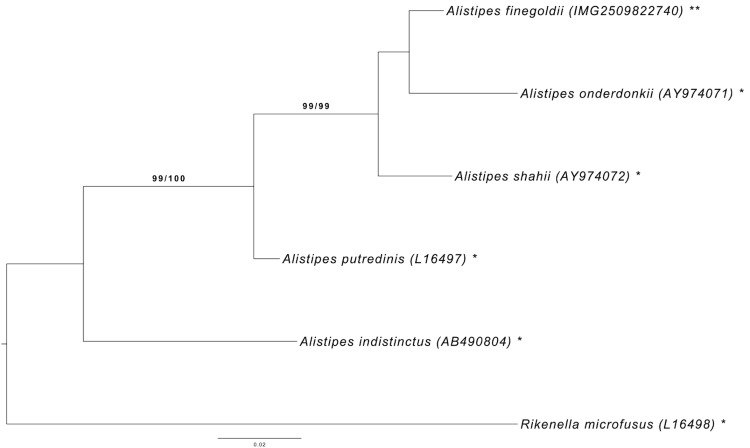
Figure 1 shows the phylogenetic neighborhood of A. finegoldii in a 16S rRNA gene based tree. The sequences of the two 16S rRNA gene copies in the genome differ from each other by ten nucleotides, and differ by up to ten nucleotides from the previously published 16S rRNA gene sequence (AY643083).
Figure 1.
Phylogenetic tree highlighting the position of A. finegoldii relative to the type strains of the other species within the family Rikenellaceae. The tree was inferred from 1,432 aligned characters [17,18] of the 16S rRNA gene sequence under the maximum likelihood (ML) criterion [19]. Rooting was done initially using the midpoint method [20] and then checked for its agreement with the current classification (Table 1). The branches are scaled in terms of the expected number of substitutions per site. Numbers adjacent to the branches are support values from 1,000 ML bootstrap replicates [21] (left) and from 1,000 maximum-parsimony bootstrap replicates [22] (right) if larger than 60%. Lineages with type strain genome sequencing projects registered in GOLD [23] are labeled with one asterisk, those also listed as 'Complete and Published' with two asterisks. See also the species the not yet validly published names described together with their genome sequences in [6].
Table 1. Classification and general features of A. finegoldii AHN2437T according to the MIGS recommendations [24].
| MIGS ID | Property | Term | Evidence code |
|---|---|---|---|
| Domain: Bacteria | TAS [25] | ||
| Phylum Bacteroidetes | TAS [12,26] | ||
| Class Bacteroidia | TAS [12,27] | ||
| Current classification | Order Bacteroidales | TAS [12,28] | |
| Family Rikenellaceae | TAS [11,12] | ||
| Genus Alistipes | TAS [1,2] | ||
| Species Alistipes finegoldii | TAS [1,2] | ||
| MIGS-12 | Reference for biomaterial | Rautio et al., 2003 | TAS [1] |
| MIGS-7 | Subspecific genetic lineage (strain) | AHN2437T | TAS [1] |
| Gram stain | negative | TAS [1] | |
| Cell shape | rod-shaped | TAS [1] | |
| Motility | non-motile | TAS [1] | |
| Sporulation | non-sporulating | TAS [1] | |
| Temperature range | mesophile | TAS [1] | |
| Optimum temperature | 37°C | TAS [1] | |
| Salinity | not reported | ||
| MIGS-22 | Relationship to oxygen | strictly anaerobe | TAS [1] |
| Carbon source | not reported | ||
| Energy metabolism | chemoorganotroph | TAS [1] | |
| MIGS-6 | Habitat | probably human gut | TAS [1] |
| MIGS-6.2 | pH | not reported | |
| MIGS-15 | Biotic relationship | unknown | |
| MIGS-14 | Known pathogenicity | none | TAS [1] |
| MIGS-16 | Specific host | Homo sapiens | TAS [1] |
| MIGS-18 | Health status of Host | unknown | |
| Biosafety level | 1 | TAS [29] | |
| MIGS-19 | Trophic level | unknown | |
| MIGS-23.1 | Isolation | human appendix tissue | TAS [1] |
| MIGS-4 | Geographic location | Helsinki, Finland | TAS [1] |
| MIGS-5 | Time of sample collection | 1988 | NAS |
| MIGS-4.1 | Latitude | not reported | |
| MIGS-4.2 | Longitude | not reported | |
| MIGS-4.3 | Depth | not reported | |
| MIGS-4.4 | Altitude | not reported |
Evidence codes: TAS: Traceable Author Statement (i.e., a direct report exists in the literature); NAS: Non-traceable Author Statement (i.e., not directly observed for the living, isolated sample, but based on a generally accepted property for the species, or anecdotal evidence). Evidence codes are from the Gene Ontology project [30].
Morphology and physiology
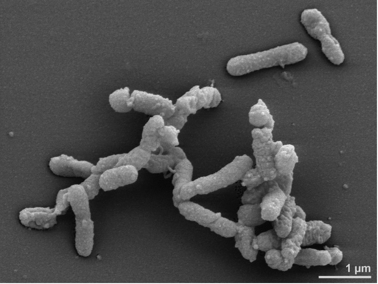
Most members of A. finegoldii were isolated on Bacteroides-bile-esculin (BBE) agar, others on kanamycin/vancomycin laked blood agar. Cells stain Gram-negative, and are non-spore forming and rod-shaped with rounded ends (0.2 x 0.8 to 2 μm), mostly occurring singly, though longer filaments are observed occasionally (Figure 2). After 4 days growth on Brucella sheep blood agar colonies are 0.3–1.0 mm in diameter, circular, gray, translucent or opaque and weakly β-hemolytic. On laked rabbit blood agar colonies are light brown after 4 days incubation, turning reddish or chocolate brown after 10 days [1,3]. Growth temperature is 37°C [31]. The organism is strictly anaerobic, indole-positive, catalase-negative and grows in peptone-yeast extract-glucose containing 20% bile [1,3]. Nitrate is not reduced to nitrite, gelatin is liquefied and esculin hydrolysis is negative. Metabolism is fermentative, however, due to scanty growth on agar media and in liquid media, carbohydrate metabolism is difficult to evaluate. In PYG broth, succinic acid is the major end product, while acetic and propionic acids are minor products; isovaleric and lactic acids are sometimes produced in very small amounts. Acid- and alkaline phosphatases, N-acetyl-β-glucosaminidase, esterase, esterase lipase, α- and β-galactosidases, and α-glucosidase are detected in the API ZYM (bioMérieux) gallery, while no activity is detected for lipase C4, leucine/valine/cystine arylamidases, trypsin, β-glucuronidase, β-glucosidase or α-mannosidase. In addition, using Rosco diagnostic tablets (Rosco, Taastrup, Denmark), α-fucosidase is detected, but not β-xylosidase or trypsin. Strains are resistant to vancomycin (5 μg), kanamycin (1,000 μg), and colistin (10 μg). Susceptibility to penicillin varies and some strains produce β-lactamase (reaction for the type strain has not been specified) [1,3].
Figure 2.
Scanning electron micrograph of A. finegoldii AHN2437T
Strain AHN2437T was isolated from a human appendiceal tissue sample. The habitat is not known but strains are probably members of the microflora of the human gut [1]. A. finegoldii-type organisms were identified by molecular methods as part of the microbiota of chicken guts [32] and they were detected in blood cultures from colon cancer patients [33].
Chemotaxonomy
The major cellular fatty acid of strain AHN2437T is iso-C15:0; smaller amounts (with 5 to 10% occurrence) are anteiso-C15:0, C15:0, C16:0, iso-C17:0, and one or both of C17:0 iso-3OH/C18:2 DMA. The mol% G+C of DNA is 57 [1,3]. No information is available for the peptidoglycan composition, isoprenoid composition, polar lipids or whole cell sugars.
Genome sequencing and annotation
Genome project history
This organism was selected for sequencing on the basis of its phylogenetic position [34], and is part of the Genomic Encyclopedia of Bacteria and Archaea project [35]. The genome project is deposited in the Genomes OnLine Database [23] and the complete genome sequence is deposited in GenBank. Sequencing, finishing and annotation were performed by the DOE Joint Genome Institute (JGI) using state of the art sequencing technology [46]. A summary of the project information is shown in Table 2.
Table 2. Genome sequencing project information.
| MIGS ID | Property | Term |
|---|---|---|
| MIGS-31 | Finishing quality | Finished |
| MIGS-28 | Libraries used | Three genomic libraries: one 454 pyrosequence standard library, one 454 PE library (11.0 kb insert size), one Illumina library |
| MIGS-29 | Sequencing platforms | Illumina GAii, 454 GS FLX Titanium |
| MIGS-31.2 | Sequencing coverage | 133.3 × Illumina; 27.8 × pyrosequence |
| MIGS-30 | Assemblers | Newbler version 2.3, Velvet version 1.0.13, Phrap version SPS - 4.24 |
| MIGS-32 | Gene calling method | Prodigal 1.4, GenePRIMP |
| INSDC ID | CP003274 | |
| GenBank Date of Release | June 8, 2012 | |
| GOLD ID | Gc02257 | |
| NCBI project ID | 440775 | |
| Database: IMG-GEBA | 2509601035 | |
| MIGS-13 | Source material identifier | DSM 17242 |
| Project relevance | Tree of Life, GEBA |
Growth conditions and DNA isolation
A. finegoldii strain AHN2437T, DSM 17242, was grown anaerobically in DSMZ medium 104 (PYG, supplemented with vitamin solution (see DSMZ medium 131)) [36] at 37°C. DNA was isolated from 1-1.5 g of cell paste using MasterPure Gram-positive DNA purification kit (Epicentre MGP04100) following the standard protocol as recommended by the manufacturer with modification st/LALM for cell lysis as described in Wu et al. 2009 [35]. DNA is available through the DNA Bank Network [37].
Genome sequencing and assembly
The genome was sequenced using a combination of Illumina and 454 sequencing platforms. All general aspects of library construction and sequencing can be found at the JGI website [38]. Pyrosequencing reads were assembled using the Newbler assembler (Roche). The initial Newbler assembly consisting of 103 contigs in four scaffolds was converted into a phrap [39] assembly by making fake reads from the consensus, to collect the read pairs in the 454 paired end library. Illumina GAii sequencing data (500.5 Mb) was assembled with Velvet [40] and the consensus sequences were shredded into 2.0 kb overlapped fake reads and assembled together with the 454 data. The 454 draft assembly was based on 160.8 Mb 454 draft data and all of the 454 paired end data. Newbler parameters are -consed -a 50 -l 350 -g -m -ml 20. The Phred/Phrap/Consed software package [39] was used for sequence assembly and quality assessment in the subsequent finishing process. After the shotgun stage, reads were assembled with parallel phrap (High Performance Software, LLC). Possible mis-assemblies were corrected with gapResolution [38], Dupfinisher [41], or sequencing cloned bridging PCR fragments with subcloning. Gaps between contigs were closed by editing in Consed, by PCR and by Bubble PCR primer walks (J.-F. Chang, unpublished). A total of 696 additional reactions and 2 shatter libraries were necessary to close gaps and to raise the quality of the finished sequence. Illumina reads were also used to correct potential base errors and increase consensus quality using a software Polisher developed at JGI [42]. The error rate of the completed genome sequence is less than 1 in 100,000. Together, the combination of the Illumina and 454 sequencing platforms provided 161.1 × coverage of the genome. The final assembly contained 324,940 pyrosequence and 13,793,104 Illumina reads.
Genome annotation
Genes were identified using Prodigal [43] as part of the DOE-JGI genome annotation pipeline [47], followed by a round of manual curation using the JGI GenePRIMP pipeline [44]. The predicted CDSs were translated and used to search the National Center for Biotechnology Information (NCBI) non-redundant database, UniProt, TIGR-Fam, Pfam, PRIAM, KEGG, COG, and InterPro databases. Additional gene prediction analysis and functional annotation was performed within the Integrated Microbial Genomes - Expert Review (IMG-ER) platform [45].
Genome properties
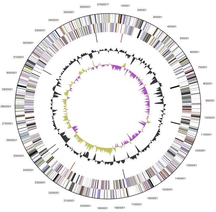
The genome statistics are provided in Table 3 and Figure 3. The genome consists of one circular chromosome with a total length of 3,734,239 bp and a G+C content of 56.6%. Of the 3,302 genes predicted, 3,234 were protein-coding genes, and 68 RNAs; 121 pseudogenes were also identified. The majority of the protein-coding genes (62.0%) were assigned a putative function while the remaining ones were annotated as hypothetical proteins. The distribution of genes into COGs functional categories is presented in Table 4.
Table 3. Genome Statistics.
| Attribute | Value | % of Total |
|---|---|---|
| Genome size (bp) | 3,734,239 | 100.00 |
| DNA coding region (bp) | 3,244,847 | 86.89 |
| DNA G+C content (bp) | 2,115,287 | 56.65 |
| Number of replicons | 1 | |
| Extrachromosomal elements | 0 | |
| Total genes | 3,302 | 100.00 |
| RNA genes | 68 | 2.06 |
| rRNA operons | 2 | |
| tRNA genes | 52 | 1.57 |
| Protein-coding genes | 3,234 | 97.94 |
| Pseudo genes | 121 | 3.66 |
| Genes with function prediction | 2,046 | 61.96 |
| Genes in paralog clusters | 1,627 | 49.27 |
| Genes assigned to COGs | 1,974 | 59.78 |
| Genes assigned Pfam domains | 2,183 | 66.11 |
| Genes with signal peptides | 967 | 29.29 |
| Genes with transmembrane helices | 642 | 19.44 |
| CRISPR repeats | 0 |
Figure 3.
Graphical map of the chromosome. From outside to the center: Genes on forward strand (color by COG categories), Genes on reverse strand (color by COG categories), RNA genes (tRNAs green, rRNAs red, other RNAs black), GC content, GC skew (purple/olive).
Table 4. Number of genes associated with the general COG functional categories.
| Code | Value | %age | Description |
|---|---|---|---|
| J | 144 | 6.8 | Translation, ribosomal structure and biogenesis |
| A | ... | ... | RNA processing and modification |
| K | 140 | 6.6 | Transcription |
| L | 214 | 10.0 | Replication, recombination and repair |
| B | ... | ... | Chromatin structure and dynamics |
| D | 36 | 1.7 | Cell cycle control, cell division, chromosome partitioning |
| Y | ... | ... | Nuclear structure |
| V | 40 | 1.9 | Defense mechanisms |
| T | 81 | 3.8 | Signal transduction mechanisms |
| M | 171 | 8.0 | Cell wall/membrane biogenesis |
| N | 7 | 0.3 | Cell motility |
| Z | ... | ... | Cytoskeleton |
| W | ... | ... | Extracellular structures |
| U | 56 | 2.6 | Intracellular trafficking and secretion, and vesicular transport |
| O | 77 | 3.6 | Posttranslational modification, protein turnover, chaperones |
| C | 127 | 6.0 | Energy production and conversion |
| G | 165 | 7.7 | Carbohydrate transport and metabolism |
| E | 141 | 6.6 | Amino acid transport and metabolism |
| F | 56 | 2.6 | Nucleotide transport and metabolism |
| H | 92 | 4.3 | Coenzyme transport and metabolism |
| I | 55 | 2.6 | Lipid transport and metabolism |
| P | 113 | 5.3 | Inorganic ion transport and metabolism |
| Q | 20 | 0.9 | Secondary metabolites biosynthesis, transport and catabolism |
| R | 259 | 12.2 | General function prediction only |
| S | 137 | 6.4 | Function unknown |
| - | 1,328 | 40.2 | Not in COGs |
Acknowledgements
We would like to gratefully acknowledge the help of Sabine Welnitz for growing A. finegoldii cultures, and Evelyne-Marie Brambilla for DNA extraction and quality control (both at DSMZ). This work was performed under the auspices of the US Department of Energy Office of Science, Biological and Environmental Research Program, and by the University of California, Lawrence Berkeley National Laboratory under contract No. DE-AC02-05CH11231, Lawrence Livermore National Laboratory under Contract No. DE-AC52-07NA27344, and Los Alamos National Laboratory under contract No. DE-AC02-06NA25396, UT-Battelle and Oak Ridge National Laboratory under contract DE-AC05-00OR22725.
References
- 1.Rautio M, Eerola E, Väisänen-Tunkelrott ML, Molitoris D, Lawson P, Collins MD, Jousimies-Somer H. Reclassification of Bacteroides putredinis (Weinberg et al., 1937) in a new genus Alistipes gen. nov., as Alistipes putredinis comb. nov., and description of Alistipes finegoldii sp. nov., from human sources. Syst Appl Microbiol 2003; 26:182-188 10.1078/072320203322346029 [DOI] [PubMed] [Google Scholar]
- 2.Validation List no. 94. Int J Syst Evol Microbiol 2003; 53:1701-1702 10.1099/ijs.0.03001-0 [DOI] [PubMed] [Google Scholar]
- 3.Song Y, Könönen E, Rautio M, Liu C, Bryk A, Eerola E, Finegold SM. Alistipes onderdonkii sp. nov. and Alistipes shahii sp. nov., of human origin. Int J Syst Evol Microbiol 2006; 56:1985-1990 10.1099/ijs.0.64318-0 [DOI] [PubMed] [Google Scholar]
- 4.Rautio M, Lönnroth M, Saxén H, Nikku R, Väisänen ML, Finegold SM, Jousimies-Somer H. Characteristics of an unusual anaerobic pigmented Gram-negative rod isolated from normal and inflamed appendices. Clin Infect Dis 1997; 25(Suppl 2):S107-S110 10.1086/516210 [DOI] [PubMed] [Google Scholar]
- 5.Rautio M, Saxén H, Siitonen A, Nikku R, Jousimies-Somer H. Bacteriology of histopathologically defined appedicitis in children. Pediatr Infect Dis 2000; 19:1078-1083 10.1097/00006454-200011000-00010 [DOI] [PubMed] [Google Scholar]
- 6.Mishra AK, Gimenez G, Lagier JC, Robert C, Raoult D, Fournier PE. Genome sequence and description of Alistipes senegalensis sp. nov. Stand Genomic Sci 2012; 6:304-314 10.4056/sigs.2625821 [DOI] [PMC free article] [PubMed] [Google Scholar]
- 7.Weinberg M, Nativelle R, Prévot AR. Les Microbes Anaérobies, Masson et Cie, Paris 1937; 1-1186. [Google Scholar]
- 8.Munoz R, Yarza P, Ludwig W, Euzéby J, Amann R, Schleifer KH, Glöckner FO, Rosselló-Móra R. Release LTPs104 of the All-Species Living Tree. Syst Appl Microbiol 2011; 34:169-170 10.1016/j.syapm.2011.03.001 [DOI] [PubMed] [Google Scholar]
- 9.Collins MD, Shah HN, Mitzuoka T. Reclassification of Bacteroides microfusus (Kaneuchi and Mitsuoka) in a new genus Rikenella, as Rikenella microfusus comb. nov. Syst Appl Microbiol 1985; 6:79-81 10.1016/S0723-2020(85)80015-1 [DOI] [Google Scholar]
- 10.Validation List N. ° 18. Int J Syst Evol Microbiol 1985; 35:375-376 [Google Scholar]
- 11.Krieg NR, Staley JT, Brown DR, Hedlund BP, Paster BJ, Ward NL, Ludwig W, Whitman WB. Family III. Rikenellaceae fam. nov. In: Krieg NR, Staley JT, Brown DR, Hedlund BP, Paster BJ, Ward NL, Ludwig W, Whitman WB. (eds) Bergey’s Manual of Systematic Bacteriology, second edition, vol. 4 (The Bacteroidetes, Spirochaetes, Tenericutes, Mollicutes), Acidobacteria, Fibrobacteres, Fusobacteria, Dictyoglomi, Gemmatimonadetes, Lentisphaerae, Verrucomicrobia, Chlamydiae, and Planctomycetes), Springer, New York, 2011 p. 54. [Google Scholar]
- 12.Validation List No 143. Int J Syst Evol Microbiol 2012; 62:1-4 10.1099/ijs.0.039487-0 [DOI] [Google Scholar]
- 13.Altschul SF, Gish W, Miller W, Myers EW, Lipman DJ. Basic local alignment search tool. J Mol Biol 1990; 215:403-410 [DOI] [PubMed] [Google Scholar]
- 14.Korf I, Yandell M, Bedell J. BLAST, O'Reilly, Sebastopol, 2003. [Google Scholar]
- 15.DeSantis TZ, Hugenholtz P, Larsen N, Rojas M, Brodie EL, Keller K, Huber T, Dalevi D, Hu P, Andersen GL. Greengenes, a chimera-checked 16S rRNA gene database and workbench compatible with ARB. Appl Environ Microbiol 2006; 72:5069-5072 10.1128/AEM.03006-05 [DOI] [PMC free article] [PubMed] [Google Scholar]
- 16.Porter MF. An algorithm for suffix stripping. Program: electronic library and information systems 1980; 14:130-137.
- 17.Lee C, Grasso C, Sharlow MF. Multiple sequence alignment using partial order graphs. Bioinformatics 2002; 18:452-464 10.1093/bioinformatics/18.3.452 [DOI] [PubMed] [Google Scholar]
- 18.Castresana J. Selection of conserved blocks from multiple alignments for their use in phylogenetic analysis. Mol Biol Evol 2000; 17:540-552 10.1093/oxfordjournals.molbev.a026334 [DOI] [PubMed] [Google Scholar]
- 19.Stamatakis A, Hoover P, Rougemont J. A rapid bootstrap algorithm for the RAxML web-servers. Syst Biol 2008; 57:758-771 10.1080/10635150802429642 [DOI] [PubMed] [Google Scholar]
- 20.Hess PN, De Moraes Russo CA. An empirical test of the midpoint rooting method. Biol J Linn Soc Lond 2007; 92:669-674 10.1111/j.1095-8312.2007.00864.x [DOI] [PMC free article] [PubMed] [Google Scholar]
- 21.Pattengale ND, Alipour M, Bininda-Emonds ORP, Moret BME, Stamatakis A. How many bootstrap replicates are necessary? Lect Notes Comput Sci 2009; 5541:184-200 10.1007/978-3-642-02008-7_13 [DOI] [Google Scholar]
- 22.Swofford DL. PAUP*: Phylogenetic Analysis Using Parsimony (*and Other Methods), Version 4.0 b10. Sinauer Associates, Sunderland, 2002. [Google Scholar]
- 23.Pagani I, Liolios K, Jansson J, Chen IM, Smirnova T, Nosrat B, Markowitz VM, Kyrpides NC. The Genomes OnLine Database (GOLD) v.4: status of genomic and metagenomic projects and their associated metadata. Nucleic Acids Res 2012; 40:D571-D579 10.1093/nar/gkr1100 [DOI] [PMC free article] [PubMed] [Google Scholar]
- 24.Field D, Garrity G, Gray T, Morrison N, Selengut J, Sterk P, Tatusova T, Thomson N, Allen MJ, Angiuoli SV, et al. The minimum information about a genome sequence (MIGS) specification. Nat Biotechnol 2008; 26:541-547 10.1038/nbt1360 [DOI] [PMC free article] [PubMed] [Google Scholar]
- 25.Woese CR, Kandler O, Wheelis ML. Towards a natural system of organisms: proposal for the domains Archaea, Bacteria, and Eucarya. Proc Natl Acad Sci USA 1990; 87:4576-4579 10.1073/pnas.87.12.4576 [DOI] [PMC free article] [PubMed] [Google Scholar]
- 26.Krieg NR, Ludwig W, Euzéby J, Whitman WB. Phylum XIV. Bacteroidetes phyl. nov. In: Krieg NR, Staley JT, Brown DR, Hedlund BP, Paster BJ, Ward NL, Ludwig W, Whitman WB. (eds) Bergey’s Manual of Systematic Bacteriology, second edition, vol. 4 (The Bacteroidetes, Spirochaetes, Tenericutes, Mollicutes), Acidobacteria, Fibrobacteres, Fusobacteria, Dictyoglomi, Gemmatimonadetes, Lentisphaerae, Verrucomicrobia, Chlamydiae, and Planctomycetes), Springer, New York, 2011 p. 25. [Google Scholar]
- 27.Krieg NR. Class I. Bacteroidia class. nov. In: Krieg NR, Staley JT, Brown DR, Hedlund BP, Paster BJ, Ward NL, Ludwig W, Whitman WB. (eds) Bergey’s Manual of Systematic Bacteriology, second edition, vol. 4 (The Bacteroidetes, Spirochaetes, Tenericutes, Mollicutes), Acidobacteria, Fibrobacteres, Fusobacteria, Dictyoglomi, Gemmatimonadetes, Lentisphaerae, Verrucomicrobia, Chlamydiae, and Planctomycetes), Springer, New York, 2011 p. 25. [Google Scholar]
- 28.Krieg NR. Order I. Bacteroidales ord. nov. In: Krieg NR, Staley JT, Brown DR, Hedlund BP, Paster BJ, Ward NL, Ludwig W, Whitman WB. (eds) Bergey’s Manual of Systematic Bacteriology, second edition, vol. 4 (The Bacteroidetes, Spirochaetes, Tenericutes, Mollicutes), Acidobacteria, Fibrobacteres, Fusobacteria, Dictyoglomi, Gemmatimonadetes, Lentisphaerae, Verrucomicrobia, Chlamydiae, and Planctomycetes), Springer, New York, 2011 p. 25. [Google Scholar]
- 29.BAuA. 2010, Classification of Bacteria and Archaea in risk groups. http://www.baua.de TRBA 466, p. 19.
- 30.Ashburner M, Ball CA, Blake JA, Botstein D, Butler H, Cherry JM, Davis AP, Dolinski K, Dwight SS, Eppig JT, et al. Gene ontology: tool for the unification of biology. Nat Genet 2000; 25:25-29 10.1038/75556 [DOI] [PMC free article] [PubMed] [Google Scholar]
- 31.DSMZ catalogue of strains, Braunschweig, Germany. (http://www.dsmz.de/catalogues/catalogue-microorganisms.html)
- 32.Torok VA, Hughes RJ, Mikkelsen LL, Perez-Maldonado R, Balding K, MacAlpine R, Percy NJ, Ophel-Keller K. Identification and characterization of potential performance-related gut microbiotas in broiler chickens across various feeding trials. Appl Environ Microbiol 2011; 77:5868-5878 10.1128/AEM.00165-11 [DOI] [PMC free article] [PubMed] [Google Scholar]
- 33.Fenner L, Roux V, Ananian P, Raoult D. Alistipes finegoldii in blood cultures from colon cancer patients. Emerg Infect Dis 2007; 13:1260-1262 10.3201/eid1308.060662 [DOI] [PMC free article] [PubMed] [Google Scholar]
- 34.Klenk HP, Göker M. En route to a genome-based classification of Archaea and Bacteria? Syst Appl Microbiol 2010; 33:175-182 10.1016/j.syapm.2010.03.003 [DOI] [PubMed] [Google Scholar]
- 35.Wu D, Hugenholtz P, Mavromatis K, Pukall R, Dalin E, Ivanova NN, Kunin V, Goodwin L, Wu M, Tindall BJ, et al. A phylogeny-driven genomic encyclopedia of Bacteria and Archaea. Nature 2009; 462:1056-1060 10.1038/nature08656 [DOI] [PMC free article] [PubMed] [Google Scholar]
- 36.List of growth media used at DSMZ: http://www.dsmz.de/catalogues/catalogue-microorganisms/culture-technology/list-of-media-for-microorganisms.html
- 37.Gemeinholzer B, Dröge G, Zetzsche H, Haszprunar G, Klenk HP, Güntsch A, Berendsohn WG, Wägele JW. The DNA Bank Network: the start from a German initiative. Biopreserv Biobank 2011; 9:51-55 10.1089/bio.2010.0029 [DOI] [PubMed] [Google Scholar]
- 38.JGI website. http://www.jgi.doe.gov/
- 39.The Phred/Phrap/Consed software package. http://www.phrap.com
- 40.Zerbino DR, Birney E. Velvet: algorithms for de novo short read assembly using de Bruijn graphs. Genome Res 2008; 18:821-829 10.1101/gr.074492.107 [DOI] [PMC free article] [PubMed] [Google Scholar]
- 41.Han C, Chain P. Finishing repeat regions automatically with Dupfinisher. In: Proceeding of the 2006 international conference on bioinformatics & computational biology. Arabnia HR, Valafar H (eds), CSREA Press. June 26-29, 2006:141-146. [Google Scholar]
- 42.Lapidus A, LaButti K, Foster B, Lowry S, Trong S, Goltsman E. POLISHER: An effective tool for using ultra short reads in microbial genome assembly and finishing. AGBT, Marco Island, FL, 2008. [Google Scholar]
- 43.Hyatt D, Chen GL, LoCascio PF, Land ML, Larimer FW, Hauser LJ. Prodigal: prokaryotic gene recognition and translation initiation site identification. BMC Bioinformatics 2010; 11:119 10.1186/1471-2105-11-119 [DOI] [PMC free article] [PubMed] [Google Scholar]
- 44.Pati A, Ivanova NN, Mikhailova N, Ovchinnikova G, Hooper SD, Lykidis A, Kyrpides NC. GenePRIMP: a gene prediction improvement pipeline for prokaryotic genomes. Nat Methods 2010; 7:455-457 10.1038/nmeth.1457 [DOI] [PubMed] [Google Scholar]
- 45.Markowitz VM, Ivanova NN, Chen IMA, Chu K, Kyrpides NC. IMG ER: a system for microbial genome annotation expert review and curation. Bioinformatics 2009; 25:2271-2278 10.1093/bioinformatics/btp393 [DOI] [PubMed] [Google Scholar]
- 46.Mavromatis K, Land ML, Brettin TS, Quest DJ, Copeland A, Clum A, Goodwin L, Woyke T, Lapidus A, Klenk HP, et al. The fast changing landscape of sequencing technologies and their impact on microbial genome assemblies and annotation. PLoS ONE 2012; 7:e48837; . 10.1371/journal.pone.0048837 [DOI] [PMC free article] [PubMed] [Google Scholar]
- 47.Mavromatis K, Ivanova NN, Chen IM, Szeto E, Markowitz VM, Kyrpides NC. The DOE-JGI Standard operating procedure for the annotations of microbial genomes. Stand Genomic Sci 2009; 1:63-67 10.4056/sigs.632 [DOI] [PMC free article] [PubMed] [Google Scholar]


