
Figure 2. Otic induction from the pre-placodal epithelium in vitro.
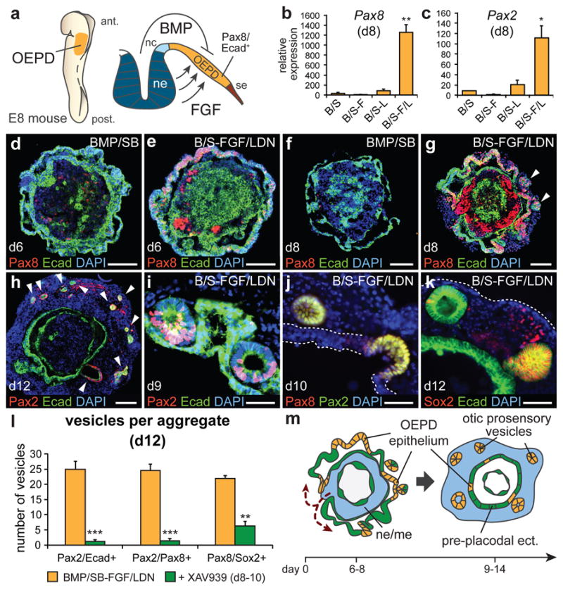
a, OEPD induction in mice. nc, neural crest; se, surface ectoderm. b, Pax8 and c, Pax2 mRNA expression on day 8 (n=3–4; **P<0.01, *P<0.05; mean ± s.e.m.) d-g, Pax8/Ecad expression in (d, f) BMP/SB and (e, g) BMP/SB-FGF/LDN aggregates on day 6 and 8. Arrowheads indicate vesicles. h, Day 12 BMP/SB-FGF/LDN aggregate with Pax2/Ecad+ vesicles (arrowheads). i, Pax2/Ecad+, (j) Pax2/8+, and (k) Pax8/Sox2+ vesicles invaginate from the inner-epithelium from day 9–12. l, XAV939 decreases the number of vesicles expressing Pax2/Ecad, Pax2/Pax8 and Pax8/Sox2 on day 12. (n=9 aggregates; ***P<0.001, **P<0.01; mean ± s.e.m.). m, Self-guided, inside-out rearrangement of BMP/SB-FGF/LDN aggregates and formation of otic vesicles. Scale bars, 100 μm (d, e, f, h), 50 μm (i-k), 25 μm (g).