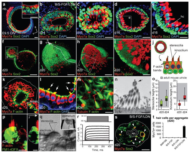
Figure 3. Stem cell-derived otic vesicles generate functional inner ear hair cells.
a, b, Expression of Myo7a in the E9.5 otic vesicle (a; OtV) and day 14 vesicles (b). nb, neuroblasts. c-e, Myo7a/Sox2+ hair cells (hc) with underlying Sox2+ supporting cells (sc) on day 15 (c) and 16 (d, e). f-i, Whole-mount immunofluorescence for Myo7a and Sox2 (f) and 3D reconstruction (g-i) of a vesicle in a day 20 BMP/SB-FGF/LDN aggregate. j, Vesicles display the hallmarks of inner ear sensory epithelia. k-m, F-actin (F-act) labels cell-cell junctions on the luminal surface and stereocilia bundles. m, Acetylated-a-Tubulin (Tublin) labels kinocilium and the cuticular plate. n, Transmission electron micrograph of stereocilia bundles and kinocilium (arrow). o, Distribution of stereocilia and kinocilium heights on days 20 and 24 compared to adult mouse utricle, range indicated by gray boxes (n>100 cells; ± max/min). p, Representative hair cell following 1 min FM1-43FX incubation, fixation and staining for F-actin. q, Representative epithelium preparation (inset) and hair cell during electrophysiological recordings. r, Representative voltage-current responses recorded from hair cells. The voltage protocol is shown at the top. s, Day 20 aggregate with Myo7a/Sox2+ vesicles. epi, epidermis (dashed outline). t, Number of hair cells on day 20 (n=12–16; mean ± s.e.m.). Scale bars, 250 μm (f, s, q-inset), 50 μm (d, g, h), 25 μm (a-c, e, i, k, l), 10 μm (q), 5 μm (m, p), 250 nm (n).

