Abstract
Background
To quantify changes in metamorphopsia and retinal contraction in eyes with idiopathic epiretinal membrane (ERM) before and after a spontaneous separation of ERM.
Methods
Among 92 eyes of 92 patients with idiopathic ERM who were followed up at our hospital, 5 eyes of 5 patients had experienced a spontaneous separation of ERM during the follow-up period. Patient's metamorphopsia was assessed horizontally and vertically by a metamorphopsia chart developed by our group, M-CHARTS, to obtain the horizontal (MH) and vertical (MV) metamorphopsia scores. Difference in the scores before and after the membrane separation represents change in patient's metamorphopsia. Changes in retinal contraction were also evaluated horizontally and vertically with our original software for fundus image analysis. The difference between M-CHARTS scores and distances of retinal vessel movements with before and after membrane separation were measured.
Results
All five subjects showed a decrease in the retinal contraction. Improved visual acuity was observed in three subjects, and no change was seen in the other two. Four subjects obtained better metamorphopsia scores after the membrane separation, while the other one was not detected with metamorphopsia by M-CHARTS either before or after the separation. In subjects with an improved MV, horizontal retinal movement was seen larger than the vertical movement. Similarly, the subjects with an improved MH indicated a larger vertical retinal movement than the horizontal movement.
Conclusions
The direction of patient's metamorphopsia closely associated with the direction of retinal contraction before and after a spontaneous separation of ERM.
Keywords: metamorphopsia, M-CHARTS, idiopathic epiretinal membrane, spontaneous separation
Introduction
Idiopathic epiretinal membrane (ERM) can cause visual symptoms such as metamorphopsia and deterioration of visual acuity.1, 2, 3 Although most patients with ERM retain stable visual acuity without surgical intervention, in cases of progressing or proliferating membranes that thicken and contract the sensory retina, patients often suffer from severe metamorphopsia. In such cases, patient's severity of metamorphopsia rather than visual acuity might be a better indicator that reveals more precise information on the progression of the disease. Amsler's charts have been the conventional method for the detection of metamorphopsia, but the charts can only qualitatively detect metamorphopsia. To quantify patient's severity of metamorphopsia, Matsumoto et al4, 5, 6 reported a new method, M-CHARTS. This simple method measures patient's metamorphopsia horizontally and vertically to obtain MH and MV M-CHARTS scores. With theses scores, it is easy to monitor patient's metamorphopsia and even the progression of ERM. Using M-CHARTS, Arimura et al7 have further examined any possible association between the directions of metamorphopsia and the retinal contraction in patients with idiopathic ERM. They have claimed significant correlations between the horizontal retinal contraction and vertical metamorphopsia, and between the vertical retinal contraction and horizontal metamorphopsia.
Spontaneous separation of ERM is a rare clinical incident. Previous studies reported that eyes with ERM (including secondary ERM) have shown improvement in visual acuity after a spontaneous membrane separation.8, 9 Although those studies have demonstrated improved visual acuity and decreased metamorphopsia in eyes with a spontaneous separation of ERM, whether a significant relationship exists between metamorphopsia and the retinal contraction in such cases has yet to be examined. In this study, we not only quantified patient's changes in metamorphopsia by M-CHARTS but also investigated the relationship between the directions of retinal contraction and metamorphopsia before and after a spontaneous separation of idiopathic ERM.
Materials and methods
Subjects
Among 92 eyes of 92 patients with idiopathic ERM who were followed up for more than 36 months (mean, 44.9±12.9 months) from 2000 to 2007 at the Kinki University School of Medicine, we observed 5 eyes of 5 patients (three males and two females; age range, 44–73 years, average, 56.6±12.5 years) with spontaneous separation of idiopathic ERM. All patients underwent a series of ophthalmic examinations including best corrected visual acuity, applanation tonometry, slit lamp biomicroscopy, dilated funduscopy, M-CHARTS tests, fundus photography, and scanning laser ophthalmoscopy (SLO; Rodenstock, Munich, Germany). No subjects were found with any systematic diseases that were likely to affect their visual functions or to cause a secondary ERM. Informed consent was obtained from all subjects. All methods adhered to the Declaration of Helsinki for research involving human subjects.
Quantification of metamorphopsia
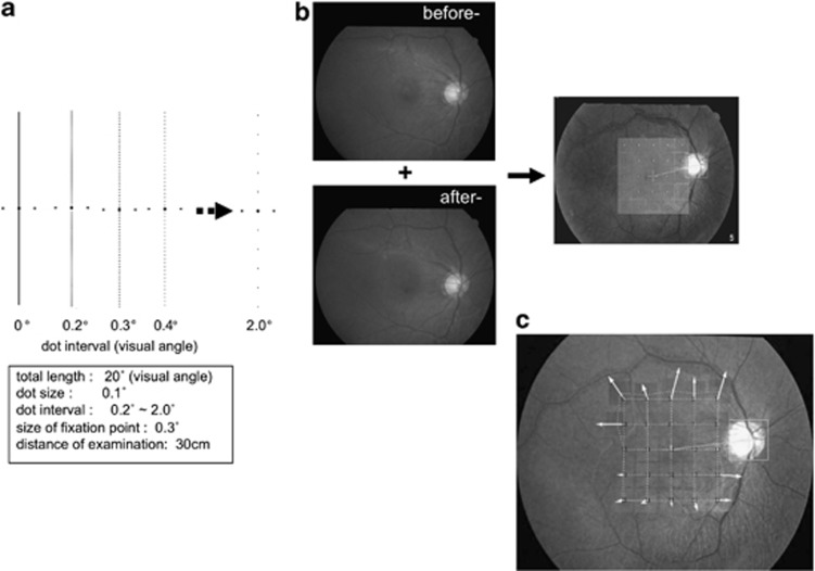
Amsler's charts and M-CHARTS were used for the evaluation of metamorphopsia during the follow-up. M-CHARTS consists of 19 dotted lines with dot intervals changing from fine to coarse (0.2°–2.0° of visual angle; Figure 1a).4, 5 Subjects viewed M-CHARTS from a distant of 30 cm through corrective lenses. At first, a vertical straight line (0°) on the first page of M-CHARTS is shown to the patient, and the patient is fixed on the center of the line. If the patients recognize the straight line as the straight, the metamorphopsia score is 0. If a patient recognizes the straight line as an irregular or curved line, more coarse dotted lines are shown to the patient in the following test. When the patients recognize a dot line as straight, its visual angle is considered as their metamorphopsia score. Also, the M-CHARTS are rotated 90° and the same test is performed using horizontal lines. M-CHARTS test was repeated three times for each eye and the average of the three scores was used. M-CHARTS test was performed with horizontal and vertical lines to obtain separate MH and MV M-CHARTS scores. The subject's fundus information was masked from the examiner during the examination.
Figure 1.
(a) Method of detecting metamorphopsia using M-CHARTS. The minimum visual angle of the dotted lines needed to cause the metamorphopsia to disappear was measured. (Quantification of retinal contraction using fundus photograph.) (b) The comparison image (after-: after spontaneous separation of ERM) was superimposed on the baseline image (before-: before spontaneous separation of ERM) so that the optic disc and choroidal vessels matched. The distance between the center of the optic disc and fovea was calibrated to be 15° (4.02 mm), and 20° macula area was divided into 25 areas. In each of the 25 areas, two overlapping fundus images were flickered back and forth at a speed of 2 Hz, and the retinal vessels were exactly matched manually. (c) The movement of retinal vessels in each of the 25 areas was a displayed as a vector and the value of each vector's vertical and horizontal components was recorded in millimeters (mm) as the index of retinal contraction. For easier visibility, the vector arrows are shown three times larger than the actual measured vectors.
Quantification of retinal contraction
Spontaneous separation of ERM releases contractions of the sensory retina and retinal vessels. To quantify retinal contraction, we compared the locations of the retinal vessels before and after the membrane separation, and measured the difference in distance. An image-analyzing software developed by us (Wellsystem Inc., Tokyo, Japan; Figures 1b and c) was used for the distance measurement in this study. Details of the application and procedure for the use of this software were described elsewhere.7 Briefly, the comparison image taken after the spontaneous separation of ERM was superimposed on the baseline image taken before the membrane separation so that the optic disc and choroidal vessels matched. The distance between the center of the optic disc and the fovea was calibrated to be 15° (4.02 mm), and the macular area within 20° was divided into 25 areas. In each of the 25 areas, two overlapping fundus images were flickered back and forth at a speed of 2 Hz, and the retinal vessels were exactly matched manually. The movement of retinal vessels in each of the 25 areas was displayed as a vector and the values of the vector's vertical and horizontal components were recorded in millimeters (mm) as the indices of the retinal contraction.
Results
Clinical characteristics of the subjects were summarized in Table 1. Three of the five patients showed improved visual acuity after the spontaneous separation. Regarding metamorphopsia, four of the five patients had improved metamorphopsia scores, while the other (patient no. 3) was not detected with either vertical or horizontal metamorphopsia by M-CHARTS. Among those four patients with improved metamorphopsia scores, two patients (patient no. 1 and 2) with a bigger improvement in the MV score than in the MH score showed a larger horizontal retinal movement than the vertical movement. Similarly, the other two patients (patient no. 4 and 5) with a bigger improvement in the MH score showed a larger vertical retinal movement.
Table 1. Changes of metamorphopsia scores and retinal movement between before and after spontaneous separation.
| Patient no. | Age (years)/gender | Refractive error (D) |
VA (logMAR) |
MV |
MH |
Retinal movement (mm) |
Follow-up period (month) | ||||
|---|---|---|---|---|---|---|---|---|---|---|---|
| Before- | After- | Before- | After- | Before- | After- | Vertical | Horizontal | ||||
| 1 | 44/Male | −4.5 | 0 | −0.1 | 1.3 | 0.2 | 0.9 | 0.4 | 0.12 | 0.16 | 58 |
| 2 | 63/Female | −4.0 | 0 | 0 | 0.4 | 0 | 0 | 0 | 0.07 | 0.16 | 22 |
| 3 | 73/Male | 0.75 | 0.1 | 0 | 0 | 0 | 0 | 0 | 0.11 | 0.07 | 27 |
| 4 | 56/Male | −7.5 | 0.3 | 0 | 0.5 | 0.2 | 1.2 | 0.3 | 0.16 | 0.10 | 43 |
| 5 | 45/Female | −4.75 | 0 | 0 | 0 | 0.2 | 0.7 | 0 | 0.14 | 0.08 | 16 |
Abbreviations: after-, after spontaneous separation of ERM; before-, before spontaneous separation of ERM; D, diopter; MH, horizontal metamorphopsia; MV, vertical metamorphopsia; VA, visual acuity.
Case report
Patient no. 1
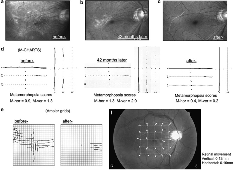
This patient was a 44-year-old male. Figure 2 shows the appearance of ERM and various test results before/after and at the membrane separation. ERM was detected in the patient's right eye by SLO with an argon blue laser beam (Figure 2a). Before the separation, Amsler's charts detected horizontal and vertical metamorphopsia in the central and upper nasal areas (Figure 2e), and M-CHARTS also showed horizontal and vertical metamorphopsia (Figure 2d). In the horizontal M-CHARTS test, decreased metamorphopsia was seen when a dotted line with an interval of 0.8° visual angle was used, and metamorphopsia completely disappeared when a dotted line of 0.9° was used. Thus, the patient's MH was determined to be 0.9. Similarly, MV of 1.3 was obtained with the vertical dotted lines (Figure 2d).
Figure 2.
SLO images obtained with an argon blue laser beam and results of Amsler charts and M-CHARTS in patient no. 1. (a) Before-, (b) 41 months later from first visit to our hospital and (c) after- SLO images. (d) The results of M-CHARTS evaluations of metamorphopsia at before, 41 months later and after spontaneous separation of ERM. (e) The results of Amsler charts evaluations of metamorphopsia at before and after spontaneous separation of ERM. (f) The fundus image with vector arrows, which was analyzed by our developing software, shows the direction and distance of retinal vessel movements.
At 41 months after the patient's first visit to our hospital, we ascertained a partial spontaneous separation of the ERM (Figure 2b). Concurrently, M-CHARTS showed that both horizontal and vertical metamorphopsia had worsened in the temporal area (Figure 2d). It appeared that the vitreoretinal traction on the central macular surface had caused the worsening metamorphopsia. When a spontaneous separation of the membrane was observed in the fundus at 58 months (Figure 2c), Amsler's charts had indicated much improved metamorphopsia (Figure 2e) and MH and MV by M-CHARTS also improved to 0.4 and 0.2, respectively (Figure 2d). MV had improved more than MH. After the membrane separation, retinal movement of 0.12 mm in the vertical direction and 0.16 mm in the horizontal direction was obtained by the image analysis (Figure 2f).
Patient no. 3
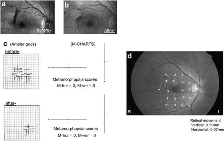
This patient was a 73-year-old male. Mild ERM was recognized in the upper nasal area of the macula (Figure 3a). Spontaneous separation of the ERM occurred at 27 months (Figure 3b). Before the spontaneous separation, neither horizontal nor vertical metamorphopsia was detected by M-CHARTS, although Amsler's charts did detect some level of metamorphopsia (Figure 3c). After the separation, Amsler's charts showed less severe metamorphopsia while M-CHARTS still did not detect any metamorphopsia. Retinal movement of 0.11 mm in the vertical direction and 0.07 mm in the horizontal direction was measured (Figure 3d).
Figure 3.
SLO images obtained with an argon blue laser beam and results of Amsler charts and M-CHARTS in patient no. 3. (a) Before- and (b) after- SLO images. (c) The results of Amsler charts and M-CHARTS evaluations of metamorphopsia at before and after spontaneous separation of ERM. (d) The fundus image with vector arrows, which was analyzed by our developing software, shows the direction and distance of retinal vessel movements.
Patient no. 5
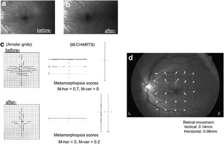
This patient was a 45-years-old female and was followed up for 16 months. The before/after SLO images clearly showed how the retinal contractions in the upper nasal area of the macula had been eased after the membrane separation (Figures 4a and b). Before the membrane separation, Amsler's charts detected severer horizontal metamorphopsia than vertical metamorphopsia, and M-CHARTS also indicated a bigger MH of 0.7 over MV of 0 (Figure 4c). After the separation of the ERM, the patient reported improved metamorphopsia in the horizontal direction. M-CHARTS also indicated that the patient's MH had improved more than MV (Figure 4c) and that the vertical retinal movement of 0.14 mm was bigger than the horizontal movement of 0.08 mm (Figure 4d).
Figure 4.
SLO images obtained with an argon blue laser beam and results of Amsler charts and M-CHARTS in patient no. 5. (a) Before- and (b) after- SLO images. (c) The results of Amsler charts and M-CHARTS evaluations of metamorphopsia at before and after spontaneous separation of ERM. (d) The fundus image with vector arrows, which was analyzed by our developing software, shows the direction and distance of retinal vessel movements.
Discussion
Our results showed that the spontaneous separation of ERM eased the retinal contractions and improved patient's metamorphopsia. In addition the improvement of metamorphopsia related to the direction of retinal movement.
Human visual systems possess an acute ability to discriminate straight lines from curved lines. This discriminating ability is thought to be within approximately one-fifth of the physical spacing between two neighboring cones and one-tenth of smallest receptive field of ganglion cells.10, 11 In addition, the cone photoreceptors are placed in a hexagonal arrangement in the fovea region and the degree of hexagonal regularity decreases as retinal eccentricity increases.12, 13, 14 Therefore, even very slight distortion or curvature will be perceived in the fovea of the human eyes. Contraction of the retina is usually caused by progression of ERM. The contraction affects the outer segments of the retina and/or photoreceptors, and disturbs the spatial arrangement of the cones. In result, patients notice and sometimes suffer from metamorphopsia.
We have previously reported the significant correlations between the horizontal retinal contraction and MV, and between the vertical retinal contraction and MH in patients with idiopathic ERM.6 Indeed our previous study showed that the stronger retinal contraction led the larger metamorphopsia, but we had not known whether the relationships of direction between retinal contractions and metamorphopsia would be fitted in the patients with spontaneous separation of ERM. Our observations suggested that the spontaneous membrane separation had eased the retinal contractions. Consequently, the patients subjectively would have noticed an improvement of seeing. We further imagined that the spontaneous membrane separation might have restored the disordered arrangement of the photoreceptor, which was caused by the retinal contractions due to progressing ERM, nearly to their original arrangement. Therefore, the patients who had larger horizontal retinal movement showed better improvement in MV than MH, and also the patients who had larger vertical retinal movement showed a better improvement in MH than MV. Although the number of the subjects in this study was small, our current results could substantiate the previous found about relationships between the directions of retinal contraction and metamorphopsia.7
It is widely known that ERM frequently associates with posterior vitreous detachment (PVD). Kishi et al15 have postulated that in cases of ERM with or without PVD, the thin premacular vitreous cortex that forms the posterior wall of the premacular liquefied pocket has a key role in the development of ERM. They had observed a spontaneous separation of ERM in a case with partial PVD and concluded that spontaneous separation of ERM, not including secondary premacular fibrosis, is clinically rare with an incidence rate of 1–2%.15, 16 We unfortunately could not verify the all patients' vitreous conditions and the incidence of PVD in this study. However, the SLO images of patient no. 1 showed the sequential process of spontaneous separation with the progression of PVD (Figures 2a–c).
In recent years, visualization of detailed morphological changes has become possible by the use of optical coherence tomography (OCT). Investigators have applied OCT to studies on the structural mechanism for macular diseases.17, 18, 19, 20 With this device, it might show the precise structural changes of macular region. However, it is regrettable that we hadn't used OCT for examining the patients in this study. It would be a limitation of this study.
Of the five cases presented here, the results of Amsler's charts agreed with the results of M-CHARTS in four cases that showed the improvement of metamorphopsia. On the other hand, in the rest of one case, Amsler's chart could only detect the metamorphopsia. This difference between the two test results might be due to the difference in the designs of the tests. Amsler's charts detect distortion on a plane, while M-CHARTS uses lines to assess distortion. Although Amsler's charts do not quantify the degree of distortion, the locations and shapes of distortion can be depicted with the subject's indications. Contrarily, M-CHARTS can quantify the degree of distortion, but it cannot give us the description of the exact shapes and locations of the subject's metamorphopsia. As indicated by Matsumoto et al,5 the dotted lines of M-CHARTS are designed to evaluate the fixation area rather than the periphery area, because metamorphopsia can be remarkably noticed in the fixation area. In addition the detection task would become more difficult for patients if the test lines in the periphery region will be used. In the patient no. 5 (Figure 4), we have speculated that the distortions, which the patient had remarkably noticed with Amsler's charts, seem to be slightly deviated from the test lines of M-CHARTS. It would be the reason that the patient did not detect any distortions with M-CHARTS.
In conclusion, our current results, in patients with a spontaneous separation of idiopathic ERM, not only have ascertained the fact that spontaneous membrane separation reduces patient's metamorphopsia, but also validated the established relationships between the directions of retinal contraction and metamorphopsia.
The authors declare no conflict of interest.
References
- Foos RY. Vitreoretinal junction. Simple epiretinal membranes. Albecht Von Grafes Arch Clin Exp Ophthalmol. 1974;189:231–250. doi: 10.1007/BF02384852. [DOI] [PubMed] [Google Scholar]
- Wise GN. Clinical features of idiopathic preretinal macular fibrosis. Am J Ophthalmol. 1975;79:349–357. doi: 10.1016/0002-9394(75)90605-4. [DOI] [PubMed] [Google Scholar]
- Sidd RJ, Fine SL, Owens SL, Patz A. Idiopathic preretinal gliosis. Am J Ophthalmol. 1982;94:44–48. doi: 10.1016/0002-9394(82)90189-1. [DOI] [PubMed] [Google Scholar]
- Matsumoto C, Arimura E, Hashimoto S, Takada S, Okuyama S, Shimomura Y. A new method for quantification of metamorphopsia using M-CHARTS (in Japanese) Rinsho Ganka. 2000;54:373–377. [Google Scholar]
- Matsumoto C, Arimura E, Hashimoto S, Takada S, Okuyama S, Shimomura Y. Quantification of metamorphopsia in patients with epiretinal membrane. Invest Opthalmol Vis Sci. 2003;44:4012–4016. doi: 10.1167/iovs.03-0117. [DOI] [PubMed] [Google Scholar]
- Arimura E, Matsumoto C, Okuyama S, Takada S, Hashimoto S, Shimomura Y. Quantification of metamorphopsia in a macular hole patient using M-CHARTS. Acta Ophthalmol Scand. 2006;85:55–59. doi: 10.1111/j.1600-0420.2006.00729.x. [DOI] [PubMed] [Google Scholar]
- Arimura E, Matsumoto C, Okuyama S, Takada S, Hashimoto S, Shimomura Y. Retinal contraction and metamorphopsia scores in eyes with idiopathic epiretinal membrane. Invest Opthalmol Vis Sci. 2005;46:2961–2966. doi: 10.1167/iovs.04-1104. [DOI] [PubMed] [Google Scholar]
- Karen DS, Lee MJ, Morton FG, Felipe UH. Spontaneous separation of epiretinal membrane. Arch Ophthalmol. 1980;98:318–320. doi: 10.1001/archopht.1980.01020030314015. [DOI] [PubMed] [Google Scholar]
- Greven CM, Slusher MM, Weaver RG. Epiretinal membrane release and posterior vitreous detachment. Ophthalmology. 1998;95:902–905. doi: 10.1016/s0161-6420(88)33077-0. [DOI] [PubMed] [Google Scholar]
- Watt RJ, Ward RM, Casco C. The detection of deviation from straightness in lines. Vis Res. 1987;27:1659–1678. doi: 10.1016/0042-6989(87)90172-6. [DOI] [PubMed] [Google Scholar]
- Hess RF, Watt RJ. Regional distribution of the mechanisms that underlie spatial localization. Vis Res. 1990;30:1021–1031. doi: 10.1016/0042-6989(90)90112-x. [DOI] [PubMed] [Google Scholar]
- Williams DR. Topography of the foveal cone mosaic in the living human eye. Vis Res. 1988;28:433–454. doi: 10.1016/0042-6989(88)90185-x. [DOI] [PubMed] [Google Scholar]
- Hirsch J, Miller WH. Does cone positional disorder limit resolution. J Opt Soc Am A. 1987;4:1841–1892. doi: 10.1364/josaa.4.001481. [DOI] [PubMed] [Google Scholar]
- Curio CA, Solan KR. Packing geometry of human cone photoreceptors: variation with eccentricity and evidence for local anisotropy. Vis Neurosci. 1992;9 (1:69–80. doi: 10.1017/s0952523800009639. [DOI] [PubMed] [Google Scholar]
- Kishi S, Shimizu K. Oval defect in detached posterior hyaloid membrane in idiopathic preretinal macular fibrosis. Am J Ophthalmol. 1994;118:451–456. doi: 10.1016/s0002-9394(14)75795-2. [DOI] [PubMed] [Google Scholar]
- Byer NE. Spontaneous disappearance of early postoperative preretinal traction. Arch Ophthalmol. 1973;90:133–135. doi: 10.1001/archopht.1973.01000050135014. [DOI] [PubMed] [Google Scholar]
- Ko TH, Fujimoto JG, Schuman JS, Paunescu LA, Kowalevicz AM, Hartl I, et al. Comparison of ultra- and standard-resolution optical coherence tomography for imaging macular pathology. Ophthalmology. 2005;112:1922–1935. doi: 10.1016/j.ophtha.2005.05.027. [DOI] [PMC free article] [PubMed] [Google Scholar]
- Schmidt-Erfurth U, Leitgeb RA, Michels S, Povazay B, Sacu S, Hermann B, et al. Three-dimensional ultra-high-resolution optical coherence tomography of macular diseases. Invest Ophthalmol Vis Sci. 2005;46:3393–3402. doi: 10.1167/iovs.05-0370. [DOI] [PubMed] [Google Scholar]
- Srinivasan VJ, Wojtkowski M, Witkin AJ, Duker JS, Ko TH, Carvalho M, et al. High-definition and 3-dementiona imaging of macular pathologies with high-speed ultra-high-resolution optical coherence tomography. Ophthalmology. 2006;113:2054–2065. doi: 10.1016/j.ophtha.2006.05.046. [DOI] [PMC free article] [PubMed] [Google Scholar]
- Koizumi H, Spaide RF, Fisher YL, Fruend KB, Klancnik JM, Jr, Yannuzzi LA. Three-dimensional evaluation of vitreomacular traction and epiretinal membrane using spectral-domain optical coherence tomography. Am J Ophthalmol. 2008;145:509–517. doi: 10.1016/j.ajo.2007.10.014. [DOI] [PubMed] [Google Scholar]


