Abstract
Background:
Sphenoid wing en plaque meningiomas are a subgroup of meningiomas defined by its particular sheet-like dural involvement and its disproportionately large bone hyperostosis. En plaque meningiomas represent 2-9% of all meningiomas and they are mainly located in the sphenoid wing. Total surgical resection is difficult and therefore these tumors have high recurrence rates.
Methods:
Eighteen patients with sphenoid wing en plaque meningiomas surgically treated between January 1998 and December 2008 were included. Clinical, surgical, and follow-up data were retrospectively analyzed.
Results:
Mean age was 52.2 years and 83% were female. Five patients presented extension of dural component into the orbit and six patients presented cavernous sinus infiltration. Adjuvant radiation therapy was performed in three patients. After a mean follow-up of 4.6 years, five patients developed tumor recurrence - two patients were submitted to surgical treatment and the other three were submitted to radiation therapy. No patient presented recurrence after radiation therapy, whether performed immediately in the postoperative period or performed after recurrence. Patients without tumor extension to cavernous sinus or orbital cavity have the best prognosis treated with surgery alone. When tumor extension involves these locations the recurrence rate is high, especially in cases not submitted to adjuvant radiation therapy.
Conclusion:
Cavernous sinus and superior orbital fissure involvement are frequent and should be considered surgical limits. Postoperative radiation therapy is indicated in cases with residual tumor in these locations.
Keywords: Cavernous sinus, meningioma, orbital tumor, proptosis, sphenoid wing
INTRODUCTION
En plaque meningiomas constitute a particular type of meningiomas that infiltrate the dura mater in a diffuse, sheet-like appearance, forming a thin layer that closely follows the contours of the inner table of the skull. The term “en plaque” was first used by Cushing and Eisenhardt [5,6] to describe this particular growing pattern, differentiating it from the most common type, which were designated “en masse” meningiomas. These tumors are mainly a bone disease, as their symptoms and prognosis are influenced mostly by the extension of bone invasion instead of intradural involvement. Although bone hyperostosis is a well known feature in all types of meningiomas, in this particular type of tumor the bone invasion is much more extensive and is responsible for the clinical manifestations. This is particular evident in sphenoid wing en plaque meningiomas that usually present as progressive proptosis. The hyperostotic bone should be regarded as part of the neoplasic process, as pathology shows meningiomatous cells invading haversian canals. [1,2,12] The diagnosis of en plaque meningioma is therefore determined by their radiological and clinical features rather than histological appearance.
En plaque meningiomas represent 2-9% of all meningiomas. [9,13,18] They are mainly located in the sphenoid wing, although they have been described in other skull regions. [7] They are three to six times more frequent in females. [17] Sphenoid wing en plaque meningiomas are also designated by spheno-orbital meningiomas, [13,18] hyperostosing meningiomas of the sphenoid wing, pterional meningioma en plaque, and invading meningioma of the sphenoid ridge.
Total removal of sphenoid wing en plaque meningiomas is difficult due to its extensive bone and dural involvement. As a result, these tumors have high recurrence rates. Cavernous sinus extension is responsible for recurrence in many circumstances.
In this study, the authors analyze the surgical results, follow-up, and recurrence patterns in a series of 18 patients.
MATERIALS AND METHODS
All patients presenting sphenoid wing en plaque meningiomas surgically treated in our hospital from January 1998 to December 2008 were included. To be classified as en plaque meningiomas the tumors had to meet the following criteria: Sheet-like dural involvement; extensive bone hyperostosis - bone invasion disproportionately large in relation to dural or intradural involvement. Nonhyperostotic sphenoid wing meningiomas, cavernous sinus meningiomas with secondary orbital involvement, primary optic nerve sheath meningiomas, and clinoidal meningiomas do not met these criteria and were excluded.
The clinical records were retrospectively reviewed and analyzed for presenting symptoms, surgical results, radiation therapy, follow-up, and recurrence rates. Computed tomography (CT) and magnetic resonance imaging (MRI) scans were also reviewed to confirm that all tumors met the criteria.
RESULTS
Clinical data
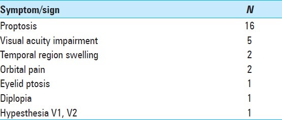
Eighteen patients presenting en plaque sphenoid wing meningiomas were surgically treated. Fifteen patients were females, representing 83% of all patients (female: male ratio 5:1). The mean age at the time of surgery was 52.2 years, ranging from 27 to 75 years. The most common presenting complaint was proptosis, referred by 16 patients. Five patients referred also visual impairment, two patients noted temporal region swelling, and two patients complained of pain in the orbital region. All signs and symptoms are listed in Table 1. In 15 patients the tumor was on the right side and only 3 patients presented left side sphenoid wing meningiomas.
Table 1.
Symptoms and signs at the time of surgery
Imaging findings
When planning the surgical treatment of an en plaque meningioma of the sphenoid wing, the extension of both tumor components - dural/intradural and bone involvement - have to be taken in consideration [Figure 1]. In the present series, the bone component was located by definition in the great sphenoid wing in all patients, therefore involving the posterolateral orbital wall. In five patients the bone invasion also extended to the superior orbital wall (lesser sphenoid wing), in two patients the pterygoid process was invaded, and in other two patients the bone component extended to the frontal and temporal bones [Figure 2].
Figure 1.
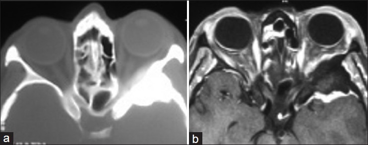
Imaging findings in a left sided sphenoid wing en plaque meningioma; a - CT scan bone window showing the bone involvement; b - T1 contrast enhanced MRI showing typical sheet-like dural involvement
Figure 2.
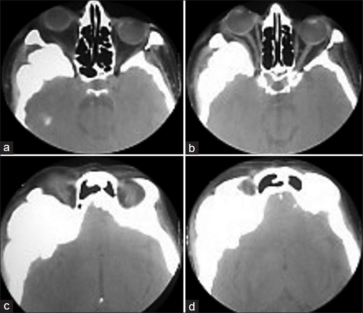
(a - d) CT scan of a sphenoid wing en plaquemeningioma showing extensive bone invasion, involving sphenoid wing, frontal bone and the squamous portion of temporal bone; note the proptosis as a result of bone invasion and not due to meningeal plaque
The dural component was located in the anterior region of the temporal fossa in 10 patients. This was the most common location of dural infiltration. Five patients presented extension of dural component into the orbit, six patients presented cavernous sinus infiltration [Figure 3], and in two cases the dural involvement extended to the sylvian fissure. We also found that in four patients there was no significant intradural extension, only a dural enhancement zone in the temporal fossa.
Figure 3.
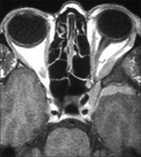
T1 contrast enhanced MRI of a left sided en plaquemeningioma; note the meningeal plaque extending into the orbit and infiltrating the cavernous sinus
Surgical treatment
The 18 patients were submitted to pterional craniotomy followed by extradural removal of the invaded bone using high speed drills, bone rongeurs and kerrison bone punches. Superolateral orbitotomy was also performed in all cases, allowing orbital decompression and removal of tumor in cases with intraorbital involvement. By removing the invaded bone we were able to decompress the superior orbital fissure as well as the orbital cavity itself, a procedure especially important in cases with proptosis. After bone removal, when needed, the dura was open and the dural/intradural component was removed. In the five cases with intraorbital extension, we also opened the periorbita and removed this tumor component. The decompression of optic canal was performed in two patients presenting visual acuity impairment preoperatively and imaging evidence of optic nerve compression due to bone invasion of the optic canal walls. Cavernous sinus tumor extension was not removed in any of the six patients who presented it, considering the high risk of postoperative neuropathy. In seven patients, the dural component was confined to the temporal fossa, presenting no extension into the orbital cavity or cavernous sinus. In these seven cases it was possible to achieve a Simpson grade II resection [Figure 4]. In all other cases we performed a subtotal resection. When the bone invasion of sphenoid wing extended too lateral, the bone removal resulted in a significant bone defect in the calvaria that had to be reconstructed. In six patients the cranioplasty was performed with titanium mesh and in one patient we used methylmethacrylate. Reconstruction of the orbital wall was performed in only two cases.
Figure 4.
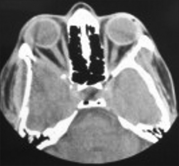
Postoperative CT scan of a sphenoid wing en plaquemeningioma without extension into the orbital cavity or cavernous sinus - complete removal of bone involvement
Histological analysis revealed that all tumors were World Health Organization (WHO) grade I meningiomas.
Follow-up
Proptosis improved after surgery in all 16 patients who presented it: 12 patients obtained a complete resolution while the other 4 improved significantly. In the postoperative period three patients presented a transient oculomotor deficit, one patient developed a transient cerebrospinal fluid (CSF) leak that was managed conservatively, and one patient complained of transient hyposthesia in V1 territory. The two patients submitted to optic canal decompression presented postoperative ipsilateral amaurosis. These two patients presented preoperative severe visual deficit (worse than 2/10). One of these patients became permanently amaurotic while the other patient presented a slight recovery of visual function in the upper visual field, resulting in a permanent unilateral inferior hemianopsia. In our series there was no mortality.
Adjuvant radiation therapy was performed in three patients presenting significant residual tumors: two patients with residual tumor in the cavernous sinus and one patient with residual tumor involving the cavernous sinus, sellar region and orbital apex. Regarding therapeutic modality, one patient was submitted to radiosurgery (RS) and two patients to fractionated stereotactic radiotherapy (FSRT). The radiation therapy modality was decided according to the following features: Size and configuration of the residual/recurrent tumor; distance to the optic nerve and quiasma.
No patient was lost to follow-up. After a mean follow-up of 4.6 years (range: 4 months to 11 years), five patients developed tumor recurrence (28%). The location of the recurrence, time to recurrence, and therapeutic decisions are listed in Table 2. Analyzing the recurrence patterns and the follow-up data we can highlight the following results:
Table 2.
Recurrence patterns and follow-up after surgical treatment of sphenoid wing en plaque meningiomas

None of the three patients submitted to immediate adjuvant radiation therapy recurred; with a mean follow-up of 3.5 years, these three patients present stable lesions, without any evidence of progression.
Regarding the location of the recurrence, two patients presented cavernous sinus recurrence, two patients presented intraorbital recurrence [Figure 5], and in one patient the recurrent tumor involved the sphenoid wing, pterygoid process, and extended to pterygomaxillary fossa.
The intraorbital tumor recurrence occurred earlier than the cavernous sinus recurrence. Intraorbital recurrence occurred after 2 and 3 years of follow-up while the cavernous sinus recurrence occurred after 4 and 5 years from initial surgery. The patient with a 3-year follow-up intraorbital recurrence, treated with surgery and tamoxifen, presented another recurrence again only 3 years after the second surgery.
Regarding therapeutic decision after diagnosis of tumor recurrence, two patients were considered surgical candidates and submitted to a surgical treatment due to tumor volume. In one of these patients, presenting a large recurrent tumor involving the sphenoid wing, pterygoid process and extending into pterygomaxillary fossa, preoperative embolization was performed. The other three patients who presented tumor recurrence were submitted to radiation therapy: RS in two cases and FSRT in one case.
After a mean follow-up of 4.6 years there was no recurrence observed after radiation therapy, whether performed immediately in the postoperative period as adjuvant therapy or performed after a recurrence.
Figure 5.
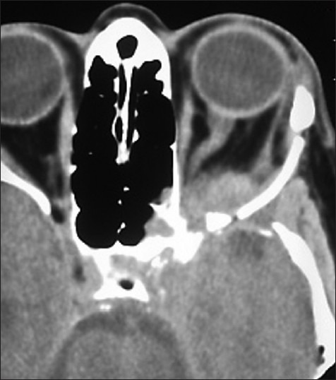
CT scan showing intraorbital recurrence 3 years after the initial treatment of a sphenoid wing en plaquemeningioma with intraorbital extension
If we analyze separately the follow-up data from three different groups: (1) patients without any tumor extension to the cavernous sinus or orbital cavity; (2) patients with cavernous sinus involvement; and (3) patients with intraorbital tumor, we can conclude that:
Group 1) - Seven patients were surgically treated for sphenoid wing en plaque meningiomas without extension to cavernous sinus or orbital cavity. None of these patients were submitted to adjuvant radiation therapy. After a mean follow-up of 3.2 years none of these patients presented a tumor recurrence.
Group 2) - Six patients presented tumor extension to the cavernous sinus [Table 3]. Three of these patients were treated with surgery and adjuvant radiation therapy and the other three patients were treated with surgery alone. None of the patients submitted to combined therapy presented tumor recurrence, while two patients submitted to surgical treatment alone presented a recurrence.
Group 3) - Five patients had intraorbital tumor extension [Table 4]. One of these patients was submitted to combined therapy (surgery and FSRT) and four patients were treated with surgery alone. The patient treated with combined therapy presents a stable lesion in the orbital apex after 6 years follow-up. Two of the four patients treated with surgery alone presented a tumor recurrence. One of these patients with intraorbital recurrent tumor presented a second recurrence after a second surgical treatment. This second recurrence was submitted to combined treatment (surgery and radiotherapy) allowing a 5-year progression-free survival since this combined treatment.
Table 3.
Follow-up of surgically treated sphenoid wing en plaque meningiomas with cavernous sinus extension
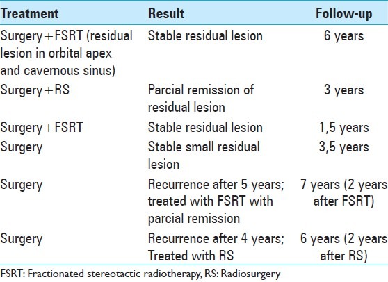
Table 4.
Follow-up of surgically treated sphenoid wing en plaque meningiomas with intraorbital extension

DISCUSSION
Sphenoid wing en plaque meningiomas are a clinical and pathological subgroup of meningiomas defined by its particular sheet-like dural involvement and its disproportionately large bone hyperostosis. Therefore, the diagnosis is determined by this particular growing pattern rather than histological appearance. [8] Meningiomas present a female predominance, with a female:male ratio of approximately 2:1. [19] In this particular type of meningiomas this ratio is higher, from 4:1 to 7:1 in some series. [13,16] In the present series the female: male ratio was 5:1. All patients with suspected en plaque meningiomas should be submitted to MRI and CT scans preoperatively. These two imaging modalities allow an accurately evaluation of the two tumor components. MRI is the best modality to assess the extension of dural/intradural involvement while CT scan allows a better visualization of bone involvement. Some CT features of hyperostosis are characteristic or suggestive of en plaque meningioma: periosteal pattern of hyperostosis, inward bulging of the vault lesion, surface irregularity of the hyperostotic bone, and intracranial changes. [10] These findings may help in differential diagnosis from other hyperostotic conditions such as osteoma, fibrous dysplasia, and Paget’s disease. In contrast, the thin layer of dural infiltration allows the distinction of en plaque meningioma from the primary intraosseous meningioma.
The first step in tumor resection is the extradural removal of all the infiltrated bone. The bone removal should be as extensive as necessary and guided by preoperative CT scan, allowing an adequate decompression of fissures and foramina of the basal cranium as well as the orbital cavity itself. This removal is performed extradurally, allowing a better protection of the underlying neural structures while the invaded bone is being drilled away. As bone infiltration is responsible for most symptoms this extensive bone removal will allow the resolution of preoperative symptoms and also prevent an early recurrence. Although bone infiltration is responsible for most symptoms, the extension of surgical resection and therefore recurrence rates are influenced mostly by the dural infiltration. Total removal of en plaque meningiomas of the sphenoid wing is very difficult to achieve. These tumors are located in a complex anatomic area and they tend to spread into foramina and fissure of the basal cranium involving the temporal fossa, the orbit, the cavernous sinus, and, more rarely, the pterygomaxillary fossa. [13] Ringel et al. [16] reported 63 patients of spheno-orbital meningioma, 76% of which had tumor residuals after surgery. Nevertheless 61% of these residuals demonstrate stability in the follow-up period, without the need for further treatments. Radical resection attempts carry a high risk of postoperative neurological morbidity, especially in cases with orbital and cavernous sinus extension. This risk has to be considered when planning the surgical treatment, especially because we are dealing with a benign tumor and there are noninvasive treatment options to deal with residual tumors, like radiation therapy. Some authors consider surgical limits the extension to cavernous sinus and superior orbital fissure. [13,17] This is our option as well. Complete tumor resection should not be achieved at the cost of increased rates of morbidity. The surgical aim should be the relief of leading symptoms rather than radical resection. [16] In contrast, resection should be as complete as possible to avoid the risk of future recurrence. As adequate resection of en plaque sphenoid wing meningiomas is difficult to achieve, recurrence rates have been as high as 35-50%. [3,13] Despite that, some authors presented encouraging total resection rates from 60% to up to 80% [13,17] with low morbidity and recent series [1,17,18] have achieved low recurrence rates of less than 10%, with early and more aggressive surgical resection. The authors believe that the goal of surgical treatment and its limits should always be planned considering patients′ signs and symptoms, tumor extension, and experience of surgical team. In our series, only in the cases without extension to the orbit or the cavernous sinus it was possible to achieve an almost total resection. In the other cases a planned subtotal resection was performed. Only the two patients submitted to optic canal decompression presented postoperative permanent morbidity. No other patient presented permanent morbidity. These two patients presented preoperative severe visual deficit (worse than 2/10). Some reports have demonstrated that when preoperative visual acuity is severely impaired, it is unlikely to improve with decompression. [4,11,13] Nevertheless some authors reported improvement in visual acuity (ranging from 27% to 79%) after decompressive surgery for meningioma involving the optic canal. [4,13] The authors believe that the indication for decompression of the optic canal should be judicious and careful because of the risk of deteriorating visual function. Initially, the senior author used to perform routinely orbital wall reconstruction after cranial approaches to remove intraorbital tumors, in order to prevent pulsating exophthalmos referred in the literature. However, based on increasing experience in orbital surgery the author progressively changed his attitude, not only because permanent pulsating exophthalmos was never experienced by any of the patients in which the orbital walls were not reconstructed but also because it allows an additional decompressive effect.
Postoperative radiation therapy is still a matter of debate. Some authors recommend postoperative radiation treatment after subtotal resection if there is dural or cavernous sinus invasion and also as soon as follow-up neuroimaging demonstrates recurrent tumor, [12] while other authors do not use postoperative radiotherapy routinely for patients with residual tumor, with the exception of patients with atypical and malignant meningioma. [13,18] Radiation therapy has been demonstrated to be effective in controlling tumor growth. In a series of 42 patients treated for residual or recurrent sphenoid wing meningiomas, none of them developed recurrence after 4.2 years. [15] Some reports have demonstrated that if the dose to the optic nerves and quiasma does not exceed 10 Gy, RS can be used safely. [14] The authors considered indications for postoperative radiation treatment the presence of significant residual tumor in the cavernous sinus and orbital apex. None of the three patients submitted to radiation therapy immediately after surgery presented tumor recurrence. In contrast, the patients presenting tumor extension to the cavernous sinus and orbital cavity treated with surgery alone presented a high incidence of tumor recurrence. The authors believe that these patients should be considered for postoperative immediate radiation therapy.
CONCLUSION
En plaque meningiomas of the sphenoid wing are challenging tumors that pose some particular issues in dealing with. Total removal is very difficult and carries high risk of postoperative morbidity. Cavernous sinus and superior orbital fissure involvement are considered surgical limits. Radiation therapy is effective in controlling tumor growth and should be considered for cases with residual tumor in cavernous sinus and orbital cavity.
Footnotes
Available FREE in open access from: http://www.surgicalneurologyint.com/text.asp?2013/4/1/86/114796
Disclaimer: The authors of this article has no conflict of interest to disclose, and has adhered to SNI's policies regarding human/animal rights, and informed consent. Advertisers in SNI did not ask for, nor did they receive access to this article prior to publication
Contributor Information
Nuno M. Simas, Email: nunosmas@hotmail.com.
João Paulo Farias, Email: jpatlfarias@gmail.com.
REFERENCES
- 1.Bikmaz K, Mrak R, Al-Mefty O. Management of bone-invasive hyperostotic sphenoid wing meningiomas. J Neurosurg. 2007;107:905–12. doi: 10.3171/JNS-07/11/0905. [DOI] [PubMed] [Google Scholar]
- 2.Charbel FT, Hyun H, Misra M, Gueyikian S, Mafee RF. Juxtaorbital en plaque meningiomas.Report of four cases and review of literature. Radiol Clin North Am. 1999;37:89–100. doi: 10.1016/s0033-8389(05)70080-4. [DOI] [PubMed] [Google Scholar]
- 3.Cophignon J, Lucena J, Clay C, Marchac D. Limits to radical treatment of spheno-orbital meningiomas. Acta Neurochir Suppl (Wien) 1979;28:375–80. [PubMed] [Google Scholar]
- 4.Cristante L. Surgical treatment of meningiomas of the orbit and optic canal: A retrospective study with particular attention to the visual outcome. Acta Neurochir (Wien) 1994;126:27–32. doi: 10.1007/BF01476490. [DOI] [PubMed] [Google Scholar]
- 5.Cushing H, Eisenhardt L. The Meningiomas: Their Classification, Regional Behavior, Life History, and Surgical End Results. Springfield: Charles C Thomas; 1938. [Google Scholar]
- 6.Cushing H. The cranial hyperostosis produced by meningeal endotheliomas. Arch Neurol Psychiatry. 1922;8:139–54. [Google Scholar]
- 7.Derome PJ, Visot A. Bony reaction and invasion in meningiomas. In: Al-Mefty O, editor. Meningiomas. New York: Raven Press; 1991. p. 169. [Google Scholar]
- 8.Honeybul S, Neil-Dwyer G, Lang DA, Evans BT, Ellison DW. Sphenoid wing meningioma en plaque: A clinical review. Acta Neurochir (Wien) 2001;143:749–58. doi: 10.1007/s007010170028. [DOI] [PubMed] [Google Scholar]
- 9.Jesus O, Toledo MM. Surgical management of meningioma en plaque of the sphenoid ridge. Surg Neurol. 2001;55:265–9. doi: 10.1016/s0090-3019(01)00440-2. [DOI] [PubMed] [Google Scholar]
- 10.Kim KS, Rogers LF, Goldblatt D. CT features of hyperostosing meningioma en plaque. AJR Am J Roentgenol. 1987;149:1017–23. doi: 10.2214/ajr.149.5.1017. [DOI] [PubMed] [Google Scholar]
- 11.Li Y, Shi JT, An YZ, Zhang TM, Fu JD, Zhang JL, et al. Sphenoid wing meningioma en plaque: Report of 37 cases. Chin Med J (Engl) 2009;122:2423–7. [PubMed] [Google Scholar]
- 12.Maroon JC, Kennerdell JS, Vidovich DV, Abla A, Sternau L. Recurrent spheno-orbital meningioma. J Neurosurg. 1994;80:202–8. doi: 10.3171/jns.1994.80.2.0202. [DOI] [PubMed] [Google Scholar]
- 13.Mirone G, Chibbaro S, Schiabello L, Tola S, George B. En Plaque Sphenoid Wing Meningiomas: Recurrence factors and surgical strategy in a series of 71 patients. Neurosurgery. 2009;65 Suppl 6:S100–8. doi: 10.1227/01.NEU.0000345652.19200.D5. [DOI] [PubMed] [Google Scholar]
- 14.Morita A, Coffey RJ, Foote RL, Schiff D, Gorman D. Risk of injury to cranial nerves after gamma knife radiosurgery for skull base meningiomas: Experience in 88 patients. J Neurosurg. 1999;90:42–9. doi: 10.3171/jns.1999.90.1.0042. [DOI] [PubMed] [Google Scholar]
- 15.Peele KA, Kennerdell JS, Maroon JC, Kalnicki S, Kazim M, Gardner T, et al. The role of postoperative irradiation in the management of sphenoid wing meningiomas.A preliminary report. Ophthalmology. 1996;103:1761–7. doi: 10.1016/s0161-6420(96)30430-2. [DOI] [PubMed] [Google Scholar]
- 16.Ringel F, Cedzich C, Schramm J. Microsurgical technique and result of a series of 63 spheno-orbital meningiomas. Neurosurgery. 2007;60 Suppl 2:S214–22. doi: 10.1227/01.NEU.0000255415.47937.1A. [DOI] [PubMed] [Google Scholar]
- 17.Schick U, Bleyen J, Bani A, Hassler W. Management of meningiomas en plaque of the sphenoid wing. J Neurosurg. 2006;104:208–14. doi: 10.3171/jns.2006.104.2.208. [DOI] [PubMed] [Google Scholar]
- 18.Shrivastava RK, Sen C, Costantino PD, Della Rocca R. Sphenoorbital meningiomas: Surgical limitations and lessons learned in their long-term management. J Neurosurg. 2005;103:491–6. doi: 10.3171/jns.2005.103.3.0491. [DOI] [PubMed] [Google Scholar]
- 19.Wiemels J, Wrensch M, Claus EB. Epidemiology and etiology of meningioma. J Neurooncol. 2010;99:307–14. doi: 10.1007/s11060-010-0386-3. [DOI] [PMC free article] [PubMed] [Google Scholar]


