

Abstract
Aim:
This in vitro study compared the coronal discoloration effect of three endodontic sealers at short-time intervals.
Materials and Methods:
The crowns of 50 extracted premolars were cut and their pulp chambers were cleaned and randomly divided into five groups (n = 10). The following materials were placed into the pulp chambers: Group I: AH Plus, group II: Apexit Plus, group III: Sultan, group IV: Amalgam, and group V: Distilled water. The color of the crowns was measured using Shadepilot™ spectrophotometer (DeguDent, Hanau, Germany) prior and after the placement of experimental materials within pulp chambers. Color changes (ΔE values) and shades were recorded in 3, 10, and 17 days. A Kruskal-Wallis and Mann-Whitney tests were used to assess significant differences between the sealers. Wilcoxon test was used to compare color changes at different periods within each group.
Results:
All sealers showed significant discoloration effect that increased over the time. At the end of the observation period, Apexit Plus showed the lowest coronal discoloration effect (ΔE = 6.38 ± 0.55).
Conclusions:
All sealers used in the current study may cause a progressive coronal discoloration effect over 10-17 days. Apexit Plus sealer showed the lowest coronal discoloration effect.
Keywords: Coronal discoloration, root canal sealers, Shadepilot, spectrophotometer
INTRODUCTION
Different types of root canal filling materials have been introduced to obturate radicular spaces, but the combination of gutta-percha as a core material with an endodontic sealer is the most widely used technique in clinical practice.[1] Regardless of core materials, root canal sealer is an essential material for any obturating technique to accomplish fluid-tight seal and to fill inaccessible areas of prepared canals.[1]
Crown discoloration after endodontic treatment is considered a common esthetic problem for the patient and dentist, particularly for anterior teeth.[2] The main causes of intrinsic crown discoloration related to endodontic treatment are: Disintegration of necrotic pulp tissue, hemorrhage into the pulp chamber, and endodontic medicaments and filling materials.[2,3,4,5]
Tooth color can be measured in several ways including visual assessment and by using instruments such as colorimeters and spectrophotometers. Spectrophotometers are considered as the reference instruments in the field of color science and have been used successfully in dentistry for tooth color measurements.[6,7] The American Dental Association recommended the use of the CIELAB color differential system to determine the color of any object.[8]
AH Plus is commonly used resin-based sealer and considered as a substitute to AH 26 sealer to overcome its discoloration effect. The staining ability of calcium-containing root canal sealer (Sealapex) was evaluated by two studies and concluded that Sealapex caused moderate to minimal discoloration of coronal dentin.[9,10] Sultan sealer is one of zinc oxide eugenol (ZOE) based sealer that contains zinc oxide powder without silver. Limited data are available about the coronal discoloration effect of AH Plus, Apexit Plus, and Sultan sealers.
This study was designed to assess crown discoloration induced by AH Plus, Apexit Plus, and Sultan sealers, using quantitative and qualitative spectrophotometric analysis at 3, 10, and 17 days.
MATERIALS AND METHODS
Samples preparation
Freshly extracted human premolar teeth were used for the current study. The teeth were free of caries, cracks, restorations, and coronal stains. The roots were resected 3 mm below the cementoenamel junction using a diamond disc. The pulp chamber of each crown was mechanically cleaned using gates glidden drills and hand k-files and then irrigated with 3% sodium hypochlorite solution and 17% ethylenediaminetetraacetic acid (EDTA) solution (Pulp dent, Watertown, MA, USA). The specimens were then randomly assigned to three experimental and two control groups, 10 samples each.
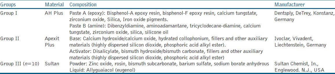
Three endodontic sealers were tested [Table 1]: AH Plus™ (Group I), Apexit Plus™ (Group II), and Sultan™ (Group III). The sealers were mixed and placed into the pulp chambers via cervical access, using a Lentulo spiral filler (Dentsply Caulk, Milford, DE, USA). In the positive and negative control groups, pulp chambers were filled with amalgam and distilled water, respectively. The apical access of samples was sealed with a sticky wax and stored in an incubator for 7 days at 37°C.
Table 1.
Experimental groups and sealers used in the current study
Tooth color measurements
Shadepilot™ spectrophotometer (DeguDent, Hanau, Germany) was used to measure the color quantitatively and qualitatively [Figure 1]. The spectrophotometer was calibrated according to the manufacturer›s instructions before taking each reading and then carefully placed at right angle to buccal surface of the crown. The resulting shades were taken directly from the digital screen of spectrophotometer device and CIE L*a*b* readings were taken using Degudent Shadepilot software version 3.01.10079. Pretreatment color shades and readings of the entire buccal surfaces were taken and considered as baseline data to which the subsequent readings at 3, 10, and 17 days were compared. The color difference (ΔE) at each time interval was calculated according to the following formula: ΔE = [(Δ*)2+ (Δa*)2+ (Δb*)2]½ where ΔL is the difference in lightness calculated from differences in the L* readings between two periods. This can be calculated for any period between baseline and at 3, 10, and 17 days. Δa and Δb refer to the difference in chroma and are also obtained in the same manner as for ΔL. A ΔE value equal or larger than 3.5 was considered a clinically perceptible color change.[11]
Figure 1.
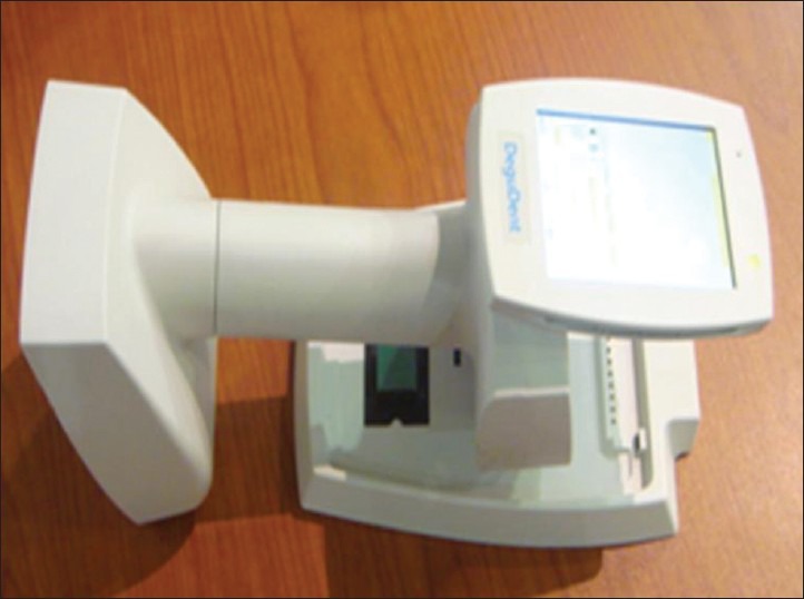
Degudent Shadepilot intraoral spectrophotometer
Statistical analysis
Delta E (ΔE) values were submitted to statistical analysis using SPSS program version 17.0 (SPSS Inc., Chicago, IL, USA). Descriptive statistics comprised calculation of means and standard deviations followed by Kruskal-Wallis and Mann-Whitney tests to assess significant differences between experimental root canal sealers at different times. Wilcoxon signed rank test was used to compare color changes at different periods within each experimental group. Results with P value less than 0.05 were considered statistically significant.
RESULTS
Descriptive statistics of color changes (∆E) from baseline (before sealer placement) for all groups are presented in Figures 2 and 3 and Tables 2 and 3. The negative controls showed no evidence of discoloration at all time intervals (∆E < 1 unit), while positive controls showed immediate discoloration (∆E > 3.5 units) at 3 days which persisted with each time interval [Figure 2].
Figure 2.
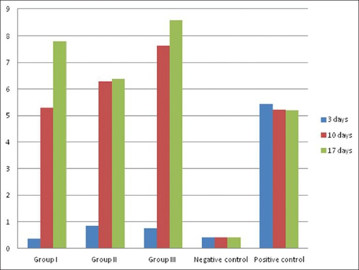
Mean color changes (ΔE mean values) for overall buccal surfaces in all groups at 3, 10, and 17 days
Figure 3.
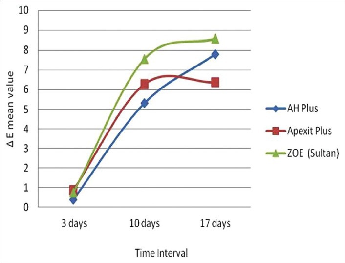
Line graph demonstrating mean color changes (ΔE mean values) induced by experimental sealers over time
Table 2.
Comparison of overall color changes (ΔE mean values) of experimental groups at each time interval
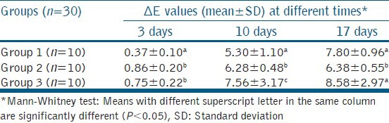
Table 3.
Comparison of overall color changes (ΔE mean values) at all time intervals for each group
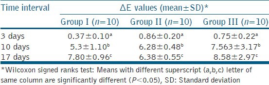
Comparison of the experimental groups (Groups I-III) regarding coronal discoloration (Kruskal-Wallis) results in significant differences (P < 0.05) [Table 2]. The results of Mann-Whitney test revealed that coronal discoloration at 17 days was significantly less for Apexit Plus (Group II) when compared to AH Plus and Sultan sealer (P < 0.05). As depicted in Table 3 and Figure 3, there is a progressive and significant color change (∆E) from days 3 to 17 within all experimental groups (P < 0.05).
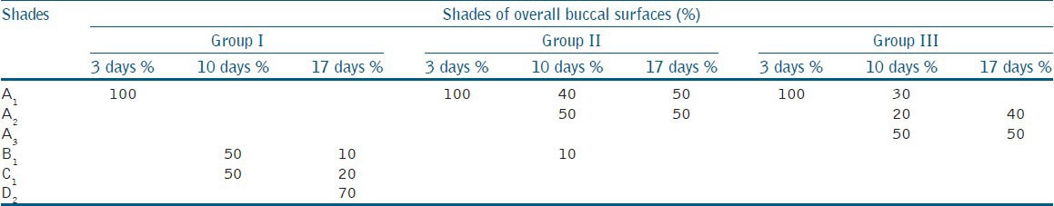
The differences in the mean color change that took place in all groups from 3 days to 10 days and 10 to 17 days are presented in Table 4. The highest significant color change from the days 3 to 10 was found in group III (∆E = 7.55), and from the day 10 to 17 was found in group I (∆E = 3.63). The shades recorded for overall buccal surfaces at different time intervals for all groups were presented in Table 5.
Table 4.
Comparison of differences in the mean overall color changes (ΔE mean values) within observation periods
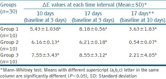
Table 5.
Percentages of the closest shade match to overall buccal surfaces in all groups at all time intervals as indicated by Shadepilot spectrophotometer
DISCUSSION
Ideal root canal sealer should have characteristics such as good adhesion, adequate seal, radiopacity, dimensional stability during setting, tissue tolerance, antibacterial effect, nonresolvability in tissue fluids and cause no discoloration for tooth structure.[12] The present study was conducted to evaluate the overall coronal discoloration effect of three of the most commonly used sealers. These sealers were AH Plus (resin-based sealer) which is a substitute for AH26, Apexit Plus (calcium hydroxide-based sealer) and Sultan (ZOE-based sealer). The literature review proved that these types of sealers have good sealing ability and good biocompatibility.[13,14]
Premolars crowns were used in the present study because of their reasonable wide pulp chambers that allow bulk placement of sealers. The pulp chamber of each sample was mechanically cleaned using and irrigated with 3% sodium hypochlorite and to eliminate any organic debris that could affect the measurement of tooth color. In addition the pulp chambers were also irrigated with 17% EDTA similar to previous studies to simulate most common clinical conditions regarding smear layer removal.[5]
In the present study, two control groups were utilized to test the ability of coronal dentin and enamel to transmit the color induced by materials that placed into the pulp chamber. The results obtained from the control groups may indicate that the dentin and enamel have the ability to transmit the color of the material placed in the pulp chamber. Application of sealer into the pulp chambers was similar to that of previous studies.[4,9,10]
Spectrophotometry is an objective and more reliable method alternative to the subjective (visual) method of assessing color.[15,16,17] Spectrophotometers are extremely sensitive devices which can determine minute color changes that are not even clinically observable. These color changes can also be detected much earlier when compared to the traditional visual assessment of tooth color.[17,18] For these reasons, it was decided to use spectrophotometric analysis in the current study for evaluating tooth discoloration determining CIE L*A*B* variables and delta E (ΔE) as well as closest shades using an intraoral spectrophotometer (Shadepilot).
The exact time interval for tooth discoloration to occur resulting from root canal therapy is still not documented. The required time for discoloration to be clinically observed depends on many factors that include the thickness of the remaining dentin, the quality and quantity of the sealer, and the presence of the smear layer.[19] Previous studies revealed that coronal tooth discoloration resulting from endodontic materials takes place from seven weeks after obturation[4] to several months.[9,10,20] Differences in the results of the previous studies could be attributed to the methodologies employed. In the present study, the discoloration effect of the sealers was determined at short periods (3, 10, and 14 days) to test prompt staining effect of sealers. In each group, the color of each sample was determined before sealer placement to act as a baseline for calculating the color change at each subsequent time interval. In addition, the color of samples at 3 and 10 days was also taken as a base to evaluate color change from 3 to 10 days and from 10 to 17 days, respectively.
The results indicated that all tested sealers did not show any evident of coronal discoloration after 3 days. This could be explained by the sealers that may penetrate into dentinal tubules, due to smear layer removal, did not show immediate chemical changes that could affect their colors. All sealers showed clinically observable coronal discoloration at 10 days (∆E > 3.5) which progressively increased with time. This was in contrast to the results of previous studies in which tooth discoloration occurred after longer times.[4,9,10,20,21] This could be largely explained by the difference in methodology utilized to prepare the experimental samples and the method of color analysis. Some of these studies did not attempt to remove the smear layer, which could reduce the rate of sealer penetration through the dentin.[9,10] In the current study, sealer penetration within dentinal tubules was not evaluated, so it was difficult to confirm the ability of the tested sealers to penetrate dentinal tubules. However, there is controversy among authors where some of them concluded that sealers[9] did not penetrate the dentinal tubules, while others[22] showed that the pathway by which staining materials can diffuse from the canal is through dentinal tubules.
The current results indicated that Sultan sealer displayed the greatest overall coronal discoloration and these were similar to the findings of previous studies.[4,9,10] The cause of severe discoloration observed with ZOE sealer may be probably due to the presence of eugenol, which forms a bond with zinc oxide. Free or even bound eugenol changes chemically (oxidizes) and darkens with time.[10,23] Although AH Plus is silver-free and advertised as nonstaining compared to its predecessor AH26, it caused more overall coronal discoloration (grayish discoloration) than Apexit Plus sealer (reddish brown discoloration). Some authors found the same results but with AH26 sealer.[24] Therefore, it can be argued that the silver ions were not the sole reason for tooth discoloration caused by AH26 and some ingredients in AH Plus could be the cause of this grayish discoloration.[20] The literature lacks evidence regarding the staining potential of AH Plus; therefore, additional research is required to investigate the constituents of AH Plus that might be responsible for the discoloration of endodontically treated teeth. The highest color changes in 3-10 days were for Sultan (∆E = 7.55), while the lowest color change was for AH Plus (∆E = 5.43), and Apexit Plus (∆ = 6.16). The color changes in 10-17 days were not clinically detectable (∆ < 3.5) except for AH Plus with a mean ∆E value 3.63. These could indicate that the main color change occurred in the period from 3 to 10 days and did not dramatically increased from 10-17 days.
CONCLUSIONS
Within the limitation of the present study, the following could be concluded:
All used sealers did not have a coronal discoloration effect after 3 days but discoloration was perceptible after 10 days
All sealers used in the current study may cause a progressive coronal discoloration effect over 10-17 days
Apexit Plus sealer showed the lowest coronal discoloration effect
Shadepilot spectrophotometer is a valuable and simple tool to detect coronal discoloration induced by endodontic materials.
Footnotes
Source of Support: Nil
Conflict of Interest: None declared
REFERENCES
- 1.Ørstavik D. Materials used for root canal obturation: Technical, biological and clinical testing. Endod Topics. 2005;12:25–38. [Google Scholar]
- 2.Sheets CG, Paquette JM, Wright RS. Tooth whitening modalities for pulpless and discoloured teeth. In: Cohen S, Burns RC, editors. Pathways of the Pulp. 8th ed. London: Mosby; 2002. p. 755. [Google Scholar]
- 3.Pittford TR. Apexification and apexogenesis. In: Walton RE, Torabinejad M, editors. Principles and Practice of Endodontics. 3rd ed. Philadelphia: WB Saunders; 1996. p. 388. [Google Scholar]
- 4.van der Burgt TP, Mullaney TP, Plasschaert AJ. Tooth discolouration induced by endodontic sealers. Oral Surg Oral Med Oral Pathol. 1986;61:84–9. doi: 10.1016/0030-4220(86)90208-2. [DOI] [PubMed] [Google Scholar]
- 5.Walton RE, Rotstein I. Bleaching discolored teeth: Internal and external. In: Walton RE, Torabinejad M, editors. Principles and practice of endodontics. 2th ed. Philadelphia: WB Saunders; 1996. p. 385. [Google Scholar]
- 6.Seghi RR, Hewlett ER, Kim J. Visual and instrumental colorimetric assessment of small color differences on translucent dental porcelain. J Dent Res. 1989;68:1760–4. doi: 10.1177/00220345890680120801. [DOI] [PubMed] [Google Scholar]
- 7.Sproull RC. Color matching in dentistry. 1. The three-dimensional nature of color. J Prosthet Dent. 1973;29:416–24. doi: 10.1016/s0022-3913(73)80019-8. [DOI] [PubMed] [Google Scholar]
- 8.Revised American Dental Association specification No 12 for denture base polymer. J Am Dent Assoc. 1975;90:451–8. doi: 10.14219/jada.archive.1975.0069. [DOI] [PubMed] [Google Scholar]
- 9.Davis MC, Walton RE, Rivera EM. Sealer distribution in coronal dentin. J Endod. 2002;28:464–6. doi: 10.1097/00004770-200206000-00012. [DOI] [PubMed] [Google Scholar]
- 10.Parsons JR, Walton RE, Ricks-Williamson L. In vitro longitudinal assessment of coronal discoloration from endodontic sealers. J Endod. 2001;27:699–702. doi: 10.1097/00004770-200111000-00012. [DOI] [PubMed] [Google Scholar]
- 11.O’Brien W. 3rd ed. Chicago: Quintessence; 2002. Dental materials and their selection; pp. 24–6. [Google Scholar]
- 12.Trope M. Trauma injuries. In: Cohen S, Burns R, editors. Pathways to the Pulp. 7th ed. St. Louis: Mosby Inc; 1998. p. 463. [Google Scholar]
- 13.Saunders EM, Saunders WP. Long-term coronal leakage of JS Quickfill root fillings with Sealapex and Apexit sealers. Endod Dent Traumatol. 1995;11:181–5. doi: 10.1111/j.1600-9657.1995.tb00484.x. [DOI] [PubMed] [Google Scholar]
- 14.McMichen FR, Pearson G, Rahbaran S, Gulabivala K. A comparative study of selected physical properties of five root-canal sealers. Int Endod J. 2003;36:629–35. doi: 10.1046/j.1365-2591.2003.00701.x. [DOI] [PubMed] [Google Scholar]
- 15.Publication CIE 1971; No. 15 (E-1.3.1) Paris, France: Bureau Central de la CIE; CIE (Commission International De L, Eclairage). Colorimetry. Official recommendations of the International Commission on illumination. [Google Scholar]
- 16.Paul S, Peter A, Pietrobon N, Hammerle CH. Visual and spectrophotometric shade analysis of human teeth. J Dent Res. 2002;81:578–82. doi: 10.1177/154405910208100815. [DOI] [PubMed] [Google Scholar]
- 17.Johnston WM, Kao EC. Assessment of appearance matches by visual observation and clinical colorimetry. J Dent Res. 1989;68:819–22. doi: 10.1177/00220345890680051301. [DOI] [PubMed] [Google Scholar]
- 18.Seghi RR, Johnston WM, O›Brien WJ. Spectrophotometric analysis of color differences between porcelain systems. J Prosthet Dent. 1986;56:35–40. doi: 10.1016/0022-3913(86)90279-9. [DOI] [PubMed] [Google Scholar]
- 19.Grossman L, Oliet S, Delrio C. 11th ed. Chicago: Lea and Febiger; 1998. Endodontic practice. [Google Scholar]
- 20.Partovi M, Al-Havvaz AH, Soleimani B. In vitro computer analysis of crown discoloration from commonly used endodontic sealers. Aust Endod J. 2006;32:116–9. doi: 10.1111/j.1747-4477.2006.00034.x. [DOI] [PubMed] [Google Scholar]
- 21.Guan YH, Lath DL, Lilley TH, Willmot DR, Marlow I, Brook AH. The measurement of tooth whiteness by image analysis and spectrophotometry: A comparison. J Oral Rehabil. 2005;32:7–15. doi: 10.1111/j.1365-2842.2004.01340.x. [DOI] [PubMed] [Google Scholar]
- 22.Kraus B, Jordan RE. 7th ed. Baltimore: Williams and Wilkins; 1976. Dental anatomy and occlusion; p. 159. [Google Scholar]
- 23.Weinberg JE, Rabinowitz RL, Zainger M, Gennaro AF. 14C-Eugenol: 1. Synthesis, polymerization, and use. J Dent Res. 1972;51:1055–61. doi: 10.1177/00220345720510041101. [DOI] [PubMed] [Google Scholar]
- 24.van der Burgt TP, Plasschaert AJ. Tooth discolouration induced by dental materials. Oral Surg Oral Med Oral Pathol. 1985;60:666–9. doi: 10.1016/0030-4220(85)90373-1. [DOI] [PubMed] [Google Scholar]