G&H How are small and diminutive colonic polyps usually managed?
FR Small (6-9 mm) and diminutive (1-5 mm) colonic polyps are commonly encountered during colonoscopy. It is estimated that at least 1 polyp is detected in roughly half of all patients undergoing screening colonoscopy, with small and diminutive polyps accounting for over 80% of all polypoid lesions. Currently, small and diminutive polyps are resected (preferably via a cold snare) and then submitted for histologic examination, except when there are multiple tiny polyps in the rectosigmoid colon that are clearly hyperplastic (even on standard white-light colonoscopy); in this setting, it is considered adequate to sample only a few lesions.
G&H What are the drawbacks of this management strategy?
FR The current practice of routinely resecting all small colonic polyps and submitting them for histopathologic assessment has several drawbacks. First, it is well known that the likelihood of hyperplastic histology inversely correlates with the size of a polyp. Approximately 40% of lesions that are 5 mm and smaller and approximately 25% of lesions that are 6—9 mm are hyperplastic. Due to the relatively high prevalence and clinical insignificance of small left-sided hyperplastic polyps, there is a substantial—and unwarranted—cost burden associated with the removal and pathologic examination of these lesions.
G&H What does the resect-and-discard strategy entail, and why is interest in this management strategy increasing?
FR In order to address the previously mentioned drawbacks of traditional polyp management, the resect-and-discard strategy has been proposed. According to this strategy—which, to date, has been suggested only for the management of diminutive colonic polyps—the histo-logic type of the polyp (adenomatous vs non-neoplastic) is predicted in vivo by an appropriate endoscopic method, and certain postpolypectomy pathologic specimens are discarded rather than sent for histologic assessment. Realtime endoscopic assessment of the polyp’s histology determines the patient’s postpolypectomy surveillance interval.
G&H You and your colleagues recently conducted a study on the resect-and-discard strategy. What was your study design and main findings?
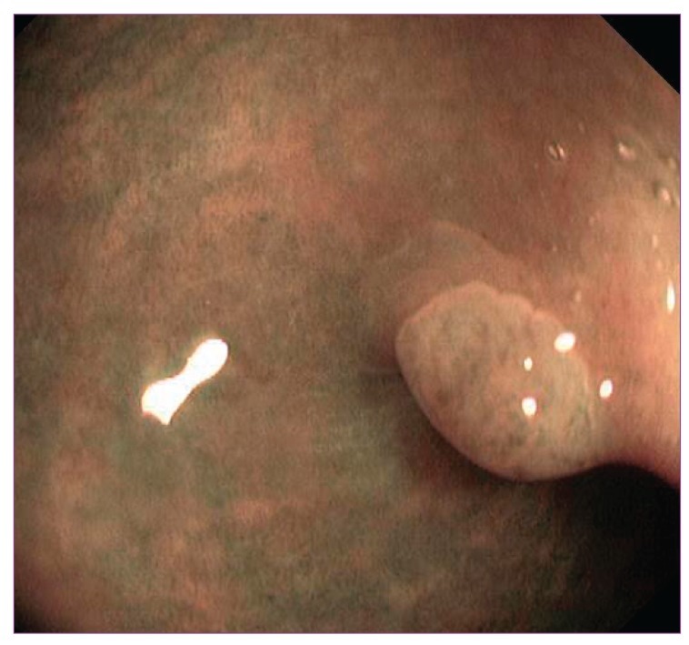
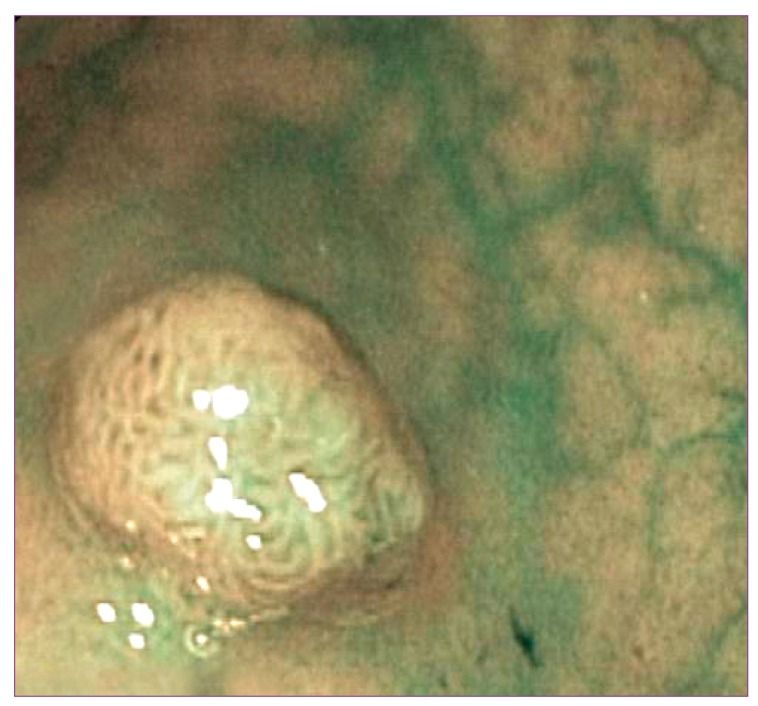
FR My colleagues and I performed a prospective single-center study with the primary aim of assessing whether the systematic use of the resect-and-discard strategy in everyday clinical practice could accurately predict postpolypectomy surveillance intervals in patients with small colonic polyps. The study population consisted of consecutive colonoscopy outpatients with at least 1 polyp smaller than 10 mm with endoscopic features that were not suspicious for malignancy (ie, depressed or ulcerated) and with morphology that could be characterized with high confidence via NBI without magnification. Patients with any lesion larger than 9 mm were excluded, regardless of the presence of small polyps. Each small or diminutive polyp was categorized as an adenoma or nonadenoma based on the overall color of the polyp, features of the vessels, and the pit pattern, according to simplified NBI criteria (Figures 1 and 2). The future postpolypectomy surveillance intervals were assigned based on guidelines from the US Multi-Society Task Force on Colorectal Cancer. Following histopathology, postpolypectomy surveillance intervals were re-assigned, and the accordance between endoscopy- and histology-directed surveillance strategies was calculated.
G&H What are the clinical implications of your study?
FR We found that approximately 30% of patients referred for colonoscopy for various indications (screening, surveillance, or symptoms) have only subcenti-metric lesions. Therefore, the application of the resect-and-discard strategy in routine clinical practice could eliminate pathology consultation in a large proportion of colonoscopy patients, which would result in a substantial economic benefit. In addition, adoption of this strategy could allow patients to learn the timing of their next colonoscopy before being discharged, thus avoiding the psychological impact of waiting for a definitive diagnosis and surveillance plan, which would further improve the efficiency of colonoscopy.
G&H Based on your study findings, is it safe to resect and discard all small colonic polyps?
FR The American Society for Gastrointestinal Endoscopy (ASGE) recently developed a Preservation and Incorporation of Valuable Endoscopic Innovations (PIVI) initiative that established thresholds for real-time endoscopic assessment of the histology of diminutive colonic polyps. A committee of experts determined that colonic polyps 5 mm or smaller could be resected and discarded without pathologic assessment if an endoscopic technique used with high confidence provided at least 90% agreement in the assignment of postpolypectomy surveillance intervals compared with decisions based on pathologic assessment.
G&H What are the drawbacks of this management approach?
FR The main criticism of real-time endoscopic prediction of polyp histology is its inability to identify advanced neoplasia, which requires more intensive postpolypec-tomy surveillance (ie, 3 years vs 5—10 years). This is the reason that the ASGE limits the use of the resect-and-discard strategy to only diminutive polyps, in which the risk of advanced neoplasia is very low (approximately 1%). Hie application of this strategy to 6—9-mm lesions is controversial because of the higher prevalence of advanced neoplasia in these lesions.
G&H Do you foresee any opposition to the adoption of this strategy?
FR It is always difficult to change paradigms and introduce new developments in medicine, and the potential adoption of the resect-and-discard strategy is not an exception. Indeed, several factors may prevent the adoption of this strategy in clinical practice.
Pathologic assessment is still considered essential to determine the timing of a patient’s next surveillance colonoscopy. This process enables the differentiation of neoplastic lesions from non-neoplastic lesions and the identification of advanced histologic features (high-grade dysplasia or villous elements), which require more intensive surveillance.
Second, small adenomas (particularly those <5 mm) rarely have advanced histologic features. Therefore, the main usefulness of pathologic assessment is to differentiate between adenomatous and non-neoplastic polyps, and endoscopic follow-up is recommended only for the former group of lesions. However, even histopathologic assessment, which is considered the gold standard for polyp characterization, may have some limitations. Indeed, studies have shown that the median kappa value for interobserver agreement for the diagnosis of adenomas versus hyperplastic polyps is not perfect (ranging from 0.84 to 0.98).
Third, compared with larger polyps, small polyps (particularly when resected via a cold snare) are associated with a higher risk of failed polyp retrieval, resulting in the complete loss of histologic information and thus preventing accurate determination of the interval to the next surveillance colonoscopy in up to 20% of cases.
The potential for translating the resect-and-discard strategy into clinical practice largely depends on the accuracy of determining the histology of a polyp immediately upon detection. Until a few years ago, the concept of real-time assessment of colonic polyp histology may have sounded absurd to many doctors. Indeed, standard white-light endoscopy was considered inaccurate for differentiating between hyperplastic and neoplastic colorec-tal lesions in vivo, as it had a sensitivity of 60-90% and a specificity of 40—90%. Disappointingly, the evolution from standard to high-definition endoscopy failed to significantly improve the former’s suboptimal accuracy in characterizing colorectal polyps. However, in recent years, imaging-enhancing technologies such as narrowband imaging (NBI; Olympus), Fujinon intelligent color enhancement (Fujinon), and i-scan (Pentax) have demonstrated promising results in several studies. These simple technologies are being incorporated into the new generation of endoscopes and can achieve the benefits of chromoendoscopy without being as expensive, cumbersome, and time consuming as using dye spray.
Most US and European studies on endoscopic prediction of histology have been conducted using NBI without optical magnification. According to many of these studies, predictions made with a high level of confidence are over 90% accurate, suggesting that expert and properly trained endoscopists may be ready to move toward endoscopic characterization of small or diminutive colorectal polyps and, thus, forgo formal pathologic evaluation.
The principal value of the resect-and-discard strategy is a substantial cost-savings due to the elimination of pathology fees. In a decision analysis that evaluated the impact of the resect-and-discard strategy for colonoscopy screening, Hassan and colleagues demonstrated that the adoption of this policy is cost-effective for eligible diminutive polyps (resulting in a savings of $25/person) and does not cause any meaningful effect on the efficacy of screening. Projected onto the US population, the adoption of this approach would result in an undiscounted annual savings of $33 million. In an era characterized by limited economic resources, it is clear that emerging strategies that have relevant economic benefits without negatively influencing the quality of care garner attention.
A total of 286 patients (about 30% of the patients evaluated) had only small polyps, and about 500 small polyps were evaluated. The sensitivity, specificity, and accuracy of NBI for in vivo diagnosis of adenomas was 95%, 66%, and 86%, respectively, and positive and negative likelihood ratios were 2.80 and 0.08, respectively. Similar diagnostic operating characteristics of NBI were obtained for diminutive polyps. Endoscopy-directed surveillance was in accordance with histology-directed surveillance in 83% of patients with small polyps. Surveillance would have been delayed or overused in 7% and 10% of patients, respectively, if it had been based on endoscopic findings.
Although we did not perform a cost analysis, the impact of the resect-and-discard strategy on procedure-related costs is evident. It could be argued that the proportion of inaccurate follow-up examinations dictated by endoscopic findings could negate the economic benefits of the resect-and-discard strategy by increasing colonos-copy overprescription. However, the cost increase related to the anticipation of follow-up colonoscopy—which occurred in approximately 10% of patients who had small or diminutive polyps in our study—appears to be negligible when taken into consideration with the reduced histology-related costs of all patients with small polyps.
Although we failed to match the 90% PIVI benchmark in our study, the resect-and-discard strategy appeared to be safe, as there was only a negligible risk of underestimating a patient’s likelihood of developing advanced neoplasia. In our study, we did not find advanced histologic features or more than 2 adenomas in any of the patients who, erroneously, did not receive a specific surveillance interval (due to the misclassification of polyps as nonadenomas). In such low-risk patients, the need for surveillance colonoscopy is still under debate, as it likely has few benefits and is not cost-effective. If a 10-year surveillance interval is recommended for a patient with 1—2 tubular subcentimetric adenomas (based on European guidelines), a false-negative result would have no effect, as adenomatous and hyperplastic lesions share the same recommended surveillance interval.
In addition, the percentage of patients who received delayed surveillance (5 years vs 3 years) due to the presence of at least 1 adenoma with advanced histologic features (villous components and/or high-grade dysplasia) was very low (approximately 5% of patients with small polyps and 0% among those with only diminutive polyps). Nevertheless, it is still controversial whether these histologic features represent consistent predictors of advanced neoplasia.
Deviating a little from the ASGE’s recommendation, we applied the resect-and-discard strategy to patients with small polyps and not solely diminutive ones in our study. We found comparable concordance rates of endoscopy-and histology-directed strategies in the small and diminutive polyp cohorts, suggesting that the impact of advanced histologic features on the resect-and-discard strategy might be marginal. Nevertheless, restricting the application of this management strategy to only diminutive polyps, as recommended by the ASGE, sounds reasonable. Indeed, this approach minimizes the risk of missing invasive cancer (which has a prevalence approaching 1% in 6—9-mm polyps in some series and, thus, is not negligible). In addition, this approach may theoretically increase the accuracy of postpolypectomy surveillance intervals and does not significantly decrease the efficiency of the strategy (due to the much lower prevalence of 6—9-mm lesions compared with lesions less than 5 mm in the general population).
Another important criticism of real-time endoscopic prediction of polyp histology involves the difficulty of differentiating between hyperplastic and serrated lesions (particularly sessile serrated polyps/adenomas) because of their similar characteristics on electronic chromoen-doscopy. It is well known that these lesions are associated with a higher risk of synchronous or metachronous advanced neoplasia, particularly when they are larger than 10 mm or located in the proximal colon. He identification of these lesions should not preclude the application of the resect-and-discard strategy. However, in the presence of a small right-sided lesion with hyperplastic-like features on electronic chromoendoscopy, the endoscopist must be aware of this potential risk, resect the lesion, and indicate an appropriate surveillance interval according to the recently published consensus update of guidelines for postpolypectomy surveillance by the US Multi-Society Task Force on Colorectal Cancer.
First, although many studies have demonstrated that experts can achieve high performance levels in realtime prediction of polyp histology, it remains to be seen whether these levels can be replicated by less experienced gastroenterologists. Several recent studies have suggested that the proficiency levels of community-based doctors are not as high as the levels of doctors from academic referral centers and, thus, do not reach the PIVI benchmark. It is unlikely that gastrointestinal societies would endorse this policy before implementing appropriate teaching and training modules and refining the credentialing process and before doctors from community practices could achieve high performance levels.
Second, foregoing pathologic assessment would shift significant responsibility from the pathologist to the endoscopist, which would have medicolegal implications. A standardized and formal reporting of in vivo histologic prediction would have to be established to prevent medical litigations. He medicolegal liability that would be taken on by gastroenterologists likely represents the major hurdle for changing the practice of sending all polyps for histologic assessment.
Finally, some resistance to the resect-and-discard strategy may arise from pathologists, as its adoption would reduce the number of specimens submitted for pathologic evaluation and would have a significant financial impact on pathologists.
However, despite all of these obstacles, it is not unrealistic to presume that the resect-and-discard strategy has the potential to enter clinical practice in the future, provided that real-world data confirm its ability to predict in vivo polyp histology with a high level of accuracy. Nevertheless, the debate on the management of diminutive and small polyps has provided the opportunity for both gastroenterologists and pathologists to improve their skills and diagnostic performances.
Figure 1.
A 2-mm hyperplastic polyp seen via narrow-band imaging without magnification. The polyp is lighter in color than the surrounding mucosa, and isolated lacy vessels can be seen across the lesion.
Figure 2.
A 3-mm adenomatous polyp seen via narrowband imaging without magnification. The polyp is brown (in contrast to the surrounding mucosa), and tubular white structures are surrounded by brown vessels.
Biography

Suggested Reading
- Paggi S, Rondonotti E, Amato A, et al. Resect and discard strategy in clinical practice: a prospective cohort study. Endoscopy. 2012;44:899–904. doi: 10.1055/s-0032-1309891. [DOI] [PubMed] [Google Scholar]
- Rex DK, Kahi C, O’Brien M, et al. He American Society for Gastrointestinal Endoscopy PIVI (Preservation and Incorporation of Valuable Endoscopic Innovations) on real-time endoscopic assessment of the histology of diminutive colorectal polyps. Gastrointest Endosc. 2011;73:419–422. doi: 10.1016/j.gie.2011.01.023. [DOI] [PubMed] [Google Scholar]
- Ignjatovic A, East JE, Suzuki N, Vance M, Guenther T. Saunders BP Optical diagnosis of small colorectal polyps at routine colonoscopy (Detect Inspect Characterise Resect and Discard; DISCARD trial): a prospective cohort study. Lancet Oncol. 2009;10:1171–1178. doi: 10.1016/S1470-2045(09)70329-8. [DOI] [PubMed] [Google Scholar]
- Hassan C, Pickhardt PJ, Rex DK. A resect and discard strategy would improve cost-effectiveness of colorectal cancer screening. Clin Gastroenterol Hepatol. 2010;8:865–869. doi: 10.1016/j.cgh.2010.05.018. [DOI] [PubMed] [Google Scholar]
- Hewett DG, Kaltenbach T, Sano Y, et al. Validation of a simple classification system for endoscopic diagnosis of small colorectal polyps using narrow-band imaging. Gastroenterology. 2012;143:599–607. doi: 10.1053/j.gastro.2012.05.006. [DOI] [PubMed] [Google Scholar]
- Ladabaum U, Fioritto A, Mitani A. Real-time optical biopsy of colon polyps with narrow band imaging in community practice does not yet meet key thresholds for clinical decisions. Gastroenterology. 2013;144:81–91. doi: 10.1053/j.gastro.2012.09.054. [DOI] [PMC free article] [PubMed] [Google Scholar]
- McGill SK, Evangelou E, Ioannidis JP, Soetikno RM, Kaltenbach T. Narrow band imaging to differentiate neoplastic and non-neoplastic colorectal polyps in real time: a meta-analysis of diagnostic operating characteristics. Gut. doi: 10.1136/gutjnl-2012-303965. 2013 Jan 7. Epub ahead of print. [DOI] [PMC free article] [PubMed] [Google Scholar]


