

Abstract
INTRODUCTION
Usually patients are admitted to hospital with a single diagnosis, but if complaints persist it is important to consider a synchronous secondary diagnosis.
PRESENTATION OF CASE
A 74-year-old woman presented with severe abdominal and back pain. On physical examination, a tender abdominal aortic aneurysm (AAA) was noted. Following endovascular treatment of the AAA, pain in the right lower abdomen persisted. Review of the pre-EVAR CT images revealed a foreign body in the terminal ileum, which was surgically removed.
DISCUSSION
Patients with foreign-body-related intestinal pain present with complaints of abdominal pain at initial presentation. The accompanied back pain and abdominal tenderness of the abdominal aorta in our case could indicate another diagnosis.
CONCLUSION
Persisting complaints post-intervention should not only arouse suspicion of an intervention-related complication, but also of a synchronous second diagnosis.
Keywords: Persisting pain, Symptomatic aortic aneurysm, Foreign body, Secondary diagnosis
1. Introduction
Usually patients are admitted to hospital with a single diagnosis, but if complaints persist it is important to consider a synchronous secondary diagnosis. We present a patient with a typical history of a symptomatic abdominal aorta aneurysm. After endovascular treatment, right lower quadrant abdominal pain persisted and a search for a secondary diagnosis was instigated.
2. Presentation of case
A 74-year-old woman, seen at another hospital for a 40 mm abdominal aortic aneurysm (AAA) one year earlier, presented to our vascular clinic. She complained of severe abdominal and back pain, which had lasted all day. Physical examination revealed a tender AAA. CT-angiography, demonstrated an infrarenal AAA with a diameter of 52 mm (Fig. 1, Fig. 4). Endovascular treatment with an Endurant® stentgraft was performed. Postoperatively, the patient initially was pain free. However, in the following days, she repeatedly experienced abdominal pain, now situated in the right lower quadrant. Review of the pre-operative CT-angiogram revealed a foreign body in the terminal ileum, which had been missed in the setting of a typical symptomatic AAA presentation. The patient could not remember having eaten anything unusual. During colonoscopy, the foreign body was seen inside the terminal ileum (Fig. 2, Fig. 5), and removal was attempted. Excessive pain was experienced during this attempt, raising concerns about possible perforation. Therefore, endoscopic retrieval was abandoned in favor of a laparotomy with enterotomy. In the terminal ileum, a plastic clip of the type normally used to close a bread bag was found (Fig. 6). The beak of the clip had attached itself to the mucosa of the small intestine (Fig. 3). Postoperatively, the patient had no abdominal complaints.
Fig. 1.
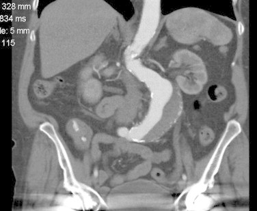
CT-image of AAA and foreign body.
Fig. 4.
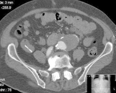
CT-image of AAA and foreign body.
Fig. 2.

Picture colonoscopy.
Fig. 5.

Picture colonoscopy.
Fig. 6.
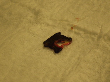
Clip.
Fig. 3.
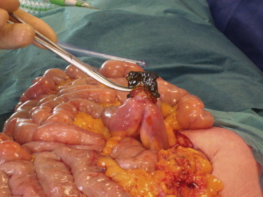
Clip in mucosa of intestine.
3. Discussion
We report a case in which there were persisting complaints after endovascular treatment of a symptomatic AAA. Our patient presented with abdominal pain and back pain. One could argue that no symptomatic aneurysm was present at initial presentation and that all symptoms could be related to the foreign body. However, previous case reports on foreign-body-related intestinal pain described only abdominal pain at initial presentation.1–4 None of these cases reported back pain or abdominal tenderness of the abdominal aorta. In our patient, both these symptoms were present at initial presentation, and all symptoms were completely relieved after endovascular treatment. Therefore we concluded that it is more likely these symptoms were due to a symptomatic aneurysm of the abdominal aorta. To our knowledge this is the first case report to describe a symptomatic aneurysm with postoperative persisting abdominal pain due to a secondary diagnosis.
4. Conclusion
This case illustrates the importance of considering a possible second, synchronous diagnosis besides an intervention-related complication if complaints persist postoperatively.
Conflict of interest statement
None.
Funding
None.
Consent
Obtained.
Author contributions
L. Mandigers: first author, written entire case, performed literature search and literature review, reviewed and improved case, obtained consent.
G.J. Lauret: second author, helped writing first version of case, reviewed case and offered critiques for case.
M.D.P. Luyer: third author, primary surgeon who performed the operation, reviewed case, offered critiques.
J.A.W. Teijink: fourth author, reviewed case, offered critiques, improved case.
References
- 1.Atila K., Güler S., Bora S., Gülay H. An unusual cause of small bowel perforation: apricot pit. Ulusal Travma ve Acil Cerrahi Dergisi. 2011;17(3):286–288. [PubMed] [Google Scholar]
- 2.Robert B., Bartoli E., Fumery M., Eoche M., Chivot C., Demasure F. Duodenal perforation due to toothpick perforation, an uncommon cause of chronic abdominal pain. Endoscopy. 2012;44(S 02):E27–E28. doi: 10.1055/s-0031-1291507. [DOI] [PubMed] [Google Scholar]
- 3.Van Ness-Otunnu R., Foster J.T., Hack J.B. Male with left lower quadrant abdominal pain. Swallowed wire brush bristle foreign body resulting in small bowel perforation. Annals of Emergency Medicine. 2012;59(June (6)) doi: 10.1016/j.annemergmed.2012.01.001. [DOI] [PubMed] [Google Scholar]
- 4.Yuki T., Ishihara S., Okada M., Kusunoki R., Moriyama I., Amano Y. Double-balloon endoscopy for treatment of small bowel penetration by fish bone. Digestive Endoscopy. 2012;24(4):281. doi: 10.1111/j.1443-1661.2011.01195.x. [DOI] [PubMed] [Google Scholar]