Abstract
The hemagglutinin-neuraminidase (HN) protein of Newcastle disease virus (NDV) plays a crucial role in the process of infection. However, the exact contribution of the HN gene to NDV pathogenesis is not known. In this study, the role of the HN gene in NDV virulence was examined. By use of reverse genetics procedures, the HN genes of a virulent recombinant NDV strain, rBeaudette C (rBC), and an avirulent recombinant NDV strain, rLaSota, were exchanged. The hemadsorption and neuraminidase activities of the chimeric viruses showed significant differences from those of their parental strains, but heterotypic F and HN pairs were equally effective in fusion promotion. The tissue tropism of the viruses was shown to be dependent on the origin of the HN protein. The chimeric virus with the HN protein derived from the virulent virus exhibited a tissue predilection similar to that of the virulent virus, and vice versa. The chimeric viruses with reciprocal HN proteins either gained or lost virulence, as determined by a standard intracerebral pathogenicity index test of chickens and by the mean death time in chicken embryos (a measure devised to classify these viruses), indicating that virulence is a function of the amino acid differences in the HN protein. These results are consistent with the hypothesis that the virulence of NDV is multigenic and that the cleavability of F protein alone does not determine the virulence of a strain.
Newcastle disease virus (NDV), the only member of the genus Avulavirus, belongs to the family Paramyxoviridae (25). NDV is an important pathogen of many species of birds; it invokes trade barriers and causes significant economic losses in the commercial poultry industry worldwide. NDV isolates display a spectrum of virulence in chickens, from a fatal to an inapparent infection (1). Strains of NDV are classified into three major pathotypes, depending on the severity of disease produced in chickens. Avirulent strains are termed lentogenic, intermediately virulent strains are termed mesogenic, and highly virulent strains are termed velogenic.
The surfaces of NDV particles contain two important functional glycoproteins: the fusion (F) and hemagglutinin-neuraminidase (HN) proteins. In general, membrane glycoproteins drive the assembly and budding of enveloped RNA viruses (41) and are the key players in determining host range and tissue tropism. The F protein mediates both virus-cell and cell-cell fusion (14). The F protein is synthesized as a nonfusogenic precursor, F0, and becomes fusogenic only after cleavage by host cell proteases into disulfide-linked F1 and F2 polypeptides (36). The cleavability of F protein is directly related to the virulence of viruses in vivo. A high content of basic amino acid residues at the F0 cleavage site is correlated with virulence (3, 47). Recent studies with recombinant NDV generated by reverse-genetics techniques showed that modification of a lentogenic F cleavage site to a velogenic cleavage site increased the virulence of the strain (32, 33) but did not reach the virulence level of velogenic strains. This result indicated that the efficiency of cleavage of the F0 protein is not the sole determinant responsible for the virulence of NDV.
The HN protein of NDV is a multifunctional protein. It possesses both the receptor recognition and neuraminidase (NA) activities associated with the virus. It recognizes sialic acid-containing receptors on cell surfaces; it promotes the fusion activity of the F protein, thereby allowing the virus to penetrate the cell surface; and it acts as an NA by removing the sialic acid from progeny virus particles to prevent self-agglutination of progeny virus (22). Thus, the HN protein plays an important role in viral infection. Although the functions of the HN protein in NDV infection have been well studied, its role in NDV pathogenesis is not known at present. It has recently been shown that the cleavability of the F protein alone does not convert an otherwise nonpathogenic strain into a highly virulent pathotype (32).
In this study, we exchanged the HN genes of the virulent recombinant NDV strain rBeaudette C (rBC) and the avirulent recombinant NDV strain rLaSota (rLaSo), thus generating the chimeric recombinant NDV viruses rBC LaSoHN and rLaSo BCHN. Here, we show that the HN gene-swapped viruses were viable and that their tropism and virulence were altered depending on the nature of the HN gene sequence. The rBC LaSoHN virus showed reduced virulence, while the rLaSo BCHN virus showed an increase in virulence.
The rBC and rLaSo BCHN viruses were distributed systemically in chicken embryos, while the rLaSota and rBC LaSoHN viruses remained in the respiratory tract and never reached systemic sites. These altered pathogenic properties evidenced by the exchange of the HN gene indicate its essential role in the tropism and virulence of NDV.
MATERIALS AND METHODS
Cells and viruses.
DF1 cells (a chicken embryo fibroblast cell line) were maintained in Dulbecco's minimal essential medium (DMEM), and Vero and HEp2 cells were maintained in Eagle's minimal essential medium, with 5% fetal bovine serum (FBS). NDV strains LaSota and BC were received from the National Veterinary Services Laboratory, Ames, Iowa. They were propagated in the allantoic cavities of embryonated chicken eggs. After 2 days, the allantoic fluid was harvested and the virus was purified (32). Briefly, the allantoic fluid was harvested and clarified by low-speed centrifugation at 1,800 × g for 30 min. The virus was pelleted by ultracentrifugation at 35,000 × g for 18 h, and the pelleted virus was resuspended in 4 ml of sterile phosphate-buffered saline (PBS). The virus was then layered on top of a discontinuous sucrose gradient made with 3 ml of 55% sucrose and 5 ml of 20% sucrose in PBS. The gradient was then ultracentrifuged at 43,000 × g for 1 h. The virus band at the interface of the 20 and 55% sucrose gradients was collected and pelleted at 35,000 × g for 18 h. The virus pellet was resuspended in 500 μl of PBS and stored at 4°C.
Construction of plasmids and recovery of chimeric viruses.
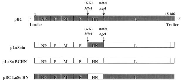
Full-length antigenomic cDNAs of NDV strains BC and LaSota, designated pBC and pLaSota, respectively, were cloned into the low-copy-number plasmid vector pBR322. These cDNA clones were used to rescue the recombinant viruses rBC and rLaSota, respectively, as described elsewhere (16, 21). A chimeric rBC virus containing the LaSota HN gene in place of its own and the reciprocal recombinant rLaSota virus containing the BC HN gene were generated for this study (Fig. 1). The unique MluI and AgeI sites in pBC and the MluI and SnaBI sites in pLaSota at the F-HN and HN-L intergenic regions were used for exchanging the HN genes between the full-length plasmids. The LaSota HN gene was amplified by PCR using the MluI (+) primer 5′ AACTACGCGTTGTAGATGACCAAAGGACGATATACGGGTAG 3′ and the AgeI (−) primer 5′ GATCACCGGTACGTATTTTGCCTTGTATCTCATTGCCACTTAC 3′. The BC HN gene was amplified by using the MluI (+) primer described above and the SnaBI (−) primer 5′ GATCTACGTATTTTGCCTTGTATCTCATTGCCACTTAC 3′. Restriction sites in each primer are indicated by boldfaced letters. The resulting full-length clones were designated pBC LaSoHN and pLaSo BCHN, respectively. These plasmids were used to recover the recombinant chimeric viruses rBC LaSoHN and rLaSo BCHN by using reverse-genetics procedures (21).
FIG. 1.
Schematic representation (not drawn to scale) of the strategy for exchange of the HN gene between the rBC and rLaSota viruses. Two unique restriction sites (MluI and AgeI in the rBC virus and MluI and SnaBI in the rLaSota virus), introduced into the HN intergenic region during construction of the full-length genome of NDV (16, 21), were used for the HN gene swap. The HN gene was exchanged as a single restriction fragment between the rBC and rLaSota viruses, by using the MluI and AgeI and the MluI and SnaBI sites, respectively. The first and last nucleotides of the entire full-length genome of the parental rBC virus are indicated. The approximate locations of the restriction sites used are indicated (arrows). NP, nucleocapsid protein; P, phosphoprotein; M, matrix protein; L, large polymerase protein.
RNA extraction and RT-PCR of recovered chimeric viruses.
The recovered recombinant chimeric viruses rBC LaSoHN and rLaSo BCHN were grown in the allantoic cavities of 9-day-old embryonated chicken eggs. After 2 days, the allantoic fluid was harvested and clarified, and the virus was purified as described above. Viral RNA was extracted from the recovered viruses by using TRIzol (Invitrogen) according to the manufacturer's instructions. Reverse transcription was carried out with the extracted RNA by using the Thermoscript RT kit (Invitrogen) to synthesize the first-strand cDNA. The genomes of recovered chimeric viruses were entirely sequenced after reverse transcription-PCR (RT-PCR) to confirm the presence of the substituted gene. The sequences of the forward and reverse primers used for RT-PCR of the substituted LaSota HN gene in the BC genome were 5′ AACTACGCGTTGTAGATGACCAAAGGACGATATACGGGTAG 3′ and 5′ GATCACCGGTACGTATTTTGCCTTGTATCTCATTGCCACTTAC 3′, respectively. The forward and reverse primers used for RT-PCR of the HN gene of pBC in the LaSota genome were 5′ AACTACGCGTTGTAGATGACCAAAGGACGATATACGGGTAG 3′ and 5′ GATCACCGGTACGTATTTTGCCTTGTATCTCATTGCCACTTAC 3′, respectively.
Growth characteristics of viruses.
The growth kinetics of rBC LaSoHN and rLaSo BCHN were determined under multiple-cycle growth conditions in DF1 cells. The virus was inoculated at a multiplicity of infection (MOI) of 0.01 into DF1 cells grown in DMEM with 5% FBS at 37°C. The medium of cells infected with rLaSo BCHN contained 1 μg of acetyl trypsin/ml. The supernatant was collected at 8-h intervals until 56 h postinfection (p.i.). The virus content in the samples was quantitated by plaque assays in DF 1 cells. Briefly, supernatants, collected from virus-inoculated samples earlier, were serially diluted, and 100 μl of each serial dilution was added per well of confluent DF1 cells. After 60 min of adsorption, cells were overlaid with DMEM (containing 2% FBS and 0.9% methylcellulose) and then incubated at 37°C for 3 to 4 days. The cells were then fixed with ethanol and stained with crystal violet for enumeration of plaques.
NA assay.
A fluorescence-based NA assay was carried out as described by Potier et al. (34). Briefly, serial twofold dilutions (undiluted to 1/1,024) of virus samples were prepared in 50-μl volumes of enzyme buffer (32.5 ml of 0.1 2-N-morpholinoethanesulfonic acid [MES; pH 6.5] and 4.0 ml of 0.1 M calcium chloride made up to a final volume of 100 ml with Milli-Q water) in a 96-well plate. Ten microliters of 12.5% (vol/vol) dimethyl sulfoxide was added to all wells of an assay plate (black 96-well plates; Microfluor, Franklin, Mass.). Ten microliters of each virus dilution was transferred in duplicate (two rows) to the assay plate, starting with the most dilute in column 11. Ten microliters of enzyme buffer alone was added to each well of column 12 (blank wells). The reaction was initiated by the addition of 30 μl of substrate mix [1 volume of 325 mM MES (pH 6.4), 3 volumes of 10 mM calcium chloride, and 2 volumes of 0.5 mM 2′-(4-methylumbelliferyl)-α-d-N-acetylneuraminic acid (MUN) (Sigma)] per well to give a final concentration of 100 μM MUN in the assay. The reaction mixture was incubated at 37°C for 15 min with shaking, and the reaction was terminated by the addition of 0.014 M sodium hydroxide in 83% (vol/vol) ethanol at 150 μl per well. The NA activities of the recovered viruses were measured as the amount of 4-methylumbelliferone released from the fluorogenic substrate, MUN. Released 4-methylumbelliferone was quantified by fluorometric determination with an excitation wavelength of 360 nm and an emission wavelength of 450 nm. Readings from the substrate blanks were subtracted from the virus sample readings, and the means of duplicate readings were calculated.
Hemadsorption (HAd) assay.
Virus was inoculated into confluent monolayers of Vero cells in 6-well plates at an MOI of 10. After 18 to 24 h, the medium was decanted and the cells were overlaid with guinea pig erythrocytes (RBCs) in PBS at a concentration of 108 cells/ml. The plates were kept at 4°C for 15 min. Unbound RBCs were removed by two washes with PBS. The RBCs bound to the virus-infected cells were lysed with 0.05 M ammonium chloride, and the released hemoglobin was measured at 549 nm in a spectrophotometer.
Fusion index assay.
The fusogenic abilities of the recombinant viruses were examined as described by Kohn (20). Virus was inoculated into confluent (106cells/ml) Vero cells in 6-well plates at an MOI of 0.1. Cells were maintained in 5% DMEM at 37°C under 5% CO2. After observation of cytopathic effects (CPE) within 48 to 72 h, the medium was removed, and cells were washed once with 0.02% EDTA and then incubated with 1 ml of EDTA for 2 min at room temperature. The cells were then washed with PBS and fixed with methanol for 20 min at room temperature. Cells were stained with hematoxylin-eosin (Hema 3). Fusion was quantitated by expressing the fusion index as the ratio of the total number of nuclei to the number of cells in which these nuclei were observed (i.e., the mean number of nuclei per cell). The NA, HAd, and fusion index values of all viruses were expressed as percentages of these values for the wild-type virus, rBC, which were considered to be 100%.
Immunohistochemistry.
The tissue tropism of recombinant NDV was examined in 9- to 11-day-old embryonated chicken eggs. Briefly, 103 PFU of each of the recombinant NDV strains was inoculated into developing chicken embryos by the chorioallantoic route. The infected embryos were chilled at 48 h p.i., and tissues such as the spleen, kidney, small intestine, large intestine, lung, heart, and brain were collected and fixed for cryosectioning and processing as described by Sheela et al. (40). The tissues were cryosectioned, and sections of 3- to 5-μm thickness were cut for immunohistochemistry. All tissues sampled were examined by immunohistochemistry using the following protocol to detect the viral HN protein. The sections were rehydrated in three changes of PBS (10 min each), followed by treatment with proteinase K (10 μg/ml) for 30 min at 37°C. The sections were then washed three times with PBS, fixed with ice-cold acetone, and washed again in PBS. To quench endogenous peroxides, the sections were flooded with 0.3% hydrogen peroxide in methanol for 30 min at room temperature. After being washed with three changes of PBS at 10-min intervals, the tissues were blocked with normal horse serum (VectaStain kit; Vector Laboratories, Burlingame, Calif.) for 30 min. The sections were then incubated with a 1:500 dilution of the primary NDV monoclonal antibody (23) cocktail against HN (10D11, AVS, 15C4, and B79) for 1 h at room temperature. After a wash, sections were incubated with the VectaStain secondary antibody (Vector Laboratories) for 30 min, as recommended by the manufacturer. After a further wash cycle, the sections were incubated with the VectaStain ABC reagent for 30 min. The substrate was NovaRed (Vector Laboratories). Sections were counterstained lightly with hematoxylin and were mounted with Vectamount (Vector Laboratories) for a permanent record.
MDT in chicken embryos.
The virulence of the recovered viruses was determined by the mean death time (MDT) in embryonated specific-pathogen-free (SPF) chicken eggs (1). A series of 10-fold dilutions of infected allantoic fluid was made in sterile PBS, and each dilution (0.1 ml) was inoculated into the allantoic cavity of five 9-day-old embryonated eggs. The eggs were incubated at 37°C and examined four times daily for 7 days. The time at which each embryo was first observed dead was recorded. The highest dilution at which all embryos died was considered the minimum lethal dose. The MDT recorded was the mean time (in hours) required for the minimum lethal dose to kill the embryos. The MDT has been used to classify NDV strains as velogenic (taking less than 60 h to kill), mesogenic (taking 60 to 90 h to kill), or lentogenic (taking more than 90 h to kill).
Pathogenicity studies in chickens.
To test the pathogenicities of the recovered viruses in vivo, an intracerebral pathogenicity index (ICPI) test was performed according to standard procedures (1). For ICPI, 103 PFU of each virus/chicken was inoculated intracerebrally into groups of 10 1-day-old SPF chicks. Inoculation was performed with a 27-gauge needle attached to a 1-ml stepper syringe dispenser that was set to dispense 0.05 ml of inoculum per inoculation. The birds were inoculated by inserting the needle up to the hub into the right or left rear quadrant of the cranium. The birds were observed for clinical symptoms and mortality once every 12 h for 8 days. Equal numbers of chickens were used in each experiment. Each experiment included mock-inoculated controls that received a similar volume of sterile PBS by the respective routes. ICPI values were calculated as described by Alexander (1). Briefly, the birds were scored daily: 0 if normal, 1 if sick, and 2 if dead. The ICPI value was the mean score per bird per observation. Highly virulent (velogenic) viruses give values approaching 2; avirulent (lentogenic) viruses give values close to 0. To assess the survival rates of birds inoculated with the recombinant viruses, 1-day-old chicks were inoculated intracerebrally with 103 PFU/chick. Infected chicks were observed daily for 8 days for signs of paralysis and death. The percent survivability of inoculated chicks was plotted over time.
RESULTS
Recovery of recombinant parental and chimeric NDV.
The recovery of recombinant NDV from infectious cDNA clones, derived from a mesogenic strain of NDV, BC, and a lentogenic NDV strain, LaSota, has been reported earlier from our laboratory (16, 21). We used the established reverse-genetics system to examine the role of the HN gene in the virulence of NDV in this study. The HN genes of the avirulent recombinant NDV strain rLaSota and a virulent NDV strain were exchanged in an otherwise identical genomic background. To ensure the presence of the intended HN gene swaps, the entire cDNA clone of each chimeric virus was sequenced by using the Big Dye terminator (Applied Biosystems) method. The supernatants from transfected HEp2 cells were passaged twice in DF1 cells to recover infectious NDV. Virus-positive supernatants were used to inoculate the allantoic cavities of 9- to 11-day-old embryonated SPF eggs. The allantoic fluid harvested after embryo deaths was analyzed in a hemagglutination assay. Allantoic fluid with a positive hemagglutination titer was used for the isolation of viral RNA, followed by sequence analysis of an RT-PCR fragment that covered the HN gene swap region. Nucleotide sequencing confirmed that the HN gene exchanges introduced in rBC LaSoHN and rLaSo BCHN were retained in the recovered viruses. The recovered rBC virus bearing the LaSota HN gene in place of its own HN gene was designated rBC LaSoHN, and the rLaSota virus with its HN gene replaced by the HN gene of BC virus was designated rLaSo BCHN. These exchanges were stable and were seen even after 5 sequential passages of these viruses at a high MOI in DF1 cells and in 9-day-old embryonated chicken eggs (data not shown).
Growth of recombinant viruses in vitro.
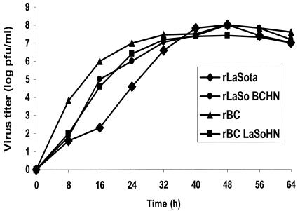
The growth characteristics of the parental and chimeric viruses were assessed by multistep growth curves (Fig. 2). The differences in the growth kinetics of HN gene swap viruses were observed until 16 h p.i. Compared to that of its parental rLaSota virus, the growth rate of the rLaSo BCHN virus was more than twofold higher at 16 h p.i., while the growth rate of the rBC LaSoHN virus was 1.4-fold lower than that of its parental rBC virus. The parental and gene swap viruses grew to similar titers after 32 h p.i. These studies showed the relevance of HN to virus growth in vitro. No differences were observed between the plaque sizes of recombinant HN gene swap viruses and their parental viruses. These results indicated that heterotypic F and HN proteins were compatible in the chimeric viruses.
FIG. 2.
Growth kinetics of parental and chimeric viruses in chicken embryo fibroblast (DF1) cells. Multiple-cycle growth conditions were used to assess the differences in the growth of these viruses. DF1 cells were infected with virus at an MOI of 0.01. Supernatants collected at 8-h intervals were used for determining virus titers by a plaque assay. Data are means from three independent experiments.
Biological activities of mutant viruses.
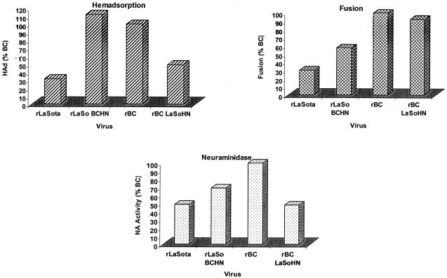
We analyzed whether the origin of the HN protein determines the biological activities of NDV. Figure 3 shows the NA, HAd, and fusion activities of the parental and chimeric viruses. The biological activity of each chimeric or parental virus is shown as a percentage of that of the rBC virus, whose biological activities were considered to be 100%. The NA and HAd activities of rBC LaSoHN were only 50% of those of the parental rBC virus, while the rLaSo BCHN virus showed an 80% increase in HAd activity and a 20% increase in NA activity over those of the rLaSota virus. The fusogenic abilities of the parental and chimeric viruses did not differ significantly. These studies reiterate the importance of the HN protein in the attachment and NA functions of NDV in the context of a viral infection. These differences in attachment and elution may translate into differences in viral growth kinetics in vivo. Furthermore, these results demonstrate that a combination of F and HN proteins derived from heterologous viruses are fully functional in inducing fusion.
FIG. 3.
In vitro biological activities of parental and HN chimeric viruses. The HAd activities, NA activities, and fusogenicities of the parental and HN chimeric viruses were examined. The biological activities of these viruses are expressed as percentages of those of the rBC virus, whose activities were considered to be 100%. HAd was measured as the percentage of the hemoglobin released from guinea pig RBCs attached to infected cells. The NA activities of purified parental and chimeric viruses were measured by a fluorometric assay. The fusion index was estimated in Vero cells infected at an MOI of 0.1. Cells were stained with hematoxylin-eosin, and the fusion index was calculated. The fusion index is the ratio of the total number of nuclei to the number of cells in which the nuclei were observed, i.e., the mean number of nuclei per cell. Data are means from three independent experiments.
Cytopathogenicity and tissue tropism.
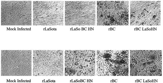
The CPE of the HN swap viruses differed significantly. At 48 h p.i., the rLaSo BCHN virus showed more extensive CPE in primary CEF cells than the rLaSota virus (Fig. 4). Similarly, the rBC LaSoHN virus showed markedly less CPE than the rBC virus. The rapid destruction of cell monolayers by the rLaSo BCHN virus was consistent with the increases in the receptor recognition and NA activities of the virus. For the rBC LaSoHN virus, the reductions in the receptor binding and NA activities may have resulted in the differential CPE observed in CEF cells.
FIG. 4.
Cytopathogenicities of parental and chimeric viruses in DF1 cells. Cells were infected with virus at an MOI of 0.1. After 24 and 48 h, the CPE of each virus-infected monolayer were examined under an inverted microscope. The differences in the CPE of each virus-infected monolayer at 24 and 48 h p.i. are shown. (Top) CPE observed at 24 h p.i. for the indicated viruses. (Bottom) CPE at 48 h p.i.
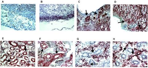
On the other hand, the tissue distribution of the chimeric viruses after chorioallantoic inoculation of 10-day-old chicken embryos showed that the HN protein probably determines tissue tropism. The chimeric rBC LaSoHN virus and the lentogenic rLaSota virus did not reach the brains of inoculated embryos, while with the rBC and rLaSo BCHN viruses; NDV-specific antigen could be demonstrated in different parts of the brain. (Fig. 5). NDV antigens were localized in the alveolar epithelium and peribronchiolar space with the rBC and rLaSo BCHN viruses, while chicken embryos inoculated with the rLaSota or rBC LaSoHN virus showed discrete staining of the bronchial and bronchiolar epithelium. The rLaSota and rBC LaSoHN viruses were also distributed in the villous epithelial cells of the small and large intestines, in addition to the lungs. There was a wide dissemination of the rBC and rLaSo BCHN viruses in chicken embryonic tissues such as the lung, brain, kidney, intestines and spleen. The extent and distribution of antigen-positive areas in these tissues were marked and extensive.
FIG. 5.
Tissue tropism of recombinant NDVs in developing chicken embryos. Groups of 9- to 11-day-old chicken embryos were inoculated with 103 PFU of each of the parental and HN chimeric NDVs by the chorioallantoic route. Frozen sections from infected embryos were processed for immunohistochemistry after 48 h. Tissue sections were stained with the primary NDV monoclonal antibody cocktail followed by VectaStain (Vector Laboratories) and the VectaStain ABC reagent. The substrate was NovaRed (Vector Laboratories). Representative sections from each virus-infected embryo are shown. While the rLaSota and rBC LaSoHN viruses showed no staining in the cerebellum (A and B), the cerebellum showed extensive antigen-specific red staining (arrows) in the molecular layer with the rBC or rLaSo BCHN virus (C and D). Viral antigens were localized in the bronchial and bronchiolar epithelium with the rLaSota (E) or rBC LaSoHN (F) virus, but diffuse interstitial staining was noticed with the rBC (G) or rLaSo BCHN (H) virus. Magnification, ×14.
Pathogenicity studies of mutant viruses.
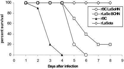
We wanted to determine whether the differences in in vitro biological characteristics of the chimeric viruses would translate into increased or decreased virulence in chickens or chicken embryos. The MDT for rLaSo BCHN was decreased to 84 h from that of the parental rLaSota virus, for which the MDT was 96 h, indicating an enhancement in the virulence of the chimeric virus. The MDT for the rBC virus was 62 h, while rBC LaSoHN took 72 h to kill embryonated SPF eggs, indicating the degree of attenuation imparted by the LaSo HN to the rBC genetic background. The ICPI values indicated altered virulence properties in chimeric viruses relative to parental viruses (Table 1). The recombinant rLaSo BCHN virus had an ICPI of 0.75 while its lentogenic parental strain, rLaSota, had an ICPI of 0.00, despite the fact that they were isogenic except for the swapped HN gene. The rBC LaSoHN virus, on the other hand, showed a reduced ICPI value of 1.02 relative to its mesogenic parental virus, rBC, with an ICPI of 1.58. Figure 6 shows the survivability of 1-day-old chicks inoculated intracerebrally with the recombinant viruses. The rLaSota virus induced 0% mortality at 8 days after inoculation. The rLaSo BCHN virus showed a dramatic increase in virulence relative to its parent, rLaSota, with only 20% of the birds surviving on day 8. After inoculation with the rBC virus, 20% of the birds survived on day 3, and by day 4 all the inoculated birds were dead. In contrast, the rBC LaSoHN virus showed decreased virulence relative to its parent, rBC, with 20% of the birds surviving on day 5 and 100% mortality on day 6. The gain or loss of virulence in these viruses, therefore, appears to be mediated solely by the HN gene.
TABLE 1.
HN chimeras of NDV and virus pathogenicity in vivoa
The virulence of the mutant and parental viruses was evaluated by the ICPI in 1-day-old-chicks and by MDT in embryonated chicken eggs.
Determined by inoculating groups of 10 1-day-old SPF chicks with 103 PFU of virus/bird via the intracerebral route. Birds were observed daily for 8 days; at each observation, they were scored 0 if normal, 1 if sick, and 2 if dead. The ICPI value is the mean score per bird per observation. Highly virulent virusees give values approaching 2, while avirulent viruses give values approaching 0.
For the minimum lethal dose to kill 9-day-old embryonated chicken eggs inoculated with virus by the allantoic route. Highly virulent viruses take less than 60 h to kill the embryos, whereas avirulent viruses take more than 90 h.
FIG. 6.
Survivability of 1-day-old chicks inoculated with parental or HN chimeric viruses. SPF 1-day-old chicks were inoculated intracerebrally with 103 PFU/chick. Infected chicks were observed daily for 8 days for signs of paralysis and death. Percent survivors for each virus is plotted over time.
DISCUSSION
The HN gene plays an important role in the pathogenesis of paramyxoviruses (11). The HN gene of NDV is a multifunctional protein with receptor recognition, NA activity on sialic acid-containing receptors, and fusion promotion (22). Transfection studies with HN mutants of NDV have highlighted the importance of the different regions of the HN protein in the biological activities of the protein (9, 26, 27). However, the contribution of the HN gene to NDV pathogenesis in the context of virus infection is not known. In this study, by use of reverse-genetics techniques, a chimeric rBC virus (a recombinant moderately virulent, mesogenic NDV strain) containing the HN gene of LaSota (a recombinant avirulent, lentogenic NDV strain) in place of its own and the reciprocal recombinant, consisting of rLaSota bearing the BC HN gene, were generated in order to assess the effect of HN gene substitution on viral pathogenesis. The hypothesis tested in the present study was that differences in biological functions of HN protein displayed by different strains of NDV in vitro are important determinants of pathogenesis in vivo. The pathogenicity of the chimeric NDV was examined in the natural host, chickens. The results of this study indicated that the in vitro biological characteristics of the HN are good indicators of in vivo pathogenicity and that the HN gene makes an important contribution to the overall virulence of NDV isolates.
Sequence analysis of the chimeric viruses confirmed the presence of the substituted HN gene. These chimeric viruses reached similar titers, with similar kinetics, compared to those viruses with homologous proteins. This finding suggested that heterologous pairs of HN and F proteins were fully functional despite being derived from different viruses. This functionality probably reflects the high level of amino acid sequence identity between these two different NDV strains (98.4% in F and 97.1% in HN). The BC and LaSota strains used in this study are phylogenetically closely related (2). Therefore, it would be interesting to examine whether the HN and F protein pairs derived from distantly related NDV strains are also functional in vitro and in vivo.
In vitro studies with these viruses yielded results that supported the hypothesis of the importance of the HN gene in NDV virulence. The parental rBC virus showed higher HAd and NA activities than the parental rLaSota virus. Interestingly, the rLaSo BCHN virus also showed higher HAd and NA activities than its parent, rLaSota. These activities were lower in the rBC LaSoHN virus than in the parental rBC virus. These results indicated that the magnitude of the HAd and NA activities of NDV are determined solely by the amino acid differences in the HN protein. The fusion promotion activity of HN appeared to be independent of the HAd and NA activities. Despite the fact that the HAd and NA activities of the BC and LaSota viruses were at different levels, the heterotypic HN proteins in the chimeric viruses were fully functional in fusion promotion. Our results are in agreement with those of Sergel et al. (38), who have shown that the fusion promotion activity of HN did not correlate with the level of NA activity.
Several reports have suggested that a type-specific functional interaction between the F and HN proteins of paramyxoviruses is required for cell fusion (10, 15, 43). Studies of hybrid HN proteins by three different laboratories have all implicated the membrane-proximal ectodomain in virus-specific fusion promotion activity (9, 44, 45). With this approach, the specificity of the NDV F protein has been mapped to amino acids 55 to 141 of the NDV HN protein, a sequence adjacent to the HN protein transmembrane domain (9). However, we have demonstrated in this study that chimeric NDVs containing heterotypic F and HN proteins were compatible, since they were derived from phylogenetically closely related virus strains (2). It has been suggested that a specific interaction occurs between the F protein HR2 domain and the HN protein domain from amino acids 124 to 152 for fusion promotion (13). There was only one amino acid difference, from phenylalanine to tyrosine (F132I), between the BC and LaSota viruses in the proposed F and HN interacting domain, which may be the reason for the heterotypic interaction of the F and HN proteins from distantly related strains of NDV.
It has been proposed that for influenza virus, the NA activity may be important for removing respiratory tract mucin sialic acids, allowing the virus to reach target cells (7). There is a precedent for decreased virulence as a result of decreased NA activity, in influenza viruses in animals (30, 46). In contrast, the C-28 variant of human parainfluenza virus, with deficient NA activity, caused more-intense disease in cotton rats (35). Examination of a temperature-sensitive NDV variant and two sequential revertant viruses revealed that alterations in NA can compensate for alterations in binding (39, 42). The original NDV variant, with an amino acid substitution at position 129, was deficient in binding erythrocytes; a second mutation, at position 175, reduced NA activity but restored binding; the third sequential mutation, at position 193, partially restored NA activity. Thus, the balance between the receptor binding and NA activities appears to be critical to the virulence of NDV. The early induction of CPE in CEF cells by rLaSo BCHN and the diminished CPE of rBC LaSoHN, relative to their parental types with homologous HN, in our study might have resulted from these changes in receptor recognition and NA activities. Increased attachment and NA activities are consistent with the early growth and CPE of those viruses containing virulent HN and with a delay in reaching peak titers and inducing CPE for those viruses containing avirulent HN.
A small amino acid motif in the fusion protein precursor (F0), termed the cleavage site, has been identified as a pathotype determinant. Thus, viruses with multibasic amino acids at the cleavage site are considered virulent, and those characterized by monobasic amino acids at the cleavage site are avirulent (3, 4, 5, 6). Cleavage of F0 polypeptides into F2 and F1 is essential for a virus particle to become infectious (31). In chickens, the F protein of pathogenic NDV is cleaved by ubiquitous intracellular furin-like proteases, and the F protein of nonpathogenic viruses is cleaved only by trypsin-like proteases secreted from a limited number of tissues (12). Thus, virulent viruses can spread rapidly throughout the host and cause disease. In comparison, the avirulent motif can be cleaved only in the respiratory tract and gut, where trypsin-like proteases are available. The replication of these viruses is, therefore, restricted to these organs (31). However, in our studies with chicken embryos, where F proteins with either mono- or multibasic amino acids at the cleavage site are cleaved with equal efficiency due to the availability of trypsin-like proteases, the HN gene sequence appears to determine the tropism of the virus to different tissues and organs. Especially, the discrete replication of viruses with avirulent HN in bronchial and bronchiolar epithelium, the widespread distribution of the viruses with virulent HN in the lung parenchyma, and the systemic distribution of the viruses with virulent type HN in several organs, including the brain, indicate that the HN protein, independently of F protein cleavability, determines viral tropism in chicken embryos. This may be the result of differences in receptor recognition and NA activities between the HN proteins of the BC and LaSota viruses. It would be interesting to study the tropism of these viruses in chickens, where F cleavability may play a major role in virulence via natural routes of infection. A set of carefully defined mutations in the HN protein may indicate the amino acid residues involved in the tropism and virulence of NDV.
The pathogenicity of the chimeric viruses was assessed by ICPI and MDT tests. Interestingly, the results showed that the chimeric rBC virus bearing the HN gene of LaSota (the rBC LaSoHN virus) was markedly less virulent than the parental rBC virus, and the reciprocal chimeric rLaSo BCHN virus was also mildly virulent, compared to its parental rLaSota virus. Chicks inoculated with rLaSo BCHN had lower survival rates than chicks inoculated with rLaSota, and chicks inoculated with rBC LaSoHN had higher survivability than those inoculated with rBC. MDT results also showed a gain of virulence for rLaSo BCHN and a loss of virulence for rBC LaSoHN, relative to rLaSota and rBC, respectively. Thus, when the HN gene of the avirulent LaSota strain replaced the HN gene of the virulent BC strain in the BC virus, the chimeric virus (rBC LaSoHN) showed reduced virulence. Exactly the opposite effect was observed for the chimeric virus rLaSo BCHN, with enhanced virulence. This “gain or loss of function” between the chimeric and parental virus strains was a further indication that the differences observed were associated with the HN protein.
It has been demonstrated previously that the cleavability of the F protein is a major determinant of NDV virulence (32, 33). By creating recombinant NDV in which the V protein has been deleted, researchers have shown that the V protein is an important determinant of virulence (17, 28). These results further support our hypothesis that the virulence of NDV is multigenic. Studies with influenza A viruses have also suggested the multifactorial nature of viral pathogenicity (24, 37).
In summary, using reciprocal HN gene swaps between avirulent and virulent NDV strains in an otherwise identical genetic background, we have demonstrated that the reduced receptor recognition and NA activities inherent to avirulent HN translate into differences in the tropism and virulence of the virus. Chimeric viruses bearing avirulent HN remained restricted in growth to limited tissues and organs, besides being attenuated in virulence relative to the parental type. On the other hand, virulent HN imparted the ability to spread systemically and enhanced the virulence of an otherwise avirulent NDV.
It will be interesting to study the molecular mechanism by which the HN gene determines tropism and virulence. The terminal globular head of the HN protein contains the active sites involved in virus attachment and NA activities (8, 18, 19, 29, 48). Recent results suggest that a specific interaction between the F protein HR2 domain and the HN protein membrane-proximal ectodomain spanning amino acids 124 to 152 occurs for fusion promotion (13). There are only 17 amino acid differences between the HN proteins of the BC and LaSota viruses. The amino acid differences are spread throughout the HN protein. However, most of the differences are present in the globular head region. With the availability of an established reverse-genetics system for avirulent and virulent NDV strains, it would be of special interest to mutate those residues on the HN protein that are involved in its biological functions and observe the effects of these mutations on virus infectivity in vitro and in vivo.
Acknowledgments
We thank Peter Savage for excellent technical assistance.
This work was partially supported by U.S. Department of Agriculture grant 2002-35204-1601.
REFERENCES
- 1.Alexander, D. J. 1989. Newcastle disease, p. 114-120. In H. G. Purchase, L. H. Arp, C. H. Domermuth, and J. E. Pearson (ed.), A laboratory manual for the isolation and identification of avian pathogens, 3rd ed. American Association for Avian Pathologists, Inc., Kennett Square, Pa.
- 2.Chare, E. R., E. A. Gould, and E. C. Holmes. 2003. Phylogenetic analysis reveals a low rate of homologous recombination in negative-sense RNA viruses. J. Gen. Virol. 84:2691-2703. [DOI] [PubMed] [Google Scholar]
- 3.Collins, M. S., J. B. Bashiruddin, and D. J. Alexander. 1993. Deduced amino acid sequences at the fusion protein cleavage site of Newcastle disease viruses showing variation in antigenicity and pathogenicity. Arch. Virol. 128:363-370. [DOI] [PubMed] [Google Scholar]
- 4.Collins, M. S., I. Strong, and D. J. Alexander. 1994. Evaluation of the molecular basis of the pathogenicity of the variant Newcastle disease viruses termed ‘pigeon PMV-1 viruses.’ Arch. Virol. 134:403-411. [DOI] [PubMed] [Google Scholar]
- 5.Collins, M. S., I. Strong, and D. J. Alexander. 1996. Pathogenicity and phylogenetic evaluation of the variant Newcastle disease viruses termed ‘pigeon PMV-1’ viruses based on the nucleotide sequence of the fusion protein gene. Arch. Virol. 141:635-647. [DOI] [PubMed] [Google Scholar]
- 6.Collins, M. S., S. J. Govey, and D. J. Alexander. 2003. Rapid assessment of the virulence of Newcastle disease virus isolates using the ligase chain reaction. Arch. Virol. 148:1851-1862. [DOI] [PubMed] [Google Scholar]
- 7.Colman, P., and C. Ward. 1985. Structure and diversity of influenza virus neuraminidase. Curr. Top. Microbiol. Immunol. 114:178-254. [DOI] [PubMed] [Google Scholar]
- 8.Crennell, S., T. Takimoto, A. Portner, and G. Taylor. 2000. Crystal structure of the multifunctional paramyxovirus hemagglutinin-neuraminidase. Nat. Struct. Biol. 7:1068-1074. [DOI] [PubMed] [Google Scholar]
- 9.Deng, R., Z. Wang, A. M. Mirza, and R. M. Iorio. 1995. Localization of a domain on the paramyxovirus attachment protein required for the promotion of cellular fusion by its homologous fusion protein spike. Virology 209:457-469. [DOI] [PubMed] [Google Scholar]
- 10.Deng, R., Z. Wang, P. Mahan, M. Marinello, A. Mirza, and R. M. Iorio. 1999. Mutations in the Newcastle disease virus hemagglutinin-neuraminidase protein that interfere with its ability to interact with homologous F protein in the promotion of fusion. Virology 253:43-54. [DOI] [PubMed] [Google Scholar]
- 11.Duprex, W. P., I. Duffy, S. McQuaid, L. Hamill, S. L. Cosby, M. A. Billeter, J. Schneider-Schaulies, V. ter Meulen, and B. K. Rima. 1999. The H gene of rodent brain-adapted measles virus confers neurovirulence to the Edmonston vaccine strain. J. Virol. 73:6916-6922. [DOI] [PMC free article] [PubMed] [Google Scholar]
- 12.Fujii, Y., T. Sakaguchi, K. Kiyotani, and T. Yoshida. 1999. Comparison of substrate specificities against the fusion glycoproteins of virulent Newcastle disease virus between a chick embryo fibroblast processing protease and mammalian subtilisin-like proteases. Microbiol. Immunol. 43:133-140. [DOI] [PubMed] [Google Scholar]
- 13.Gravel, K. A., and T. G. Morrison. 2003. Interacting domains of the HN and F proteins of Newcastle disease virus. J. Virol. 77:11040-11049. [DOI] [PMC free article] [PubMed] [Google Scholar]
- 14.Hernandez, L. D., L. R. Hoffmann, T. G. Wolfsberg, and J. M. White. 1996. Virus-cell and cell-cell fusion. Annu. Rev. Cell Biol. 12:627-661. [DOI] [PubMed] [Google Scholar]
- 15.Hu, X. L., R. Ray, and R. W. Compans. 1992. Functional interactions between the fusion protein and hemagglutinin-neuraminidase of human parainfluenza viruses. J. Virol. 66:1528-1534. [DOI] [PMC free article] [PubMed] [Google Scholar]
- 16.Huang, Z., S. Krishnamurthy, A. Panda, and S. K. Samal. 2001. High-level expression of a foreign gene from the most 3′-proximal locus of a recombinant Newcastle disease virus. J. Gen. Virol. 82:1729-1736. [DOI] [PubMed] [Google Scholar]
- 17.Huang, Z., S. Krishnamurthy, A. Panda, and S. K. Samal. 2003. Newcastle disease virus V protein is associated with viral pathogenesis and functions as an alpha interferon antagonist. J. Virol. 77:8676-8685. [DOI] [PMC free article] [PubMed] [Google Scholar]
- 18.Iorio, R. M., R. J. Syddall, R. L. Glickman, A. M. Riel, J. P. Sheehan, and M. A. Bratt. 1989. Identification of amino acid residues important to the neuraminidase activity of the HN glycoproteins of Newcastle disease virus. Virology 173:196-204. [DOI] [PubMed] [Google Scholar]
- 19.Iorio, R. M., R. L. Glickman, A. M. Riel, J. P. Sheehan, and M. A. Bratt. 1989. Functional and neutralization profile of seven overlapping antigenic sites on the HN glycoproteins of Newcastle disease virus: monoclonal antibodies to some sites prevent viral attachment. Virus Res. 13:245-261. [DOI] [PubMed] [Google Scholar]
- 20.Kohn, A. 1965. Polykaryocytosis induced by Newcastle disease virus in monolayers of animal cells. Virology 26:228-245. [DOI] [PubMed] [Google Scholar]
- 21.Krishnamurthy, S., Z. Huang, and S. K. Samal. 2000. Recovery of a virulent strain of Newcastle disease virus from cloned cDNA: expression of a foreign gene results in growth retardation and attenuation. Virology 278:168-182. [DOI] [PubMed] [Google Scholar]
- 22.Lamb, R. A., and D. Kolakofsky. 1996. The paramyxoviruses, p. 577-604. In B. N. Fields, D. M. Knipe, and P. M. Howley (ed.), Fields virology, 3rd ed. Lippincott-Raven Publishers, Philadelphia, Pa.
- 23.Lana, D. P., D. B. Snyder, D. J. King, and W. W. Marquardt. 1988. Characterization of a battery of monoclonal antibodies for differentiation of Newcastle disease virus and pigeon paramyxovirus-1 strains. Avian Dis. 32:273-281. [PubMed] [Google Scholar]
- 24.Mayer, V., J. L. Schulman, and E. D. Kilbourne. 1973. Nonlinkage of neurovirulence exclusively to viral hemagglutinin or neuraminidase in genetic recombinants of A/NWS (H0N1) influenza virus. J. Virol. 11:272-278. [DOI] [PMC free article] [PubMed] [Google Scholar]
- 25.Mayo, M. A. 2002. A summary of taxonomic changes recently approved by ICTV. Arch. Virol. 147:1655-1656. [DOI] [PubMed] [Google Scholar]
- 26.McGinnes, L. W., and T. G. Morrison. 1994. Modulation of the activities of HN protein of Newcastle disease virus by non-conserved cysteine residues. Virus Res. 34:305-316. [DOI] [PubMed] [Google Scholar]
- 27.McGinnes, L. W., and T. G. Morrison. 1995. The role of individual oligosaccharide chains in the activities of the HN glycoprotein of Newcastle disease virus. Virology 212:398-410. [DOI] [PubMed] [Google Scholar]
- 28.Mebatsion, T., S. Verstegen, L. T. C. de Vaan, A. Romer-Oberdorfer, and C. C. Schrier. 2001. A recombinant Newcastle disease virus with low-level V protein expression is immunogenic and lacks pathogenicity for chicken embryos. J. Virol. 75:420-428. [DOI] [PMC free article] [PubMed] [Google Scholar]
- 29.Mirza, A. M., R. T. Deng, and R. M. Iorio. 1994. Site-directed mutagenesis of a conserved hexapeptide in the paramyxovirus hemagglutinin-neuraminidase glycoproteins: effects on antigenic structure and function. J. Virol. 68:5093-5099. [DOI] [PMC free article] [PubMed] [Google Scholar]
- 30.Muster, T., E. K. Subbarao, M. Enami, B. R. Murphy, and P. Palase. 1991. An influenza A virus containing influenza B virus 5′ and 3′ noncoding regions on the neuraminidase gene is attenuated in mice. Proc. Natl. Acad. Sci. USA 88:5177-5181. [DOI] [PMC free article] [PubMed] [Google Scholar]
- 31.Nagai, Y. 1995. Virus activation by host proteinases. A pivotal role in the spread of infection, tissue tropism and pathogenicity. Microbiol. Immunol. 39:1-9. [DOI] [PubMed] [Google Scholar]
- 32.Panda, A., Z. Huang., S. Elankumaran, D. D. Rockemann, and S. K. Samal. 2004. Role of fusion protein cleavage site in the virulence of Newcastle disease virus. Microb. Pathog. 36:1-10. [DOI] [PMC free article] [PubMed] [Google Scholar]
- 33.Peeters, B. P., O. S. de Leeuw, G. Koch, and A. L. Gielkens. 1999. Rescue of Newcastle disease virus from cloned cDNA: evidence that cleavability of the fusion protein is a major determinant for virulence. J. Virol. 73:5001-5009. [DOI] [PMC free article] [PubMed] [Google Scholar]
- 34.Potier, M., L. Mameli, M. Belishem, L. Dallaire, and S. B. Melancon. 1979. Fluorometric assay of neuraminidase with a sodium (4-methylumbelliferyl-α-d-N-acetylneuraminate) substrate. Anal. Biochem. 94:287-296. [DOI] [PubMed] [Google Scholar]
- 35.Prince, G. A., M. G. Ottolini, and A. Moscona. 2001. Contribution of the human parainfluenza virus type 3 HN-receptor interaction to pathogenesis in vivo. J. Virol. 75:12446-12451. [DOI] [PMC free article] [PubMed] [Google Scholar]
- 36.Rott, R., and H. D. Klenk. 1988. Molecular basis of infectivity and pathogenicity of Newcastle disease virus, p. 98-112. In D. J. Alexander (ed.), Newcastle disease. Kluwer Academic Publishers, Boston, Mass.
- 37.Schlesinger, R. W., P. J. Husak, G. L. Bradshaw, and P. P. Panayotov. 1998. Mechanisms involved in natural and experimental neuropathology of influenza viruses: evidence and speculation. Adv. Virus Res. 50:289-379. [DOI] [PubMed] [Google Scholar]
- 38.Sergel, T., L. W. McGinnes, and T. G. Morrison. 2000. A single amino acid change in the Newcastle disease virus fusion protein alters the requirement for HN protein in fusion. J. Virol. 74:5101-5107. [DOI] [PMC free article] [PubMed] [Google Scholar]
- 39.Sheehan, J., and R. Iorio. 1992. A single amino acid substitution in the hemagglutinin-neuraminidase of Newcastle disease virus results in a protein deficient in both functions. Virology 189:778-781. [DOI] [PubMed] [Google Scholar]
- 40.Sheela, R. R., U. Babu, J. Mu, S. Elankumaran, D. A. Bautista, R. B. Raybourne, R. A. Heckert, and W. Song. 2003. Immune responses against Salmonella enterica serovar Enteritidis infection in virally immunosuppressed chickens. Clin. Diagn. Lab. Immunol. 10:670-679. [DOI] [PMC free article] [PubMed] [Google Scholar]
- 41.Simons, K., and H. Garoff. 1980. The budding mechanisms of enveloped animal viruses. J. Gen. Virol. 50:1-21. [DOI] [PubMed] [Google Scholar]
- 42.Smith, G. W., and L. E. Hightower. 1983. Biological consequences of neuraminidase deficiency in Newcastle disease virus. J. Virol. 47:385-391. [DOI] [PMC free article] [PubMed] [Google Scholar]
- 43.Stone-Hulslander, J., and T. G. Morrison. 1997. Detection of an interaction between the HN and F proteins in Newcastle disease virus-infected cells. J. Virol. 71:6287-6295. [DOI] [PMC free article] [PubMed] [Google Scholar]
- 44.Tanabayashi, K., and R. W. Compans. 1996. Functional interaction of paramyxovirus glycoproteins: identification of a domain in Sendai virus HN which promotes cell fusion. J. Virol. 70:6112-6118. [DOI] [PMC free article] [PubMed] [Google Scholar]
- 45.Tsurudome, M., M. Kawano, T. Yuasa, M. Nishio, H. Komada, and Y. Ito. 1995. Identification of regions on the hemagglutinin-neuraminidase protein of human parainfluenza virus type 2 important for promoting cell fusion. Virology 213:190-203. [DOI] [PubMed] [Google Scholar]
- 46.von Itzstein, M., W. Y. Wu, G. B. Kok, M. S. Pegg, J. C. Dyason, B. Jin, T. V. Phan, M. L. Smythe, H. F. White, S. W. Oliver, P. M. Colman, J. N. Varghese, D. M. Ryan, J. M. Woods, R. C. Bethel, V. J. Hothman, J. M. Cameron, and C. R. Penn. 1993. Rational design of potent sialidase-based inhibitors of influenza virus replication. Nature 363:418-423. [DOI] [PubMed] [Google Scholar]
- 47.Werner, O., A. Romer-Oberdorfer, B. Kollner, R. J. Manvell, and D. J. Alexander. 1999. Characterization of avian paramyxovirus type I strains isolated in Germany during 1992 to 1996. Avian Pathol. 28:79-88. [DOI] [PubMed] [Google Scholar]
- 48.Yusoff, K., M. Nesbit, H. McCartney, P. T. Emmerson, and A. C. R. Samson. 1988. Mapping of three antigenic sites on the hemagglutinin-neuraminidase protein of Newcastle disease virus. Virus Res. 11:319-333. [DOI] [PMC free article] [PubMed] [Google Scholar]


