Abstract
Nonmissile penetrating intracranial injuries are uncommon events in modern times. Most reported cases describe trajectories through the orbit, skull base foramina, or areas of thin bone such as the temporal squama. Patients who survive such injuries and come to medical attention often require foreign body removal. Critical neurovascular structures are often damaged or at risk of additional injury resulting in further neurological deterioration, life-threatening hemorrhage, or death. Delayed complications can also be significant and include traumatic pseudoaneurysms, arteriovenous fistulas, vasospasm, cerebrospinal fluid leak, and infection. Despite this, given the rarity of these lesions, there is a paucity of literature describing the management of neurovascular injury and skull base repair in this setting. The authors describe three cases of nonmissile penetrating brain injury and review the pertinent literature to describe the management strategies from a contemporary cerebrovascular and skull base surgery perspective.
Keywords: Penetrating trauma, brain, skull base, pseudoaneurysm
Penetrating nonmissile stab wounds to the brain are relatively uncommon due to the protective effect of the skull.1 The majority of cases involve penetration through the orbit, temporal squama, or large foramina or areas of thin bone within the skull base. Although these injuries are rare, they were first described in the literature as early as 1806.2 Penfield reexamined these injuries by studying pathological features of experimental stab wounds using a cannula.2,3 Pilcher in 1936 compiled a list of objects that had penetrated the brain, including knives, pitchforks, crochet hooks, knitting needles, breech pins, umbrella bibs, crowbars, and iron rods.4 The current list of objects reported has expanded and now includes a toilet brush handle,5 arrows,6 chopsticks,7 flatware,8 screwdrivers,1 keys,9 car antenna aerials,10 and scissors.11
Due to the rarity of these events, there is not substantial literature on the management and outcome of these patients. We report three cases of nonmissile penetrating brain injuries at our institution and review the pertinent literature to highlight the proper management of these cases with the goal to improve outcome and minimize short- and long-term complications that may result from these injuries.
Description of Cases
Case Report 1
History and Presentation
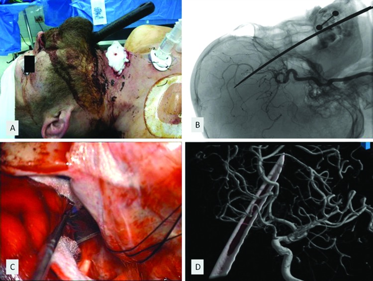
A 31-year-old man presented to our emergency room after a failed suicide attempt with a transverse neck laceration, exposing the trachea and larynx, and retained intracranial knife entering through the submental area into the cranium (Fig. 1A and 1B). On neurological examination, the patient's pupils were equal and reactive to light. He had full strength and purposeful movements in all extremities. A noncontrast head computed tomography (CT) scan showed intraventricular and subarachnoid hemorrhage in the basal cisterns adjacent to the retained knife. There was no large hematoma. A CT angiogram of his head and neck showed partial occlusion of the distal right A2. He underwent digital subtraction angiography to better assess the intracranial vasculature in relation to the knife's edge, and injury was again seen to the distal right A2 branch of the anterior cerebral artery (ACA) without evidence of further vascular injury (Fig. 1D).
Figure 1.
(A) Preoperative photograph of a patient with retained intracranial knife, zone 2 neck laceration, and formal tracheostomy placement. (B) Unsubtracted lateral angiogram during a selective right internal carotid artery (ICA) injection showing the trajectory of the retained knife. (C) Intraoperative photomicrograph of anterior skull base with retained knife located immediately anterior to optic apparatus. (D) 3-D reconstructed angiography of right ICA injection showing relationship of the retained knife edge to the adjacent vasculature.
Operation
The patient was taken to the operating room and underwent formal tracheostomy placement followed by combined right pterional and interhemispheric craniotomies for knife extraction (Fig. 1C). Proximal vascular control was obtained through a transsylvian approach with exposure of the A1/A2 complex. Subfrontal dissection followed with visualization of the knife blade as it transgressed the floor of the anterior fossa. A simultaneous interhemispheric approach was undertaken to dissect the distal ACA vessels off the knife's edge, protecting them with cottonoids. A small laceration was noted in the right A2, prior to its bifurcation, which was controlled with Surgicel (Johnson & Johnson, Somerville, NJ) and temporary compression, salvaging the vessel. The knife was cautiously removed along the same trajectory to minimize new injury. The anterior fossa floor was repaired with an onlay dural substitute (Duragen, Integra, Plainsboro, NJ), a dural sealant (Tissel, Baxter, Deerfield, IL), and a pedicled pericranial flap. Finally, a ventriculostomy was placed. Our ear, nose, and throat team explored and repaired injuries to the larynx and oro- and nasopharynx.
Postoperative Course
The patient was managed in the intensive care unit (ICU) and required percutaneous gastrostomy placement. He underwent a CT angiogram 2 weeks after his operation, which showed a patent distal right A2 and a small area of encephalomalacia within the injury bed. There was no evidence of distal ACA stroke or pseudoaneurysm. He was ultimately weaned from the ventilator and cerebrospinal fluid (CSF) diversion. After extensive psychiatric evaluation, he was discharged home. At 3-month follow-up, he was ambulatory, had undergone tracheostomy and gastrostomy removal, and was cleared to return to work. A follow-up CT angiography at that time did not demonstrate development of a pseudoaneurysm.
Case Report 2
History and Presentation
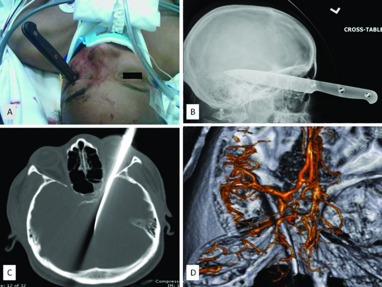
A 21-year-old woman presented to our emergency room (ER) in transfer, following a love triangle dispute that resulted in a steak knife being inserted into the patient's left eye (Fig. 2A and 2B). The patient was initially unresponsive with emesis and questionable seizure activity. She was intubated and treated with antibiotics and mannitol. On neurological examination, the patient's pupils were equal and reactive to light. She had minimal movement to noxious stimuli. A skull X-ray showed the blade of the knife extending from the orbit along the middle fossa floor posteromedially (Fig. 2B). A noncontrast CT scan of the head did not show any significant hemorrhage and confirmed the location of the retained knife extending into the left cavernous sinus and perimesencephalic cistern (Fig. 2C). Reconstructed CT angiography showed the knife blade adjacent to the petrous carotid and posterior cerebral arteries with no large-vessel vascular occlusion (Fig. 2D). A 3-D reconstructed digitally subtracted cerebral angiogram confirmed there were no large-vessel abnormalities.
Figure 2.
(A) Preoperative photograph of a patient with retained transorbital knife. (B) Preoperative lateral X-ray showing the retained knife. (C) Noncontrast head computed tomography (CT) showing the trajectory of the knife through the superior orbital fissure. The knife ends within the perimesencephalic cistern. (D) 3-D reconstructed CT angiogram shows the relationship of the knife edge to the left cavernous internal carotid artery and perimesencephalic vasculature.
Operation
The patient was taken to the operating room and underwent a left cranio-orbito-zygomatic approach for knife extraction with direct visualization of the adjacent neurovasculature. Proximal vascular control was obtained through exposure of the cervical carotid. Additionally, a lumbar drain was placed to maximize intraoperative brain relaxation. The proximal knife blade was directly visualized transversing the superior orbital fissure extradurally during initial exposure. Dissection proceeded through a transsylvian approach with exposure and protection of the proximal vasculature with cottonoids. The distal knife blade was clearly visualized exiting the cavernous sinus and lying lateral to the brain stem within the perimesencephalic cistern, consistent with preoperative imaging. The knife was removed along its initial trajectory under direct vision without significant hemorrhage. Intraoperative Doppler confirmed filling along the vascular tree left internal carotid artery (ICA), M1, and A1. At the time of closure, the brain appeared edematous, requiring CSF diversion and posterior extension of the craniectomy. An intraparenchymal intracranial pressure monitor was placed, and closure proceeded with an onlay duraplasty substitute (Duragen, Integra, Plainsboro, NJ). Our ophthalmology team explored the globe and closed the entry wound primarily, with the globe intact.
Postoperative Course
The patient was managed postoperatively in the ICU and required tracheostomy and gastrostomy placement. A follow-up CT scan showed minimal hemorrhage along the knife tract and a hypodensity in the left midbrain felt to be related to initial injury. A 2-week postoperative angiogram showed complete filling of the vascular tree without pseudoaneurysm formation. She was discharged to a skilled nursing facility and at 1-year follow-up has since undergone tracheostomy and gastrostomy removal as wells as a cranioplasty. Her follow-up CT angiogram shows no abnormalities.
Case Report 3
History and Presentation
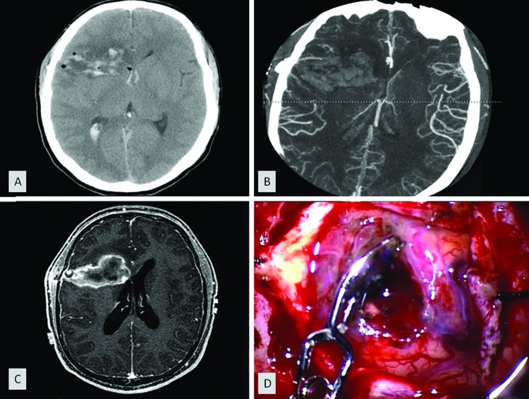
A 24-year-old right-handed man presented to Saint Louis University ER 1 day after an altercation where he was stabbed with a corkscrew in the right temporal region. There was no reported loss of consciousness immediately after the event, but he did experience nausea, vomiting, right temporal region headache, and multiple syncopal episodes over the following 19 hours. The patient initially presented to an outside hospital, where a noncontrast head CT showed a right temporal skull fracture, intraparenchymal and intraventricular hemorrhages, pneumocephalus, and midline shift (Fig. 3A). The patient received broad-spectrum antibiotics and anticonvulsants and was transferred to our institution. On neurological examination, he was confused, with intact speech and no focal neurological deficits. There was a small puncture wound in the right temporal region upon close examination. He was managed conservatively in the ICU. A CT angiogram 2 days postinjury was negative (Fig. 3B). On hospital day 21, an MRI with contrast demonstrated a traumatic pseudoaneurysm in the distal middle cerebral artery (MCA) vasculature and enhancement in the right frontal injury bed (Fig. 3C).
Figure 3.
(A) Initial noncontrast head computed tomography (CT) showing right frontal injury bed and intraventricular hemorrhage. (B) Initial CT angiogram showing no evidence of aneurysmal formation of the distal middle cerebral artery vessels at the cortical entry site. (C) Contrasted axial T1-weighted magnetic resonance image showing a partially thrombosed pseudoaneurysm at the cortical entry site of the right frontal injury bed. (D) Intraoperative photomicrograph showing clip reconstruction of the pseudoaneurysm at the right frontal entry site.
Operation
The patient was taken to the operating room for a stereotactic right frontal craniotomy for aneurysm reconstruction and exploration of the injury bed. A right frontal craniotomy was created and dissection proceeded to gain proximal vascular control of the right frontal M2 branch of the MCA within the sylvian fissure. A thin-walled traumatic pseudoaneurysm was identified on a distal frontal M4 branch at the site of cortical entry (Fig. 3D). Circumferential dissection was undertaken, and the aneurysm was successfully reconstructed with preservation of the parent vessel. The injury bed was explored and no evidence of infection was seen. Cultures from the cavity were negative for organisms.
Postoperative Course
The patient did well postoperatively, his neurological exam improved, and he was ultimately discharged home. The patient was lost to follow-up.
Discussion
Penetrating cerebral (craniocerebral or orbitocerebral) injuries can be divided between missile and nonmissile injuries. The missile injuries are caused by shrapnel or bullets, which carry a high incidence of morbidity and mortality5,12 and are associated with shock waves, cavitations,13 and additional concentric zones of injury related to blast effect.14 The nonmissile (stabbing) injuries are, by comparison, relatively rare and usually caused by objects such as knives, with an impact velocity of less than 100 m/s and pathophysiology related to tissue laceration and maceration restricted to the wound tract.15 They are, therefore, more amenable to treatment and carry a better prognosis than missile injuries.
The adult calvarium in most instances provides an effective barrier of protection against stab wounds. However, there are specific areas of weakness, such as the orbit, skull base foramina, anterior fossa floor, and temporal squama where the skull is more penetrable.1,11 The amount of force needed to penetrate the skull in the temporal region is estimated to be 5 times greater than that of the skin, as compared with 11 times that of the skin in the parietal region.16
Transorbital skull penetration follows two main pathways: either medially into the superior orbital fissure through the cavernous sinus and toward the brain stem (as seen in case 2) or superiorly through the thin orbital plate into the frontal lobe. In one report, the orbital roof was observed as the entry point in 89% of cases and could be explained by the tendency of the victims to extend their heads at the time of the injury, exposing the orbital roof to the direction of the penetrating object.13 A third, least common transorbital route includes the optic canal.17
Early recognition of these injuries is essential to ensure the best possible outcome. This can sometimes be challenging in cases of nonretained objects where the entry point may appear trivial or be hidden by hair (as seen in case 3). Additionally, the patients may present with good initial Glasgow coma scale (GCS) presentation in many cases, making it difficult to appreciate the extent of the injury, especially in children.13,18,19 A high index of suspicion is needed in patients presenting with scalp injuries because the actual amount of injury may be more extensive than what appear on the surface. A meticulous examination of the scalp and thorough neurological and radiological assessment are required to evaluate the extent of the damage.18
An initial head CT is the most effective tool for initial investigation of penetrating injuries to the brain, but it fails to identify plastic, wood, or soil.18 Given the high association of vascular abnormalities with such injuries, a CT angiogram is recommended in all cases, although the presence of metallic artifacts from the retained foreign body may limit visualization of intracranial contents.20 Therefore, when a high index of suspicion for a vascular injury exists, an angiogram should be performed. For the same reason, a second head CT should be considered after removing the foreign body to look for missed brain contusion or hemorrhage obscured by artifact in the original scan.
Situations in which cerebral angiography is indicated in the literature, given very high likelihood of associated vascular injury, include: orbitofacial or pterional entry point, presence of intracranial hematoma on CT, injuries with fragments crossing two or more dural compartments, delayed or unexplained subarachnoid hemorrhage, or delayed intraparenchymal hematoma.20 The treatment goals are to treat and prevent both short- and long-term complications associated with these injuries. In the short term, the objective is to remove the foreign object, avoiding further neurological injury, hemorrhage, and possibly death. Long-term management includes prevention and treatment of vascular abnormalities, persistent CSF leakage, infection, and seizures.
The literature is inconsistent in respect to the safety of blindly removing retained objects from the skull. Some authors suggest that blind removal of a retained blade is acceptable,21 others claim that the safety of blind removal is unknown due to the lack of evidence-based studies,5 and some authors suggest that blind removal is unacceptable.22 In our experience, we feel blind removal is unacceptable, and removing the retained blade prior to obtaining high-resolution imaging imposes undue risk on the patient, including possibly death. We routinely use high-resolution CT scan, CT angiogram, and formal four-vessel cerebral angiogram to assess both brain parenchyma injury as well as potential vascular injury prior to surgical planning. As we reported in our first and second cases, the high-resolution imaging protocol was critical to understanding the relationship of the knife blades to intracranial vascular structures, allowing us to successfully remove the retained blades without causing further injury and to plan the necessary craniotomies to visualize the removal of the offending object around the vascular structures at risk under direct vision with proximal control.
The most common vascular complications that can result from these injuries are usually traumatic pseudoaneursym formation or carotid-cavernous fistula.23 Traumatic aneurysms comprise less than 1% of intracranial aneurysms.24,25 The most common aneurysm associated with penetrating brain injuries is actually a false or pseudoaneurysm, rather than a true aneurysm. These lesions usually do not have a neck, are irregularly shaped, and have delayed filling and emptying.26 Kieck and de Villiers reviewed vascular lesions secondary to penetrating trauma and found abnormalities in 26 of 74 patients, with the largest proportion of lesions being aneurysm formation (42%).27 Traumatic aneurysms may occur in a delayed fashion and carry a 50% mortality rate if left untreated.26,27,28,29 Not all these lesions are visible on initial angiography and therefore a second angiogram should be performed 2 to 3 weeks later in all patients with penetrating brain injuries. Our third case exemplifies this phenomenon. In Kieck and de Villiers' series, 80% of the untreated aneurysms ruptured with subsequent death of the patient, and 100% of the aneurysms that were treated had good outcomes.27
Pseudoaneurysms following transcranial or transorbital stab wounds have a tendency to form in the peripheral vessels—sylvian candelabra and median hemispheric vessels—as opposed to the circle of Willis. They are usually found along the course of the arteries as opposed to vascular bifurcations. Haddad et al30 found that these lesions range in size from 2 mm to 15 mm and are multiple in 20% of the patients. Their natural history is more malignant than congenital aneurysms, with greater propensity for intraoperative rupture. Surgical mortality of these lesions varies from 15 to 20%.31 Therefore, aggressive management of traumatic pseudoaneurysms associated with penetrating brain injuries is warranted to prevent life-threatening hemorrhage and/or thromboembolic phenomenon. Oftentimes, these aneurysms cannot be safely clipped, and therefore trapping or occlusion of the parent vessel may be necessary. Preliminary results suggest that placement of stent grafts through endovascular techniques associated with appropriate antiplatelet therapy may be a safe and effective method to treat such traumatic ICA pseudoaneurysms.32 The main question still unanswered is the long-term efficacy of stenting in this setting.
Infectious complications, such as brain abscesses and meningitis, are also associated with penetrating brain injury, being found in 48 to 64% of cases7; however, in 20% of cases, microbiological culture remains sterile,33 and this also happened to our case 3. Due to the incidence and potential severity of this complication, it is essential to use broad-spectrum antibiotic prophylaxis in these patients for at least 7 to 14 days.23,34 Other factors associated with higher likelihood of infectious complications include CSF leaks, air sinus wounds, transventricular injuries, or wounds crossing the midline.19 Whenever an intracranial abscess occurs, presence of a retained foreign body should be suspected and investigated.13 About 30 to 50% of patients suffering penetrating brain injuries will develop seizures, with up to 10% of them appearing early (first 7 days after trauma). Prophylactic antiseizure drugs are recommended during the first week after injury to reduce the incidence of early posttraumatic seizures.19
Stabbing injuries to the brain are more frequently seen as a cause of emergent neurosurgical admissions in South Africa. The largest report in the literature regarding nonmissile penetrating injury is from a neurosurgical group in South Africa where cranial stab wounds are a frequent cause of emergent admission. In this study, the presenting GCS score was identified as the most indicative prognostic factor of long-term outcome.35 Other predictive factors included presence of intraventricular hemorrhage, intracranial hemorrhage, and number of surgical procedures.35 Additionally, there was a marked association between vascular abnormalities (41% cases) and mortality (76.5% cases), especially when brain stem injury was involved.
Other reports confirmed the presenting GCS score as the strongest prognostic indicator of outcome after penetrating brain injury.16,19 Such injuries in the vast majority of the Western world remain uncommon among civilians and are usually related to violence, accidents, or suicide attempts in association with mental disorders.19
Conclusion
In our experience, the management of direct penetrating brain injuries requires high-resolution imaging to assess the relationship of the surrounding vasculature to the foreign object. Immediate surgical exploration, in our opinion, is essential when compared with blind removal of the object. Skull base approaches are often required and can improve the safety profile of foreign object removal. Craniotomies should be tailored to optimally visualize the involved vascular and neural structures, provide access to proximal vascular control, and offer a direct route to skull base reconstruction for prevention of CSF leaks. Vascular studies must be obtained 2 to 3 weeks postinjury to evaluate for delayed pseudoaneurysm formation.
References
- 1.Dempsey L C, Winestock D P, Hoff J T. Stab wounds of the brain. West J Med. 1977;126:1–4. [PMC free article] [PubMed] [Google Scholar]
- 2.Mason F. Case of a young man who had a pitchfork driven into his head four inches who speedily got well. Lancet. 1870;1:700–701. [Google Scholar]
- 3.Penfield W, Buckley R C. Punctures of the brain. Arch Neurol Psychiatry. 1928;20:1–13. [Google Scholar]
- 4.Pilcher C. Penetrating wounds of the brain: an experimental study. Ann Surg. 1936;103:173–198. doi: 10.1097/00000658-193602000-00003. [DOI] [PMC free article] [PubMed] [Google Scholar]
- 5.Farhadi M R, Becker M, Stippich C, Unterberg A W, Kiening K L. Transorbital penetrating head injury by a toilet brush handle. Acta Neurochir (Wien) 2009;151:685–687. doi: 10.1007/s00701-009-0221-9. [DOI] [PubMed] [Google Scholar]
- 6.Kurt G, Börcek A O, Kardeş O, Aydincak O, Ceviker N. Transcranial arrow injury: a case report. Ulus Travma Acil Cerrahi Derg. 2007;13:241–243. [PubMed] [Google Scholar]
- 7.Matsuyama T, Okuchi K, Nogami K, Hata M, Murao Y. Transorbital penetrating injury by a chopstick—case report. Neurol Med Chir (Tokyo) 2001;41:345–348. doi: 10.2176/nmc.41.345. [DOI] [PubMed] [Google Scholar]
- 8.Vaicys C, Hunt C D, Heary R F. Successful recovery after an orbitocranial injury. J Trauma. 2000;49:788. doi: 10.1097/00005373-200010000-00037. [DOI] [PubMed] [Google Scholar]
- 9.Tiwair S M, Singh R G, Dharker S R, Chaurasia B D. Unusual craniocerebral injury by a key. Surg Neurol. 1978;9:267. [PubMed] [Google Scholar]
- 10.Markham J W, McCleve D E, Lynge H N. Penetrating craniocerebral injuries. Report of two unusual cases. J Neurosurg. 1964;21:1095–1097. doi: 10.3171/jns.1964.21.12.1095. [DOI] [PubMed] [Google Scholar]
- 11.Dooling J A, Bell W E, Whitehurst W R Jr. Penetrating skull wound from a pair of scissors. Case report. J Neurosurg. 1967;26:636–638. doi: 10.3171/jns.1967.26.6.0636. [DOI] [PubMed] [Google Scholar]
- 12.Lichter H, Snir M, Segal K, Yassur Y. Penetrating orbitocranial knife injury. J Pediatr Ophthalmol Strabismus. 1999;36:44–46. doi: 10.3928/0191-3913-19990101-11. [DOI] [PubMed] [Google Scholar]
- 13.Chibbaro S Tacconi L Orbito-cranial injuries caused by penetrating non-missile foreign bodies. Experience with eighteen patients Acta Neurochir (Wien) 2006148937–941., discussion 941–942 [DOI] [PubMed] [Google Scholar]
- 14.Campbell E, Kuhlenbeck H. Mortal brain wounds; a pathologic study. J Neuropathol Exp Neurol. 1950;9:139–149. doi: 10.1097/00005072-195004000-00002. [DOI] [PubMed] [Google Scholar]
- 15.MacEwen C J, Fullarton G. A penetrating orbitocranial stab wound. Br J Ophthalmol. 1986;70:147–149. doi: 10.1136/bjo.70.2.147. [DOI] [PMC free article] [PubMed] [Google Scholar]
- 16.Saint-Martin P, Prat S, Bouyssy M, Sarraj S, O'Byrne P. An unusual death by transcranial stab wound: homicide or suicide? Am J Forensic Med Pathol. 2008;29:268–270. doi: 10.1097/PAF.0b013e318183456f. [DOI] [PubMed] [Google Scholar]
- 17.Smely C, Orszagh M. Intracranial transorbital injury by a wooden foreign body: re-evaluation of CT and MRI findings. Br J Neurosurg. 1999;13:206–211. doi: 10.1080/02688699944014. [DOI] [PubMed] [Google Scholar]
- 18.Gupta P K, Thajjuddin B A, Al Sikri N E, Bangroo A K. Penetrating intracranial injury due to crochet needle. Pediatr Neurosurg. 2008;44:493–495. doi: 10.1159/000180306. [DOI] [PubMed] [Google Scholar]
- 19.Gutiérrez-González R, Boto G R, Rivero-Garvía M, Pérez-Zamarrón A, Gómez G. Penetrating brain injury by drill bit. Clin Neurol Neurosurg. 2008;110:207–210. doi: 10.1016/j.clineuro.2007.09.014. [DOI] [PubMed] [Google Scholar]
- 20.Martin S, Raup G H, Cravens G, Arena-Marshall C. Management of embedded foreign body: penetrating stab wound to the head. J Trauma Nurs. 2009;16:82–86. doi: 10.1097/JTN.0b013e3181ac91e1. [DOI] [PubMed] [Google Scholar]
- 21.Grobbelaar A, Knottenbelt J D. Retained knife blades in stab wounds of the face: is simple withdrawal safe? Injury. 1991;22:29–31. doi: 10.1016/0020-1383(91)90156-9. [DOI] [PubMed] [Google Scholar]
- 22.Nath F P, Teasdale E, Mendelow A D. Penetrating injury of the tuberculum sellae. Neurosurgery. 1984;14:598–600. doi: 10.1227/00006123-198405000-00016. [DOI] [PubMed] [Google Scholar]
- 23.Okay O, Dağlioğlu E, Ozdol C, Uckun O, Dalgic A, Ergungor F. Orbitocerebral injury by a knife: case report. Neurocirugia (Astur) 2009;20:467–469. doi: 10.1016/s1130-1473(09)70145-9. [DOI] [PubMed] [Google Scholar]
- 24.Benoit B G, Wortzman G. Traumatic cerebral aneurysms. Clinical features and natural history. J Neurol Neurosurg Psychiatry. 1973;36:127–138. doi: 10.1136/jnnp.36.1.127. [DOI] [PMC free article] [PubMed] [Google Scholar]
- 25.Parkinson D, West M. Traumatic intracranial aneurysms. J Neurosurg. 1980;52:11–20. doi: 10.3171/jns.1980.52.1.0011. [DOI] [PubMed] [Google Scholar]
- 26.Horiuchi T Nakagawa F Miyatake M Iwashita T Tanaka Y Hongo K Traumatic middle cerebral artery aneurysm: case report and review of the literature Neurosurg Rev 200730263–267., discussion 267 [DOI] [PubMed] [Google Scholar]
- 27.Kieck C F, de Villiers J C. Vascular lesions due to transcranial stab wounds. J Neurosurg. 1984;60:42–46. doi: 10.3171/jns.1984.60.1.0042. [DOI] [PubMed] [Google Scholar]
- 28.Acosta C, Williams P E Jr, Clark K. Traumatic aneurysms of the cerebral vessels. J Neurosurg. 1972;36:531–536. doi: 10.3171/jns.1972.36.5.0531. [DOI] [PubMed] [Google Scholar]
- 29.Litvack Z N, Hunt M A, Weinstein J S, West G A. Self-inflicted nail-gun injury with 12 cranial penetrations and associated cerebral trauma. Case report and review of the literature. J Neurosurg. 2006;104:828–834. doi: 10.3171/jns.2006.104.5.828. [DOI] [PubMed] [Google Scholar]
- 30.Haddad F S, Haddad G F, Taha J. Traumatic intracranial aneurysms caused by missiles: their presentation and management. Neurosurgery. 1991;28:1–7. doi: 10.1097/00006123-199101000-00001. [DOI] [PubMed] [Google Scholar]
- 31.Levy M L, Rezai A, Masri L S. et al. The significance of subarachnoid hemorrhage after penetrating craniocerebral injury: correlations with angiography and outcome in a civilian population. Neurosurgery. 1993;32:532–540. doi: 10.1227/00006123-199304000-00007. [DOI] [PubMed] [Google Scholar]
- 32.Maras D, Lioupis C, Magoufis G, Tsamopoulos N, Moulakakis K, Andrikopoulos V. Covered stent-graft treatment of traumatic internal carotid artery pseudoaneurysms: a review. Cardiovasc Intervent Radiol. 2006;29:958–968. doi: 10.1007/s00270-005-0367-7. [DOI] [PubMed] [Google Scholar]
- 33.Heininger A D, Will B E, Krueger W A, Kottler B M, Unertl K E, Stark M. Detection and identification of the pathogenic cause of a brain abscess by molecular genetic methods. Anaesthesist. 2004;53:830–835. doi: 10.1007/s00101-004-0729-6. [DOI] [PubMed] [Google Scholar]
- 34.van Dellen J R, Lipschitz R. Stab wounds of the skull. Surg Neurol. 1978;10:110–114. [PubMed] [Google Scholar]
- 35.Nathoo N Boodhoo H Nadvi S S Naidoo S R Gouws E Transcranial brainstem stab injuries: a retrospective analysis of 17 patients Neurosurgery 2000471117–1122., discussion 1123 [DOI] [PubMed] [Google Scholar]


