Abstract
Background:
This study aimed to evaluate the effect of two weeks oral administration of pomegranate seed extract (PGSE) on active and passive avoidance memories after permanent bilateral common carotid arteries occlusion (2CCAO) to induce permanent cerebral ischemia in adult female rats.
Methods:
Seventy adult female Wistar rats (250 ± 20 g) were used. Animals were divided randomly into seven groups with 10 in each: 1) Sham-operated; 2) Ischemic; 3–6) Ischemic received PGSE (100, 200, 400, and 800 mg/2mL/kg, orally) for 14 days; 7) Ischemic received vehicle. In order to create 2CCAO, carotid arteries were ligatured and then cut bilaterally. Active and passive avoidance task were measured using criterion condition responses (CCRs) in Y-maze and step-through latency (STL) in two-way shuttle box in all female rats.
Results:
Both active and passive avoidance memories were significantly impaired in rats after cerebral hypoxia-ischemia (CHI) (P < 0.001). PGSE treatment significantly improved passive and active memory impairments with 2CCAO (P < 0.05, P < 0.01, and P < 0.001). No toxicity was observed even with high-dose PGSE consumption (800 mg/kg, for 14 days).
Conclusion:
PGSE exhibits therapeutic potential for avoidance memories, which is most likely related at least in part to its antioxidative and free radical scavenging actions.
Keywords: cerebral ischemia, pomegranate, memory, female, rats
Introduction
Stroke is a major cause of adult-onset disability and dependency (1). Neuropsychological impairment after stroke when no motor, sensory or language deficits are left remains understudied (2). The outcome for patients with hypoxic-ischemic brain injury (HIBI) is often poor. It is important to establish an accurate prognosis as soon as possible after the insult to guide management (3). Cerebral ischemia resulting from low oxygen and glucose supply evidently decreases the formation of adenosine triphosphate (ATP) (4,5). Damage to brain tissue resulting from cerebral ischemia is a major cause of adult disability. It can lead to cognition problems, seizures, and death (6,7). Transient global cerebral ischaemia (forebrain ischaemia), occurring during cardiorespiratory arrest induces selective and delayed neuronal cell death (7,10). The hippocampus plays a central role in the brain network that is essential for memory function. Paradoxically, the hippocampus is also the brain structure that is most sensitive to hypoxic-ischemic episodes (11). Pyramidal neurons in the CA1 region of the hippocampus are particularly vulnerable and die after global ischemia. Hippocampal CA1 sector injury is observed a few days after untreated forebrain ischemia in the rat (5–7,9,10), gerbil and human (7,12).
Treatments for protection against neuronal cell death induced by hypoxia-ischemia (HI) and reperfusion have been developed in recent years, but none has been highly successful. A fundamental process believed to be responsible for HI damage to neurons is excitotoxicity, triggered mainly by elevated extracellular glutamate concentration (13). This ischemia-induced release of glutamate likely occurs in man as well (14), and possibly underlies selective damage to the human hippocampus. Glutamate may cause ischemic neuronal death by acting at excitatory N-methyl-D-aspartate (NMDA) receptors (15) which play an important physiological role in memory (16). Thus, the high concentration of NMDA excitatory receptors on the dendrite trees of hippocampal CA1 pyramidal cells (17) likely explains the longknown selective vulnerability of the CA1 zone of the hippocampus to ischemic brain damage.
Reactive oxygen species (ROS) are generated within brain tissue during HI and play a role in the development of cerebral damage. They may be directly involved in glutamate release (18) and more importantly, they may participate in the excitotoxic process itself. ROS are extremely reactive and attack lipids, proteins, and nucleic acids, which eventuates in tissue injury and cell death (19). Although there is strong evidence that total destruction of hippocampal CA1 neurons is sufficient to cause a memory deficit (20), it is still presently unclear to what degree subtotal ischemic hippocampal damage may occur without an ensuing memory deficit (21). The free radicals can be neutralized by an elaborate antioxidant defense system consisting of enzymes and numerous non-enzymatic antioxidants, including vitamins A, E and C, glutathione, ubiquinone, and flavonoids (22,23).
Extracts from natural substances have the ability to protect neurons from ischemic damage (24,25). The extracts have several functions including antioxidant effects in neuroprotection from ischemic insults (26). Polyphenols have been found to possess antioxidant properties as well as having effects on gene expression (27). Recent studies indicate that among foods which contain polyphenols, juice extracted from the pomegranate has the highest concentration of measurable polyphenols (28,29). The pharmacologic actions of pomegranate juice include anti-atherosclerotic, antibacterial, and anti-proliferative properties (30,31). Studies of dietary supplementation with pomegranate juice have shown protective effects against atherogenesis and atherosclerosis as well as reductions in serum angiotensin-converting enzyme activity with subsequent reductions in systolic blood pressure (32–34). Also, maternal dietary supplementation with pomegranate juice results in protection against neonatal brain injury (35).
During recent years, phytoestrogens have been receiving an increasing amount of interest, as several lines of evidence suggest a possible role in preventing a range of diseases. The presence of these phytoestrogenic compounds in pomegranate has been shown to exert suppressive effects on disease (36). Another study via biochemical analysis revealed that pomegranate with highest antioxidant capacity was found in leaves followed by peel, pulp, and seed (37). In our previous work (38), the beneficial effects of PGSE on adult male rats suffering with cerebral hypoxia-ischemia (HI) and Parkinson’s disease were determined. With regard to the several beneficial effects mentioned for pomegranate, it seems that administration of its seed extract (PGSE) can be effective for the improvement of post-ischemic injuries. On the other hand, female sex hormones such as estrogen are neuroprotective and PGSE also contains phytoestrogenic substances with antioxidative properties, we decided therefore to test the effect of PGSE on cognition deficiency induced by HI in adult female rats. The present study investigated the effects of different doses of PGSE on passive and active avoidance memories following cerebral hypoxia-ischemia induced via bilateral common carotid artery occlusion.
Materials and Methods
Animals
Seventy healthy adult female albino rats of Wistar strain (250 ± 20 g, 3–4 months) obtained from Ahvaz Jundishapur University of Medical Sciences (AJUMS) Laboratory Animal Centre were used in this study. Animals were housed in standard cages under controlled room temperature (20 ± 2 °C), humidity (55–60%) and light exposure conditions 12:12 h light–dark cycle (light on at 07:00 am). All experiments were carried out during the light phase of the cycle (8:00 am to 6:00 pm). Access to food and water were ad libitum except during the experiments. Animal handling and experimental procedures were performed under observance of the University and Institutional legislation, controlled by the Local Ethics Committee for the Purpose of Control and Supervision of Experiments on Laboratory Animals. All efforts were made to minimize animal suffering and to reduce the number of animals used. Prior to the onset of behavioural testing, all rats were handled for 5 days (5 minutes daily). Animals were divided randomly into seven groups consisting of 10 animals in each. Simple randomization was performed by an examiner who was blind to the grouping.
1. Group 1: Sham-operated (Sh) with manipulation of both common carotid arteries without occlusion.
2. Group 2: Untreated ischemic (I) group with occlusion of bilateral common carotid arteries.
3. Group 3: Ischemic rats received 100 mg/kg, po. PGSE for 14 days (I+P100).
4. Group 4: Ischemic rats received 200 mg/kg, po. PGSE for 14 days (I+P200).
5. Group 5: Ischemic rats received PGE 400 mg/kg, po. for 14 days (I+P400).
6. Group 6: Ischemic rats received PGE 800 mg/kg, po. for 14 days (I+P800).
7. Group 7: Ischemic rats received same volume of normal saline as PGSE vehicle (2 mL/kg, po.) for 14 days as Sham group (I+Veh).
CCAO procedure
We used Cechetti’s method (2010) with little modification (12). Briefly, rats were anaesthetized with ketamine/xylazine (50/5 mg/kg, i.p). A neck ventral midline incision was made and the common carotid arteries were then exposed and gently separated from the vagus nerve. Carotids were occluded with a one week interval between interventions, the right common carotid being the first to be assessed and the left one being occluded one week later. Sham-operated controls (SI) went under same surgical procedures without carotid artery ligation and occlusion (39).
Behavioural assessment to demonstrate ischemic brain damage consisted of sensorimotor (spontaneous activity, and Symmetry in the movement of four limbs), gait, and cognitive (passive and active avoidance tasks) evaluations. All the sham-operated and CHI rats were assessed on sensorimotor tasks and gait before surgery (at baseline) and five days after surgery by an examiner who was blind to the surgical procedure.
Sensorimotor evaluation consisted of two tests developed and described by Garcia et al. (40) with some modifications. Spontaneous activity: The animal is observed for five minutes in its normal cage. Scores indicate the following: (1) rat moves around, explores the environment, and approaches at least three walls of the cage; (2) rat moves around in the cage but does not approach all sides and hesitates to move, although it eventually reaches at least one upper rim of the cage (height = 10 cm); (3) rat dose not rise up at all and barely moves in the cage; (4) rat does not move at all. Symmetry in the movement of four limbs: The rat is held in the air by the tail to observe symmetry in the movement of the four limbs. Scores indicate the following: (1) all four limbs extended symmetrically; (2) limbs on one side extended less or more slowly than those on the other side; or slow extension of the four limbs; (3) limbs on one or both sides show minimal movements; (4) forelimbs on one or both sides do not move at all. The scores assigned to each rat at the end of the examination is the sum of the two test scores. The minimum neurological score is three and the maximum is fifteen.
Gait performance evaluation: This was carried out by means of the elevated platform test (41). Each rat was positioned at the beginning of a 5 cm wide, 60 cm long wooden bridge suspended between two platforms. The rats were tested for their ability to remain on the bridge during a single 3 minutes trial. The number of rats falling from the bridge and the length of the bridge covered by each animal, either falling or not, was recorded.
PGSE preparation
Pomegranate fruits (Punica granatum L) were used. The seeds of this plant were collected from Shaivand Granatum Gardens district in Izeh, Iran, and its voucher number identified (Ref. No. AJUMS/DP-SP/AS-2/2011) by Pharmacognosy Division, School of Pharmacy, Ahvaz Jundishapur University of Medical Sciences (AJUMS) - Khuzestan, Iran. They were shade dried, pulverized, and passed through a 40 mesh sieve. The powdered seeds were extracted with soxhlet extractor with ethanol 70% (v/v). On evaporation a greyish residue was obtained (yield 17.2% w/w). Prior to use it was suspended as a 2% (w/v) aqueous solution and administered (p.o.) (42).
Treatment
Different doses of extract were administrated to each animal in separate groups via forced oral administration (i.g.) every day 8:00–9:00 am for 2 weeks, starting on the fifth day post-ischaemic injury. Sham-treated animals (I+Veh) received the same volume of normal saline for same period.
Passive avoidance task
The apparatus used for the passive avoidance task was the two-way shuttle box (Borj Sanaat Co. Tehran, Iran), which consisted of two adjacent Plexiglas compartments of identical dimensions (27 cm × 14.5 cm × 14 cm) with grid floors. The floor of the two compartments has been covered with stainless steel bars (2 mm diameter) spaced 1 cm apart. The compartment was illuminated by a 5 W lamp mounted on its wall just below a movable transparent Plexiglas ceiling. Tamburella’s procedure with little modification was used for the passive avoidance memory test (43). Twenty days after surgery (6 days post-surgery recovery +14 days PGSE or vehicle gavage) each rat was allowed a 10-minute adaptation period with free access to either the light or dark compartment of the avoidance training box after being placed in a shuttle-box (in order to familiarise with instruments). Two days after this adaptation period, rats were placed into the illuminated compartment and 30 seconds later the sliding door was raised (initial latency was recorded as learning phase). Upon entering the dark compartment, the door was closed and a 1.5 mA constant-current was applied to the fore and hind paws for 3 seconds. After 20 seconds, the rat was removed from the dark compartment and placed into the home cage. In order to test short-term learning, 24 hours after receiving foot shock, the rats were placed in the illuminated chamber again and 30 seconds later the sliding door was raised and latency of entering the dark compartment was recorded again (as step-through latency). The maximum time considered in this procedure was 300 seconds (44–46).
Active avoidance task
The apparatus used for evaluating active avoidance was the 3-equal arms Y-maze (Made in Ahvaz, Iran). The Yu and Besnard procedures with little modifications were used for active avoidance, memory (47,48). Sham-operated, ischemic and all treated rats were trained in the maze using an A/D converter, special software on a PC for active avoidance learning. Training involved 30 trials daily for 4 days. Animals were conditioned using a 12-watt light as conditioned stimulus (CS) and 20–25 volts, 3 mA electrical foot shock as unconditioned stimulus (UCS). Inter-trial interval (ITI) and inter-stimulus interval (ISI) were 60 seconds and 5 seconds respectively. Trained animals left the dark arms and entered into the light arm. If this occurred within the 5 second ISI, the effort was counted as a conditioned response. Criterion condition response (CCR) was 90% correct responses.
Statistics
Data were expressed as mean ± SD of values for initial latency and memory tests. Statistical analysis was performed using t test and one-way analysis of variance (ANOVA) to compare the initial and step-through latencies for the passive avoidance task and repeated measurements ANOVA followed by LSD post-hoc test. P values less than 0.05 were assumed to denote a significant difference.
Results
Passive avoidance memory
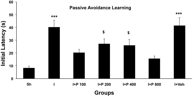
As shown in Figure 1 the initial latency (learning) for leaving rats from light to dark compartment of shuttle box before exposing them to any serious stimulus (electrical shock) was decreased significantly (P < 0.001) in ischemic rats two weeks after 2CCAO when compared with sham group. Treatment of 2CCAO rats with oral administration of 100 mg/kg of PGSE for 14 days improved initial latency significantly when compared with Sh and I groups (P < 0.001 for I+P100 vs. I and P < 0.05 for I+P100 vs. Sh respectively). Initial latency of I+P100 after treatment with PGSE was less than Sh groups. On the other hand, initial latency of ischemic animals did not change after treatment with administration of the vehicle (normal saline). Ischemic rats with other doses of PGSE (200, 400, and 800 mg/kg, ig. for 14 days) improved initial latency significantly so that there was no difference between these and the Sh group (P < 0.001 for each one of treated groups vs I respectively). Data show that dose 400 mg/kg of PGSE has same effect of a higher dose (800 mg/kg). Short-term passive avoidance memory (24 hours after exposing the electrical shock to paws of rats) in all groups was evaluated with same procedure.
Figure 1:
Mean ± SD of initial latency (s) just before exposing to electrical shock to paws in shuttle box (as passive avoidance learning) of sham ischemic (Sh), ischemic (I), I+P100, I+P200, I+P400, I+P800, and sham treated (I+Veh) groups. Data were analyzed by one-way ANOVA followed by LSD post hoc test. Levels of significance are indicated by symbols: * and $ P < 0.05, $$ P < 0.01, *** P < 0.001. Stars symbols indicate difference with SI and $ for difference with I group.
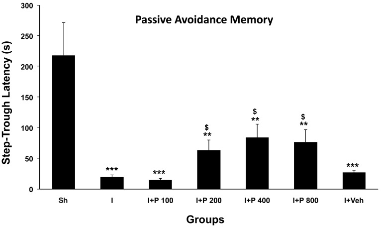
As shown in Figure 2, step-through latency (memory) was impaired significantly in both ischaemic and I+Veh groups (P < 0.001). Treatment of ischemic groups with different doses of extract higher than 100 mg improved the memory significantly when compared with ischemic rats (P < 0.05 for treated groups with PGSE vs I), so step-through latency in treated ischemic groups has reversed but it is lower than control levels.
Figure 2:
Mean ± SD of step-trough latency (s), 24 h after shock delivery to paws in shuttle box (as passive avoidance memory) of sham ischemic (Sh), ischemic (I), I+P100, I+P200, I+P400, I+P800 and sham treated (I+Veh). Data were analyzed by oneway ANOVA followed by LSD post hoc test. Levels of significance are indicated by symbols: $ P < 0.05, ** P < 0.01, *** P < 0.001. Stars symbols indicate difference with SI and $ for difference with I group.
Active avoidance memory
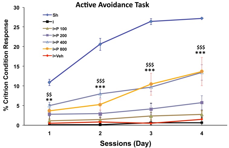
Figure 3 shows that the proportion of criterion condition responses (%CCRs) was significantly lowered after ischemia induction (P < 0.001). Ischemic groups with different doses of PGSE showed that only doses 400 and 800 mg/kg improved %CCRs during 1–4 sessions significantly (P > 0.01 and P < 0.001), but it is still significantly lower than that of the SI group (P < 0.01 and P < 0.001).
Figure 3:
Effect of cerebral hypoperfusion/ischemia on active avoidance learning in Y-maze test. Data were expressed as Mean ± SD of criterion condition responses in different groups including sham operated ischemic (Sh), ischemic (I), I+P100, I+P200, I+P400, I+P800 and sham treated (I+Veh). Data were analyzed by one way ANOVA (to compare CCR in different groups for each session) and repeated measurements ANOVA (RM) followed by LSD post hoc test (to compare CCR in different times for each group). Levels of significance are indicated by symbols: ** and $$ P < 0.01, *** and $$$ P < 0.001. Stars symbols indicate difference with SI and $ for difference with I group.
Discussion
We have found that 14 days oral administration of PGSE can improve both active and passive avoidance learning and memory in ischemic rats. PGSE at 400 mg/kg was the best effective dose for significantly improving initial latency and short-term (24 h) memory as measured by the step-trough latency paradigm and condition training on the Y-maze test. However, 14 days treatment at this dosage did not change either initial latency nor short-term memory in healthy intact rats (data not shown).
Phytochemical investigation of ethanol extract for the presence of phenolic compounds, flavonoids, tannins, anthocyanins, sugars and saponins was also carried out. Phytochemical screening and measurement of reducing power revealed the central nervous system (CNS) activity of ethanol extract of PGSE may be due to its antioxidative profile (49,50). Another study also showed that pomegranate seed extract contains several compounds such as linolenic acid, ellagic acid, punic acid, alphaeleostearic acid (AEA), flavonoids, polyphenols, ellagitannin (37% punicalagins), and other useful contents. Pomegranate seed linolenic acid isomers were evaluated as selective estrogen receptor modulators (SERMs). Punicic acid (PA) inhibited (IC-50) estrogen receptor (ER) alpha at 7.2 microM, ERbeta at 8.8 microM; AEA inhibited ERalpha/ERbeta at 6.5/7.8 microM. PA (not AEA) agonized ERalpha/ERbeta (EC-50) at 1.8/2 microM, antagonizing at 101/80 microM (38). About the ellagitannin in pomegranate, recently, potent anti-tumorigenic effects of pomegranate juice and extracts have also been reported. In other words, pomegranate has potential not only as a treatment but also as a preventive against certain types of cancer, including prostate cancer (51). Another study via biochemical analysis revealed that pomegranate with highest antioxidant capacity was found in leaves followed by peel, pulp, and seed. Pomegranate seed had an average lipid content of 19.2% with punicic acid as the predominant fatty acid. Pomegranate seed has high contents of alpha-tocopherol (161.2–170.1 mg/100 g) and gamma-tocopherol (80.2–92.8 mg/100 g) (53).
Twenty different varieties of pomegranate (Punica granatum) from Turkey were analyzed for vitamin C level, lipid content, sterol determination, anthocyanin content, and elemental analysis (calcium, magnesium, phosphorus, iron, sodium, and potasium studies). Vitamin C content range of 312–1050 mg/100 g, oil content range of 2.41–3.73%, sterol content range of 5.78–8.43%, anthocyanin content range of 2100–4400 mg/L, potassium range of 250–1200 ppm, calcium range of 35–326 ppm, magnesium range of 176–427 ppm, iron range of 21–46 ppm, sodium range of 35–76 ppm, and phosphorus range of 12–43 ppm were observed in these varieties (22,23).
Although in the literature studied, we have found few specific references to effects of PGSE on brain damage due to HI or degeneration. On the basis of the current finding, PGSE with these compounds could act as an antioxidant against the free radicals in brain after HI. PGSE had no toxicity after administration of different doses (100–800 mg/kg, orally) for 14 days in healthy and ischaemic rats. Mortality of 30–35% in our rats occurred during recovery from ischemia surgery in all ischemic groups and the same range of mortality has been reported previously by others. The acute oral toxicity study revealed no significant findings at 2000 mg (PSO)/kg body weight. In the 28 days dietary toxicity study, PSO was dosed at concentrations of 0, 10 000, 50 000 and 150 000 ppm, which would mean an intake of 0–0, 825–847, 4 269–4 330 and 13 710–14 214 mg PSO/kg body weight per day in malesfemales, respectively (53,54).
During the period of ischemia large quantities of stimulatory amino acids and calcium are released, leading to an increase in free radicals that is the signature of exitoxicity (55). Both a great production of free radicals and the deficiency or depletion of many antioxidant systems may exacerbate oxidative cellular injury, while the supplementation of many antioxidants generates diverse outcomes (56,57). Free radicals are highly reactive molecules that may be formed during various biochemical reactions in the cell. Many of these free radicals contain oxygen and are called ROS. Typically, the levels of ROS and other free radicals are controlled by various scavenger molecules, known as antioxidants, that are normally found within the cell and which eliminate free radicals. The antioxidant defense mechanisms include antioxidant enzymes like SOD, GPx and several non-enzymatic free radical scavengers (58,59). The nervous tissue has a high content of polyunsaturated fatty acids (60), which are easy targets for oxidative damage by free radicals due to the unsaturated bonds they contain (61). On the other hand, it has been revealed that brain structures supporting memory are uniquely sensitive to oxidative stress due to their elevated demand for oxygen (62,63). The hippocampus is a brain area particularly susceptible to ischaemia-induced oxidative stress. However, its anti-oxidative activity in the central nervous system and its effects on spatial memory deficits induced by ischemia have not been scientifically documented so far (64). Behavioural studies in animals have demonstrated that hippocampal damage can produce learning and memory impairments (20,65), particularly on tasks that involve place learning (66). It has been shown that modulation of nitric oxide (NO) availability is an important determinant of ischemic stroke risk (67). Thus, optimal NO/ROS balance in the brain seems to be a crucial parameter in the prevention of brain damage including ischaemic stroke and neurodegenerative diseases as well. The neuroprotective effects of many polyphenols present in PGSE rely on their ability to permeate brain barrier and here directly scavenge pathological concentration of reactive oxygen and nitrogen species and chelate transition metal ions (68). Different polyphenolic compounds were shown to have scavenging activity and the ability to activate key antioxidant enzymes in the brain and thus breaking the vicious cycle of oxidative stress and tissue damage (42,69). There is a growing interest in the potential of natural polyphenols to improve memory, learning and general cognitive ability. Recent evidence has indicated that flavonoids may exert especially powerful actions on mammalian cognition and may reverse age-related declines (22,70). Since the behavioral impairments were observed after cerebral hypoperfusion/ischemia, and PGSE improved those deficits, therefore, it may be concluded that the PGSE bio-active compounds has been passed through blood brain barrier (BBB) and improved the behaviour of ischemic rats.
Pomegranate fruit consumption has long history and advises in many literatures and different religions. For example it was mentioned three times in the Islam holey Quran (Anaam 99, 141, and Al-Rahman 68). It grows in many countries with moderate to relatively warm climates and its fruit is juicy highly enriched beneficial biochemical substances. So we advise its juice, seed oil and extract, fruit peel and even stem skin should consider more and more as supplemental food and medicinal uses.
Conclusion
PGSE improves active and passive memory deficits due to HI in female rats, possibly because its contents act as antioxidants in injured brain tissue after HI. However, the exact mechanisms behind the effect of PGSE on cognition require further investigation.
Acknowledgments
This article was partially extracted from Mr Moslem Rezaiei’s MSc thesis, Kazeroon Islamic Azad University, Fars, Iran. Thesis was supported financially by Ahvaz Jundishapur of Medical Sciences and Moslem Rezaiei. This research was done in Ahvaz Physiology Research Center, Neurosciences Lab (PRC–82).
Footnotes
Conflict of interest
Nil.
Funds
Nil.
Authors’ contributions
Conception and design, critical revision of the article for the important intellectual content and final approval of the article: AS
Statistical expertise and administrative, technical or logistic support: MKG
Analysis and interpretation of the data and drafting of the article: MRR
Provision of study materials or patient and collection and assembly of data: MR
References
- 1.Sheorajpanday RV, Nagels G, Weeren AJ, Van Putten MJ, De Deyn PP. Quantitative EEG in ischemic stroke: correlation with functional status after 6 months. Clin Neurophysiol. 2011;122(5):874–883. doi: 10.1016/j.clinph.2010.07.028. [DOI] [PubMed] [Google Scholar]
- 2.Planton M, Peiffer S, Albucher JF, Barbeau EJ, Tardy J, Pastor J, et al. Neuropsychological outcome after a first symptomatic ischaemic stroke with ‛good recovery’. Eur J Neurol. 2012;19(2):212–219. doi: 10.1111/j.1468-1331.2011.03450.x. [DOI] [PubMed] [Google Scholar]
- 3.Howard RS, Holmes PA, Siddiqui A, Treacher D, Tsiropoulos I, Koutroumanidis M. Hypoxicischaemic brain injury: imaging and neurophysiology abnormalities related to outcome. QJM. 2012;105(6):551–561. doi: 10.1093/qjmed/hcs016. [DOI] [PubMed] [Google Scholar]
- 4.Aquilano K, Baldelli S, Rotilio G, Ciriolo MR. Role of nitric oxide synthases in Parkinson’s disease: a review on the antioxidant and anti-inflammatory activity of polyphenols. Neurochem Res. 2008;33(12):2416–2426. doi: 10.1007/s11064-008-9697-6. [DOI] [PubMed] [Google Scholar]
- 5.Aviram M, Dornfeld L, Kaplan M, Coleman R, Gaitini D, Nitecki S, et al. Pomegranate juice flavonoids inhibit low-density lipoprotein oxidation and cardiovascular diseases: studies in atherosclerotic mice and in humans. Drugs Exp Clin Res. 2002;28(2–3):49–62. [PubMed] [Google Scholar]
- 6.Aviram M, Dornfeld L. Pomegranate juice consumption inhibits serum angiotensin converting enzyme activity and reduces systolic blood pressure. Atherosclerosis. 2001;158(1):195–198. doi: 10.1016/s0021-9150(01)00412-9. [DOI] [PubMed] [Google Scholar]
- 7.Aviram M, Dornfeld L, Rosenblat M, Volkova N, Kaplan M, Coleman R, et al. Pomegranate juice consumption reduces oxidative stress, atherogenic modifications to LDL, and platelet aggregation: studies in humans and in atherosclerotic apolipoprotein E-deficient mice. Am J Clin Nutr. 2000;71(5):1062–1076. doi: 10.1093/ajcn/71.5.1062. [DOI] [PubMed] [Google Scholar]
- 8.Ben Nasr C, Ayed N, Metche M. Quantitative determination of the polyphenolic content of pomegranate peel. Z Lebensm Unters Forsch. 1996;203(4):374–378. doi: 10.1007/BF01231077. [DOI] [PubMed] [Google Scholar]
- 9.Banerjee AK, Mandal A, Chanda D, Chakraborti S. Oxidant, antioxidant and physical exercise. Mol Cell Biochem. 2003;253(1–2):307–312. doi: 10.1023/a:1026032404105. [DOI] [PubMed] [Google Scholar]
- 10.Block F. Global ischemia and behavioural deficits. Prog Neurobiol. 1999;58(3):279–295. doi: 10.1016/s0301-0082(98)00085-9. [DOI] [PubMed] [Google Scholar]
- 11.Lavenex P, Sugden SG, Davis RR, Gregg JP, Lavenex PB. Developmental regulation of gene expression and astrocytic processes may explain selective hippocampal vulnerability. Hippocampus. 2011;21(2):142–149. doi: 10.1002/hipo.20730. [DOI] [PubMed] [Google Scholar]
- 12.Cechetti F, Worm PV, Pereira LO, Siqueira IR, C AN. The modified 2VO ischemia protocol causes cognitive impairment similar to that induced by the standard method, but with a better survival rate. Braz J Med Biol Res. 2010;43(12):1178–1183. doi: 10.1590/s0100-879x2010007500124. [DOI] [PubMed] [Google Scholar]
- 13.Choi DW, Rothman SM. The role of glutamate neurotoxicity in hypoxic-ischemic neuronal death. Annu Rev Neurosci. 1990;13:171–182. doi: 10.1146/annurev.ne.13.030190.001131. [DOI] [PubMed] [Google Scholar]
- 14.Chun HS, Kim JM, Choi EH, Chang N. Neuroprotective effects of several korean medicinal plants traditionally used for stroke remedy. J Med Food. 2008;11(2):246–251. doi: 10.1089/jmf.2007.542. [DOI] [PubMed] [Google Scholar]
- 15.Colbourne F, Li H, Buchan AM, Clemens JA. Continuing postischemic neuronal death in CA1: influence of ischemia duration and cytoprotective doses of NBQX and SNX-111 in rats. Stroke. 1999;30(3):662–668. doi: 10.1161/01.str.30.3.662. [DOI] [PubMed] [Google Scholar]
- 16.Collingridge GL, Kehl SJ, McLennan H. The antagonism of amino acid–induced excitations of rat hippocampal CA1 neurones in vitro. J Physiol. 1983;334:19–31. doi: 10.1113/jphysiol.1983.sp014477. [DOI] [PMC free article] [PubMed] [Google Scholar]
- 17.Esposito E, Rotilio D, Di Matteo V, Di Giulio C, Cacchio M, Algeri S. A review of specific dietary antioxidants and the effects on biochemical mechanisms related to neurodegenerative processes. Neurobiol Aging. 2002;23(5):719–735. doi: 10.1016/s0197-4580(02)00078-7. [DOI] [PubMed] [Google Scholar]
- 18.Floyd RA. Antioxidants, oxidative stress, and degenerative neurological disorders. Proc Soc Exp Biol Med. 1999;222(3):236–245. doi: 10.1046/j.1525-1373.1999.d01-140.x. [DOI] [PubMed] [Google Scholar]
- 19.Gil MI, Tomas-Barberan FA, Hess-Pierce B, Holcroft DM, Kader AA. Antioxidant activity of pomegranate juice and its relationship with phenolic composition and processing. J Agric Food Chem. 2000;48(10):4581–4589. doi: 10.1021/jf000404a. [DOI] [PubMed] [Google Scholar]
- 20.Greenamyre JT, Olson JM, Penney JB, Jr, Young AB. Autoradiographic characterization of N–methyl- D-aspartate-, quisqualate- and kainate-sensitive glutamate binding sites. J Pharmacol Exp Ther. 1985;233(1):254–263. [PubMed] [Google Scholar]
- 21.Hashimoto T, Yonetani M, Nakamura H. Selective brain hypothermia protects against hypoxic-ischemic injury in newborn rats by reducing hydroxyl radical production. Kobe J Med Sci. 2003;49(3-4):83–91. [PubMed] [Google Scholar]
- 22.Pilsakova L, Riecansky I, Jagla F. The physiological actions of isoflavone phytoestrogens. Physiol Res. 2010;59(5):651–664. doi: 10.33549/physiolres.931902. [DOI] [PubMed] [Google Scholar]
- 23.Dumlu MU, Gurkan E. Elemental and nutritional analysis of Punica granatum from Turkey. J Med Food. 2007;10(2):392–395. doi: 10.1089/jmf.2006.295. [DOI] [PubMed] [Google Scholar]
- 24.Meyer FB, Sundt TM, Jr., Yanagihara T, Anderson RE. Focal cerebral ischemia: pathophysiologic mechanisms and rationale for future avenues of treatment. Mayo Clin Proc. 1987;62(1):35–55. doi: 10.1016/s0025-6196(12)61523-7. [DOI] [PubMed] [Google Scholar]
- 25.McCarty MF. Up-regulation of endothelial nitric oxide activity as a central strategy for prevention of ischemic stroke - just say NO to stroke! Med Hypotheses. 2000;55(5):386–403. doi: 10.1054/mehy.2000.1075. [DOI] [PubMed] [Google Scholar]
- 26.Evans PH. Free radicals in brain metabolism and pathology. Br Med Bull. 1993;49(3):577–587. doi: 10.1093/oxfordjournals.bmb.a072632. [DOI] [PubMed] [Google Scholar]
- 27.Ito U, Spatz M, Walker JT, Jr., Klatzo I. Experimental cerebral ischemia in mongolian gerbils. I. Light microscopic observations. Acta Neuropathol. 1975;32(3):209–223. doi: 10.1007/BF00696570. [DOI] [PubMed] [Google Scholar]
- 28.Jee YS, Ko IG, Sung YH, Lee JW, Kim YS, Kim SE, et al. Effects of treadmill exercise on memory and c-Fos expression in the hippocampus of the rats with intracerebroventricular injection of streptozotocin. Neurosci Lett. 2008;443(3):188–192. doi: 10.1016/j.neulet.2008.07.078. [DOI] [PubMed] [Google Scholar]
- 29.Takeda A, Tamano H, Tochigi M, Oku N. Zinc homeostasis in the hippocampus of zinc-deficient young adult rats. Neurochem Int. 2005;46(3):221–225. doi: 10.1016/j.neuint.2004.10.003. [DOI] [PubMed] [Google Scholar]
- 30.Kim ND, Mehta R, Yu W, Neeman I, Livney T, Amichay A, et al. Chemopreventive and adjuvant therapeutic potential of pomegranate (Punica granatum) for human breast cancer. Breast Cancer Res Treat. 2002;71(3):203–217. doi: 10.1023/a:1014405730585. [DOI] [PubMed] [Google Scholar]
- 31.Kaplan M, Hayek T, Raz A, Coleman R, Dornfeld L, Vaya J, et al. Pomegranate juice supplementation to atherosclerotic mice reduces macrophage lipid peroxidation, cellular cholesterol accumulation and development of atherosclerosis. J Nutr. 2001;131(8):2082–2089. doi: 10.1093/jn/131.8.2082. [DOI] [PubMed] [Google Scholar]
- 32.Kostrzewa RM, Segura-Aguilar J. Novel mechanisms and approaches in the study of neurodegeneration and neuroprotection. a review. Neurotox Res. 2003;5(6):375–383. doi: 10.1007/BF03033166. [DOI] [PubMed] [Google Scholar]
- 33.Kirino T. Delayed neuronal death. Neuropathology. 2000;20(Suppl):S95–S97. doi: 10.1046/j.1440-1789.2000.00306.x. [DOI] [PubMed] [Google Scholar]
- 34.Kirino T. Delayed neuronal death in the gerbil hippocampus following ischemia. Brain Res. 1982;239(1):57–69. doi: 10.1016/0006-8993(82)90833-2. [DOI] [PubMed] [Google Scholar]
- 35.Lau FC, Shukitt-Hale B, Joseph JA. The beneficial effects of fruit polyphenols on brain aging. Neurobiol Aging. 2005;26(1 Suppl):128–132. doi: 10.1016/j.neurobiolaging.2005.08.007. [DOI] [PubMed] [Google Scholar]
- 36.Van Elswijk DA, Schobel UP, Lansky EP, Irth H, Van Der Greef J. Rapid dereplication of estrogenic compounds in pomegranate (Punica granatum) using on-line biochemical detection coupled to mass spectrometry. Phytochemistry. 2004;65(2):233–241. doi: 10.1016/j.phytochem.2003.07.001. [DOI] [PubMed] [Google Scholar]
- 37.O’Keefe J, Conway DH. Hippocampal place units in the freely moving rat: why they fire where they fire. Exp Brain Res. 1978;31(4):573–590. doi: 10.1007/BF00239813. [DOI] [PubMed] [Google Scholar]
- 38.Alireza S, Fatemah NZ, Yaghoub F, Ali AP. Impaired movements in 6-OHDA induced Parkinson’s rat model improves by pomegranate seed hydroalcoholic extract. HealthMED. 2013;7(2):348–358. [Google Scholar]
- 39.Leeuwenburgh C, Heinecke JW. Oxidative stress and antioxidants in exercise. Curr Med Chem. 2001;8(7):829–838. doi: 10.2174/0929867013372896. [DOI] [PubMed] [Google Scholar]
- 40.Garcia JH, Wagner S, Liu KF, Hu XJ. Neurological deficit and extent of neuronal necrosis attributable to middle cerebral artery occlusion in rats. Statistical validation. discussion 35Stroke. 1995;26(4):627–634. doi: 10.1161/01.str.26.4.627. [DOI] [PubMed] [Google Scholar]
- 41.Wallace JE, Krauter EE, Campbell BA. Motor and reflexive behavior in the aging rat. J Gerontol. 1980;35(3):364–370. doi: 10.1093/geronj/35.3.364. [DOI] [PubMed] [Google Scholar]
- 42.Koyama S, Cobb LJ, Mehta HH, Seeram NP, Heber D, Pantuck AJ, et al. Pomegranate extract induces apoptosis in human prostate cancer cells by modulation of the IGF-IGFBP axis. Growth Horm IGF Res. 2010;20(1):55–62. doi: 10.1016/j.ghir.2009.09.003. [DOI] [PMC free article] [PubMed] [Google Scholar]
- 43.Tamburella A, Micale V, Mazzola C, Salomone S, Drago F. The selective norepinephrine reuptake inhibitor atomoxetine counteracts behavioral impairments in trimethyltin-intoxicated rats. Eur J Pharmacol. 2012;683(1–3):148–154. doi: 10.1016/j.ejphar.2012.02.045. [DOI] [PubMed] [Google Scholar]
- 44.Lipton SA, Rosenberg PA. Excitatory amino acids as a final common pathway for neurologic disorders. N Engl J Med. 1994;330(9):613–622. doi: 10.1056/NEJM199403033300907. [DOI] [PubMed] [Google Scholar]
- 45.Mahut H, Zola-Morgan S, Moss M. Hippocampal resections impair associative learning and recognition memory in the monkey. J Neurosci. 1982;2(9):1214–1220. doi: 10.1523/JNEUROSCI.02-09-01214.1982. [DOI] [PMC free article] [PubMed] [Google Scholar]
- 46.Levy DE, Caronna JJ, Singer BH, Lapinski RH, Frydman H, Plum F. Predicting outcome from hypoxic-ischemic coma. JAMA. 1985;253(10):1420–1426. [PubMed] [Google Scholar]
- 47.Yu X, Wang LN, Du QM, Ma L, Chen L, You R, et al. Akebia Saponin D attenuates amyloid beta-induced cognitive deficits and inflammatory response in rats: Involvement of Akt/NF-kappaB pathway. Behav Brain Res. 2012;235(2):200–209. doi: 10.1016/j.bbr.2012.07.045. [DOI] [PubMed] [Google Scholar]
- 48.Besnard S, Machado ML, Vignaux G, Boulouard M, Coquerel A, Bouet V, et al. Influence of vestibular input on spatial and nonspatial memory and on hippocampal NMDA receptors. Hippocampus. 2012;22(4):814–826. doi: 10.1002/hipo.20942. [DOI] [PubMed] [Google Scholar]
- 49.Nazam Ansari M, Bhandari U, Islam F, Tripathi CD. Evaluation of antioxidant and neuroprotective effect of ethanolic extract of Embelia ribes Burm in focal cerebral ischemia/reperfusion-induced oxidative stress in rats. Fundam Clin Pharmacol. 2008;22(3):305–314. doi: 10.1111/j.1472-8206.2008.00580.x. [DOI] [PubMed] [Google Scholar]
- 50.Kumar S, Maheshwari KK, Singh V. Central nervous system activity of acute administration of ethanol extract of Punica granatum L. seeds in mice. Indian J Exp Biol. 2008;46(12):811–816. [PubMed] [Google Scholar]
- 51.Petito CK, Feldmann E, Pulsinelli WA, Plum F. Delayed hippocampal damage in humans following cardiorespiratory arrest. Neurology. 1987;37(8):1281–1286. doi: 10.1212/wnl.37.8.1281. [DOI] [PubMed] [Google Scholar]
- 52.Pellegrini-Giampietro DE, Cherici G, Alesiani M, Carla V, Moroni F. Excitatory amino acid release from rat hippocampal slices as a consequence of free-radical formation. J Neurochem. 1988;51(6):1960–1963. doi: 10.1111/j.1471-4159.1988.tb01187.x. [DOI] [PubMed] [Google Scholar]
- 53.Pulsinelli WA, Brierley JB, Plum F. Temporal profile of neuronal damage in a model of transient forebrain ischemia. Ann Neurol. 1982;11(5):491–498. doi: 10.1002/ana.410110509. [DOI] [PubMed] [Google Scholar]
- 54.Meerts IA, Verspeek-Rip CM, Buskens CA, Keizer HG, Bassaganya-Riera J, Jouni ZE. Toxicological evaluation of pomegranate seed oil. Food Chem Toxicol. 2009;47(6):1085–1092. doi: 10.1016/j.fct.2009.01.031. [DOI] [PubMed] [Google Scholar]
- 55.Qian ZJ, Jung WK, Kim SK. Free radical scavenging activity of a novel antioxidative peptide purified from hydrolysate of bullfrog skin, Rana catesbeiana Shaw. Bioresour Technol. 2008;99(6):1690–1698. doi: 10.1016/j.biortech.2007.04.005. [DOI] [PubMed] [Google Scholar]
- 56.Reiter RJ. Oxidative processes and antioxidative defense mechanisms in the aging brain. FASEB J. 1995;9(7):526–533. [PubMed] [Google Scholar]
- 57.Renis M, Calabrese V, Russo A, Calderone A, Barcellona ML, Rizza V. Nuclear DNA strand breaks during ethanol-induced oxidative stress in rat brain. FEBS Lett. 1996;390(2):153–156. doi: 10.1016/0014-5793(96)00647-3. [DOI] [PubMed] [Google Scholar]
- 58.Murray L, Stein A. The effects of postnatal depression on the infant. Baillieres Clin Obstet Gynaecol. 1989;3(4):921–933. doi: 10.1016/s0950-3552(89)80072-0. [DOI] [PubMed] [Google Scholar]
- 59.Simon RP, Swan JH, Griffiths T, Meldrum BS. Blockade of N-methyl-D-aspartate receptors may protect against ischemic damage in the brain. Science. 1984;226(4676):850–852. doi: 10.1126/science.6093256. [DOI] [PubMed] [Google Scholar]
- 60.Sun AY, Simonyi A, Sun GY. The "French Paradox" and beyond: neuroprotective effects of polyphenols. Free Radic Biol Med. 2002;32(4):314–318. doi: 10.1016/s0891-5849(01)00803-6. [DOI] [PubMed] [Google Scholar]
- 61.Taati M, Moghadasi M, Dezfoulian O, Asadian P, Kheradmand A, Abbasi M, et al. The effect of ghrelin pretreatment on epididymal sperm quality and tissue antioxidant enzyme activities after testicular ischemia/reperfusion in rats. J Physiol Biochem. 2012;68(1):91–97. doi: 10.1007/s13105-011-0122-2. [DOI] [PubMed] [Google Scholar]
- 62.Urso ML, Clarkson PM. Oxidative stress, exercise, and antioxidant supplementation. Toxicology. 2003;189(1-2):41–54. doi: 10.1016/s0300-483x(03)00151-3. [DOI] [PubMed] [Google Scholar]
- 63.Vannucci RC, Vannucci SJ. A model of perinatal hypoxic-ischemic brain damage. Ann N Y Acad Sci. 1997;835:234–249. doi: 10.1111/j.1749-6632.1997.tb48634.x. [DOI] [PubMed] [Google Scholar]
- 64.Wickens AP. Ageing and the free radical theory. Respir Physiol. 2001;128(3):379–391. doi: 10.1016/s0034-5687(01)00313-9. [DOI] [PubMed] [Google Scholar]
- 65.Wigstrom H, Gustafsson B, Huang YY. Mode of action of excitatory amino acid receptor antagonists on hippocampal long-lasting potentiation. Neuroscience. 1986;17(4):1105–1115. doi: 10.1016/0306-4522(86)90080-1. [DOI] [PubMed] [Google Scholar]
- 66.Yoo KY, Li H, Hwang IK, Choi JH, Lee CH, Kwon DY, et al. Zizyphus attenuates ischemic damage in the gerbil hippocampus via its antioxidant effect. J Med Food. 2010;13(3):557–563. doi: 10.1089/jmf.2009.1254. [DOI] [PubMed] [Google Scholar]
- 67.Zola-Morgan S, Squire LR. Memory impairment in monkeys following lesions limited to the hippocampus. Behav Neurosci. 1986;100(2):155–160. doi: 10.1037//0735-7044.100.2.155. [DOI] [PubMed] [Google Scholar]
- 68.Tran HN, Bae SY, Song BH, Lee BH, Bae YS, Kim YH, et al. Pomegranate (Punica granatum) seed linolenic acid isomers: concentration-dependent modulation of estrogen receptor activity. Endocr Res. 2010;35(1):1–16. doi: 10.3109/07435800903524161. [DOI] [PubMed] [Google Scholar]
- 69.Pande G, Akoh CC. Antioxidant capacity and lipid characterization of six Georgia-grown pomegranate cultivars. J Agric Food Chem. 2009;57(20):9427–9436. doi: 10.1021/jf901880p. [DOI] [PubMed] [Google Scholar]
- 70.Sarkaki A, Rafieirad M, Hossini SE, Farbood Y, Mansouri SMT, Motamedi F. Cognitive deficiency induced by cerebral hypoperfusion/ischemia improves by exercise and grape seed extract. HealthMED. 2012;6(4):7. [Google Scholar]


