Abstract
This case report describes the effectiveness of mechanical diagnosis and therapy (MDT) in the management of a patient referred with a diagnosis of shoulder tendonitis. The patient was a 56-year-old male with a 3-month history of left anterior shoulder pain. Upon initial assessment, he presented with a positive open-can test, lift-off test, and Hawkins–Kennedy impingement test. A MDT assessment quickly ruled out cervical involvement and identified a loss of end-range shoulder mobility and pain during active shoulder movement. After the patient underwent a repeated movement examination and treatment based on responses to end-range movements over three visits, his shoulder pain was abolished and motion was fully restored. Despite having positive rotator cuff and impingement signs, this patient was effectively treated with repeated end-range movements over a short period of 2 weeks. This case demonstrates that treatment based on MDT sub-classification principles may be an effective way to manage shoulder pain as it is in the spine.
Keywords: Mechanical diagnosis and therapy, Shoulder pain, Classification, Derangement, Directional preference
Background
Patients with shoulder pain are commonly encountered in healthcare settings1,2 and a number of diagnostic labels have been used traditionally, such as tendonitis, bursitis, rotator cuff strain, and impingement syndrome.3 However, the pathophysiology underlying shoulder disorders is still controversial4–6 as the tests used to establish a diagnosis have limited reliability,7 and the diagnostic validity of most tests is moderate at best.8,9 While these diagnoses are developed from a pathophysiological model; not all shoulder pain can be classified with a specific pathoanatomical diagnosis.1 Previous attempts of using non-specific classification systems, derived by sub-grouping patients based on specific criteria, have been successful in the classification and treatment of pain in non-specific spinal origins.10–12 The potential value of this approach has also been proposed for shoulder pain in Robin McKenzie’s book, ‘The human extremities: mechanical diagnosis and therapy’13 and has been supported by previous case studies.14,15
The McKenzie method of mechanical diagnosis and therapy (MDT)13,16 is well known and commonly applied in the management of spinal disorders. This system utilizes a mechanical evaluation that involves single and repeated active, passive, or resisted movements that are performed to end-range while evaluating symptomatic and mechanical responses. The effects of repeated end-range movements are used to classify patients into mutually exclusive mechanical syndromes such as: derangement, dysfunction, postural syndrome, or other category. Treatment is then provided based on the diagnostic classification. A derangement syndrome is caused by displacement of tissue that disrupts the normal resting position of the joint surfaces; it can present as constant or intermittent pain. The presentation is inconsistent and rapid changes in symptomatic and mechanical baselines are often observed during the examination.13,16 Pain from the dysfunction syndrome is intermittent and caused by stretch or loading of structurally impaired tissues (e.g. scarring, adherence, imperfect repair, and tendonopathy). The presentation is consistent and no rapid changes occur during the examination. Pain from posture syndrome is caused by mechanical deformation of normal soft tissues arising from prolonged end-range loading affecting the involved structure, pathology is not present in posture syndrome.13,16 The other category is for conditions that do not fit the three mechanical syndromes (e.g. acute trauma, post-surgical, frozen shoulder, and sinister pathology). One common examination finding that has been studied as criteria for classifying patients with spinal pain is directional preference. Directional preference has been defined as either (1) a specific direction of movement or posture noted during physical examination or (2) a specific aggravating and easing factor reported by the patient that alleviated or decreased patient’s pain, with or without the pain having changed location and/or increased the patient’s range of motion.12,16
The MDT evaluation when used with spinal patients has demonstrated good reliability amongst trained clinicians17–19 and prognostic validity.20–22 This also includes reliability when assessing disorders of the extremities by trained clinicians.23
The purpose of this case report is to demonstrate and describe the method of sub-classification using the principles of MDT as applied to a patient with non-specific shoulder pain.
Patient Characteristics
History
A 56-year-old male corporate executive presented with a chief complaint of left anterior shoulder pain, which had been present for 3 months. The condition had been worsening for 3 weeks, as the pain was more severe and more frequent. On initial examination, he rated his pain on a numerical pain rating (0 being no pain, 10 being worst pain) as varying between 0 and 7. He denied a traumatic event or specific activity that initiated his current symptoms. He reported a past history of shoulder problems though those were completely resolved prior to this episode. His symptoms were intermittent and occurred while raising his arm overhead, lying on the affected shoulder, and lifting objects to the shoulder height or above. His symptoms were decreased with rest and by avoiding aggravating activities. Functionally, he reported that he was unable to perform yard work or exercise at the gym, as these would consistently make his symptoms worse for several days. The patient denied a history of previous or current cervical symptoms. Shoulder radiographs were negative for evidence of fracture or dislocation but did show bone spurring. Magnetic resonance imaging (MRI) findings were positive for a partial thickness supraspinatus tear. He completed a CareConnections upper extremity questionnaire on his initial visit and scored 64%; indicating moderate functional limitations. CareConnections outcome survey is a standardized outcome measure used by the clinic; it is proven to be a valid and reliable measurement tool.24
Examination
Initially, the patient underwent a full cervical examination as symptoms in the shoulder can often emanate from the cervical spine.25 The examination utilized repeated and sustained end-range movements based on MDT principles of evaluation.16 This revealed no restriction of movement and no reproduction of symptomatic or mechanical responses. Based on these findings, cervical spine involvement was deemed to be unlikely. A shoulder examination consisting of single movements with and without end-range overpressure was conducted to gain an understanding of his symptomatic and mechanical presentation. He reported no pain at rest. Internal rotation, extension, and horizontal abduction were full and had no effect on his symptoms. Flexion was limited to 150° and produced pain at end-range. Abduction was limited to 160° with pain produced from 90° to end-range. External rotation was full but pain was produced during the motion. Results of the passive movement testing replicated active movement testing. Resisted testing exhibited weakness graded (3+/5) due to concordant pain reproduction on both abduction and external rotation. Three common orthopedic special tests with previously established reliability and validity were utilized as baselines only for the purpose of the case, ‘empty can’, Gerber ‘lift-off’, and Hawkins–Kennedy impingement.26,27 All three tests were positive, indicating specific pathoanatomical structure involvement. Based on his examination, active shoulder flexion, abduction, and resisted external rotation were utilized for his mechanical baselines prior to his repeated movement examination.
Mechanical diagnosis and therapy utilizes mechanical and symptomatic baselines to assess change and identify directional preference. For the purpose of this case report, symptomatic baselines included intensity, frequency, and location of pain. Mechanical baselines included the range of motion, quality of movement, strength, and functional limitation.
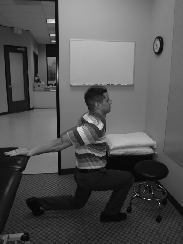
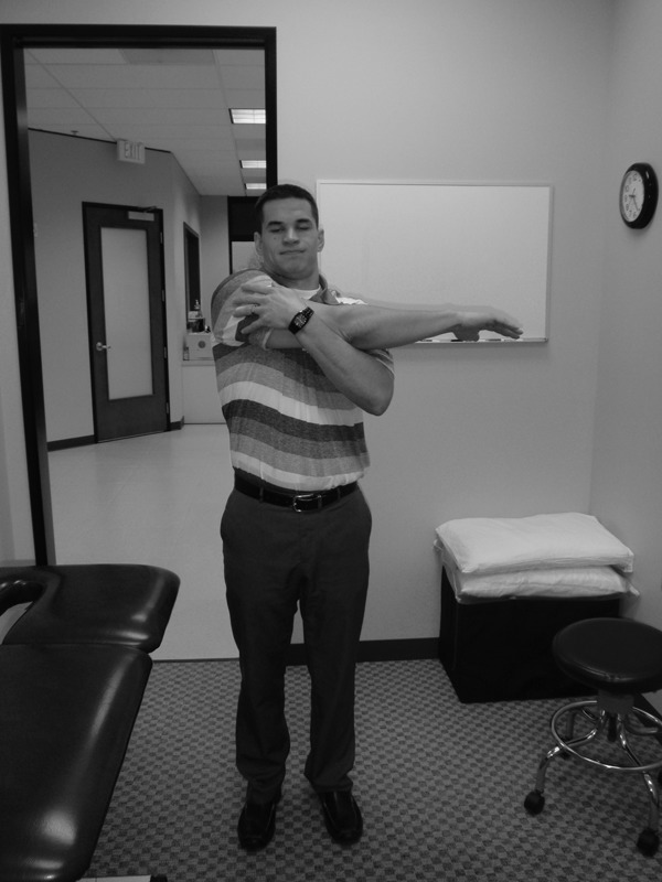
As internal rotation and extension were full and had no effect on his primary complaint, it was decided to explore repeated end-range flexion first, as this was his most restricted and painful movement. Performing this passively with assistance from a doorway was demonstrated to him (Fig. 1). His baseline symptoms were recorded and then he was asked to perform a series of 10 repetitions. During the shoulder flexion movement, he reported that the movement got increasingly painful with repetition. He was instructed to stop and baseline symptoms were re-examined. Shoulder flexion and abduction were now more obstructed confirming a worsening of symptoms and this movement was aborted. Based on his response, it was decided to explore repeated movements in the opposite direction according to the principles of MDT management.13,16 The patient was instructed on how to perform passive shoulder extension with overpressure (Fig. 2). This movement produced no appreciable change in his symptomatic or mechanical baselines. Based on these findings of sagittal plane movements, a trial of lateral movements in the transverse plane were conducted, as is commonly performed in the treatment of spinal disorders.16 The patient was instructed to perform passive shoulder horizontal adduction at 90° of shoulder flexion (Fig. 3). Once again the therapist demonstrated to the patient how to perform this passively with assistance from the other hand. His baselines were again recorded and then he was asked to perform a series of 10 repetitions. During the horizontal adduction exercise, he initially reported concordant pain and restriction at the end of the movement that improved with repetition. Upon completion of the exercise, the patient reported decreased pain with flexion, abduction, and external rotation. It was determined that this movement was his directional preference, as the range of movement in all the directions improved, resistance testing for external rotation and abduction had improved to 5/5 and the pain reproduced with resistive testing was abolished. Special testing of the shoulder was also conducted again for the purpose of the case and all three tests were now negative.
Figure 1.

Repeated shoulder flexion in the doorway.
Figure 2.

Repeated shoulder extension with overpressure.
Figure 3.

Repeated shoulder horizontal adduction.
Clinical Impression
Based on the results of the subjective and objective examinations, and the identification of directional preference, the provisional classification was a derangement of the shoulder. The most significant findings supporting this were loss of end-range shoulder mobility, pain with active and passive shoulder mobility, worsening of his symptoms with repeated flexion exercises, and a decrease followed by abolishment of his symptoms during the application of specific loading strategies that remained better, as well as a concurrent improvement in his mechanical presentation.
His presentation according to the principles of MDT was similar to that of a derangement syndrome and could not be classified as a posture or dysfunction syndrome. As previously described, pain due to posture syndrome is produced by sustained loading on normal tissue with no loss of shoulder mobility and no pain during movement.16 This patient exhibited both limited range of shoulder mobility and pain with resisted testing. With respect to extremity disorders, dysfunction syndrome is further subcategorized into an articular and contractile dysfunctions. Contractile dysfunction is determined by the presence of shoulder pain provoked through an arc of motion, particularly resisted mid-range movements, with a largely well preserved range of movements. In contrast, articular dysfunction is determined by the presence of restricted active and passive range of movements; with shoulder pain provoked at the end of the available range but not with resisted movements.16 Consequently, the patient’s clinical presentation did not match either of the dysfunction syndromes. Treatment of dysfunction syndromes involves remodeling of adaptively shortened tissue and this requires more time to resolve.13 Given that pain and mobility were rapidly improved over the course of a single treatment, only a diagnosis of a derangement was plausible.
Intervention
Treatment one: initial session
Based on the principles of MDT, the repeated movements used in the examination also serve as a treatment program. The patient reported a progressive decrease in shoulder symptoms with a concurrent improvement in mechanical baselines with repeated shoulder horizontal adduction, thus confirming a derangement. He was able to independently improve his primary complaints with self-generated forces thus negating the need for additional manual forces.13 Based on his response to the self-treatment strategies during the assessment; his self-treatment prescription for the next 48 hours was 10 repetitions of shoulder horizontal adduction with overpressure every 2 hours, or as needed to keep his baselines improved. The patient was instructed to move the affected shoulder as far as possible with every repetition. He was also precautioned and given indications to stop the exercise if he felt a worsening of his symptoms (i.e., a lasting increase in pain after the exercise or decreased shoulder mobility).
Follow up visits
The patient was examined 48 hours later. He reported that he had performed the exercises regularly every 2 hours, as instructed, and demonstrated the appropriate technique. He reported that he had no pain while resting and was able to sleep on his shoulder without complaints the previous night. He did report that reaching overhead was still painful, but improved. Previously established range of motion and strength baselines were examined and he exhibited flexion up to 170° with pain at end-range, abduction up to 170° with no pain, and external rotation was now pain free. Based on his improvement over the last 48 hours, we began with the horizontal adduction exercise that was previously prescribed. The patient performed four sets of 10 repetitions and was encouraged to add more overpressure with the unaffected hand. Four sets were indicated at this time because of progressively improved symptoms. Upon completion of the final set, the shoulder movements were now pain free with full mobility restored. Once again clinician techniques were not indicated at this time as he was still able to improve his symptoms independently. He was then instructed to continue with his current stretch, with more over pressure as this provided a greater improvement in his baselines. He was instructed to follow up after 1 week.
The patient followed up 7 days later for his third and final visit. He again reported that he had performed the exercises regularly, as instructed. He reported that he had no pain for the last 5 days and that he had been able to reach and lift overhead with restriction. On examination, all shoulder movements remained full and pain free, strength testing was 5/5 with no pain, and all special tests remained negative. To confirm that the derangement was reduced and maintained, the patient was put through a series of previously painful movements and reaching activities; which were now asymptomatic.
The MDT principles of management for a derangement syndrome are (1) reduction of the derangement, (2) maintenance of the reduction, (3) recovery of function, and (4) prophylaxis.16 In this case, the mechanical diagnosis of derangement syndrome was confirmed by a rapid change in pain and range of motion. Reduction of the derangement syndrome was confirmed by the patient reporting full pain free movement (including passive, active, and resistive) and the reduction continued over 5 days. Recovery of function was confirmed by the patient’s ability to perform all previously painful activities including; raising his arm overhead, lying on the affected shoulder, lifting weights at the gym, and performing yard work without complaints. Prophylaxis was addressed by instructing the patient to perform the prescribed exercise as frequently as needed in order to maintain full pain free mobility over the next 2 weeks and return to therapy only if needed.
The patient was contacted 2 weeks later. He reported no further symptoms and had full, pain free shoulder mobility. He stated that all daily and recreational activities were pain free and his CareConnections upper extremity outcome measure was 100%; indicating no functional limitation. He was instructed to continue with his reductive stretches as needed.
Discussion
Patients with shoulder problems are frequently encountered in health care.1,2 The natural history is not always good with only 50% of new cases in primary care recovering completely in 6 months.1,28 Various pathoanatomical mechanisms may give rise to shoulder symptoms, but the reliability for the examination process, by which a diagnosis is reached has been shown to be weak,29 and the diagnostic validity of specific tests has been shown to be only moderate at best.30–32
When treating patients with shoulder problems, specific pathoanatomical diagnosis are frequently used,1,29 but the prevalence of non-specific symptoms at the shoulder has not been explored as we continue to search for pathoanatomic origins of pain. The concept of non-specific musculoskeletal pain and sub-grouping patients based on specific inclusion criteria is well established in the field of low back pain.10–12,33,34 McKenzie and May13 proposed the idea of sub-grouping patients with non-specific shoulder pain based on distinct inclusion criteria into one of the three mechanical syndromes: derangement, dysfunction, or posture syndrome. Classification is based on response to repeated end-range movements and not on pathoanatomical diagnosis. Treatment is then applied according to that specific classification. In the derangement syndrome, repeated end-range loading in the appropriate direction progressively decreases pain, with a simultaneous improvement in the range of motion.16 Likewise, movements in the opposite direction may increase symptoms and limitations in movement. This is known as directional preference and previous studies on spinal pain have demonstrated that the presence of directional preference can improve prognosis for function and pain outcomes.35 This case report provides an example of such a response. Not only were his symptoms quickly resolved with a self-instructed regimen based on MDT principles of management, but previously used special tests, indicating specific pathoanatomical involvement, were also rapidly changed. These tests were used for the purpose of this case report as a mechanical baseline to demonstrate a rapid change and not as a diagnostic test to rule in/out a pathoanatomical structure/tissue.
From a theoretical perspective, a variety of structures could have been the source of the patient’s symptoms, including, but not limited to the bursa, tendon, muscle belly, labrum, acromion, or capsule. The examination of this patient may have been prolonged by an exhaustive search of the involved pathoanatomical source, including a plethora of special tests that have unproven validity and uncertain reliability.29–32 Instead, a complete evaluation utilizing a thorough history followed by a concise physical examination was used to sub-classify the condition and quickly improve this patient’s symptomatic and mechanical baselines once a directional preference was identified. The evaluation and treatment methods used in this case allowed active patient involvement, without the use or need for passive modalities or manual techniques, which the patient stated improved his self-confidence in addressing the problem independently and preventing further recurrence.
The application of MDT in the extremities is growing as there is more published literature regarding its utilization. There are now reports on its reliability23 and effectiveness14,15 with specific regards to the extremities. Aina14 and Littlewood15 previously described the principles of MDT management for the shoulder extremity and researchers have since expanded beyond the shoulder and discussed MDT’s usefulness in treating the sacro-iliac joint36 and temporomandibular joint.37 The purpose of this case report was not only to expand upon the previous literature by further explanation of the use of MDT principles in the management of a shoulder derangement syndrome but also to emphasize the importance of moving beyond a pathoanatomical model toward a classification system with the use of MDT principles and directional preference.
Conclusion
This case report details the history and assessment of a patient who presented with non-specific shoulder pain. During physical examination, repeated movements were utilized to sub-classify his condition; this provided a rapid decrease in his symptoms and improvement in his range of motion and pain. The self-management strategy was an individualized program that arose directly from the clinical assessment emphasizing repeated end-range movements and the identification of directional preference. It demonstrates that sub-grouping patients based on symptomatic and mechanical responses may be beneficial in the treatment of shoulder pain.
References
- 1.Van der Windt DA, Koes BW, de Jong BA, Bouter LM. Shoulder disorders in general practice: incidence, patient characteristics, and management. Ann Rheum Dis. 1995;54:959–64 [DOI] [PMC free article] [PubMed] [Google Scholar]
- 2.May S. An outcome audit for musculoskeletal patients in primary care. Physiother Theory Pract. 2003;19:189–98 [Google Scholar]
- 3.Cyriax J. Textbook of orthopaedic medicine, volume 1: diagnosis of soft tissue lesions. London: Bailliere Tindall; 1982 [Google Scholar]
- 4.Lewis JS, Green AS, Dekel S. The aetiology of subacromial impingement syndrome. Physiotherapy. 2001;87:458–69 [Google Scholar]
- 5.Carette S. Adhesive capsulitis – research advances frozen in time? J Rheumatol. 2000;27:1329–31 [PubMed] [Google Scholar]
- 6.Khan KM, Cook JL, Taunton JE, Bonar F. Overuse tendinosis, not tendonitis: part 1: a new paradigm for a difficult clinical problem. Phys Sportsmed. 2000;28:38–46 [DOI] [PubMed] [Google Scholar]
- 7.Hanchard N, Cummins J, Jeffries C. Evidence-based clinical practice guidelines for the diagnosis, assessment and physical therapy management of shoulder impingement syndrome. London: Chartered Society of Physiotherapy; 2004 [Google Scholar]
- 8.Hegedus EJ, Goode A, Campbell S, Morin A, Tamaddoni M, Moorman CT, et al. Physical examination tests of the shoulder: a systematic review with meta-analysis of individual tests. Br J Sports Med. 2008;42:80–92 [DOI] [PubMed] [Google Scholar]
- 9.Hughes PC, Taylor NF, Green RA. Most clinical tests cannot accurately diagnose rotator cuff pathology: a systematic review. Aust J Physiother. 2008;54:159–70 [DOI] [PubMed] [Google Scholar]
- 10.Brennan GP, Fritz JM, Hunter SJ, Thackeray A, Delitto A, Erhard RE. Identifying subgroups of patients with acute/subacute ‘non-specific’ low back pain: results of a randomized clinical trial. Spine. 2006;31:623–31 [DOI] [PubMed] [Google Scholar]
- 11.Long A, Donelson R, Fung T. Does is matter which exercise? A randomized control trial of exercise for low back pain. Spine. 2004;29:2593–602 [DOI] [PubMed] [Google Scholar]
- 12.May S, Aina A. Centralizations and directional preference: a systematic review. Man Ther. 2012;17:497-506 [DOI] [PubMed] [Google Scholar]
- 13.McKenzie R, May S. The human extremities: mechanical diagnosis and therapy. Waikanae: Spinal Publications; 2000 [Google Scholar]
- 14.Aina A, May S. Case report: a shoulder derangement. Man Ther. 2005;10:159–63 [DOI] [PubMed] [Google Scholar]
- 15.Littlewood C, May S. Case report: a contractile dysfunction. Man Ther. 2007;12:80–3 [DOI] [PubMed] [Google Scholar]
- 16.McKenzie R, May S. The Lumbar Spine: mechanical diagnosis and therapy. Waikanae: Spinal Publications; 2003 [Google Scholar]
- 17.Razmjou H, Kramer JF, Yamada R X. Intertester reliability of the McKenzie evaluation in assessing patients with mechanical low back pain. J Orthop Sports Phys Ther. 2000;30:368–89 [DOI] [PubMed] [Google Scholar]
- 18.Fritz JM, Delitto A, Vignovic M, Busse RGX. Interrater reliability of judgments of the centralisation phenomenon and status change during movement testing in patients with low back pain. Arch Phys Med Rehabil. 2000;81:57–61 [DOI] [PubMed] [Google Scholar]
- 19.Kilpikoski S, Airaksinem O, Kankaanpaa M, Lemine P, Videman T, Alen M. Interexaminer reliability of low back pain assessment using the McKenzie method. Spine. 2002;E207–14 [DOI] [PubMed] [Google Scholar]
- 20.Sufka A, Hauger B, Trenary M, Bishop B, Hagen A, Lozon R, et al. Centralization of low back pain and perceived functional outcome. J Orthop Sports Phys Ther. 1998;27:205–12 [DOI] [PubMed] [Google Scholar]
- 21.Werneke M, Hart DL, Cook D. A descriptive study of the centralization phenomenon. A prospective analysis. Spine. 1999;24:676–83 [DOI] [PubMed] [Google Scholar]
- 22.Werneke M, Hart DL. Centralization phenomenon as a prognostic factor for chronic pain or disability. Spine. 2001;26:758–65 [DOI] [PubMed] [Google Scholar]
- 23.May S, Ross J. The McKenzie classification system in the extremities: a reliability study using McKenzie assessment forms and experienced clinicians. J Manipulative Physiol Ther. 2009;32:556–63 [DOI] [PubMed] [Google Scholar]
- 24.Schunk C, Rutt R. TAOS Functional Index: orthopaedic rehabilitation outcomes tool. J Rehabil Outcomes Measure. 1998;2:55–61 [Google Scholar]
- 25.Mannifold SG, McCann PD. Cervical radiculitis and shoulder disorders. Clin Orthop. 1999;368:105–13 [PubMed] [Google Scholar]
- 26.Nada R, Gupta S, Kanapathipilai P, Loi RYL, Rangan A. An assessment of the interexaminer reliability of clinical tests for subacromial impingement and rotator cuff integrity. Eur J Orthop Surg Traumatol. 2008;18:495–500 [Google Scholar]
- 27.Fodor D, Poanta L, Felea I, Rednic S, Bolosiu H. Shoulder impingement syndrome: correlations between clinical tests and unltrasonographic findings. Ortop Traumatol Rehabil. 2009;11:120–26 [PubMed] [Google Scholar]
- 28.Croft P, Pope D, Silmand The clinical course of shoulder pain: prospective cohort study in primary care. BMJ. 1996;313:601–2 [DOI] [PMC free article] [PubMed] [Google Scholar]
- 29.Liesdek C, van der Windt DA, Koes BW, Bouter LM. Soft-tissue disorders of the shoulder. Phys Ther. 1997;83:2–17 [Google Scholar]
- 30.Calis M, Akgun K, Birtane M, Karacan I, Calis H, Tuzun F. Diagnostic values of clinical diagnostic tests in subacromial impingement syndrome. Ann Rheum Dis. 2000;59:44–7 [DOI] [PMC free article] [PubMed] [Google Scholar]
- 31.Dessaur WA, Margarey ME. Diagnostic accuracy of clinical tests for superior labral anterior posterior lesions: a systematic review. J Orthop Sports Phys Ther. 2008;38:341–52 [DOI] [PubMed] [Google Scholar]
- 32.Schellingerhout JM, Verhagen AP, Thomas S, Koes BW. Lack of uniformity in diagnostic labeling of shoulder pain: time for a different approach. Man Ther. 2008;13(6):478–83 [DOI] [PubMed] [Google Scholar]
- 33.Van Dillen LR, Sahrmann SA, Norton BJ, Caldwell CA, McDonnell MK, Bloom NJ. Movement system impairment-based categories for low back pain: Stage 1 validation. J Orthop Sports Phys The. 2003;33:126–42 [DOI] [PubMed] [Google Scholar]
- 34.O’Sullivan P. Classification of lumbopelvic pain disorders-why is it essential for management. Man Ther. 2006;11:169–70 [DOI] [PubMed] [Google Scholar]
- 35.Werneke MW, Hart DL, Cutrone G, Oliver D, McGill T, Weinberg J. Association between directional preference and centralization in patients with low back pain. J Orthop Sports Phys Ther. 2011;41:22–31 [DOI] [PubMed] [Google Scholar]
- 36.Horton SJ, Franz A. Mechanical diagnosis and therapy approach to assessment and treatment of derangement of the sacro-iliac joint. Man Ther. 2007;12:26–32 [DOI] [PubMed] [Google Scholar]
- 37.Krog C, May S. Derangement of the temporomandibular joint: a case study using mechanical diagnosis and therapy. Man Ther. 2012;17:483–86 [DOI] [PubMed] [Google Scholar]


