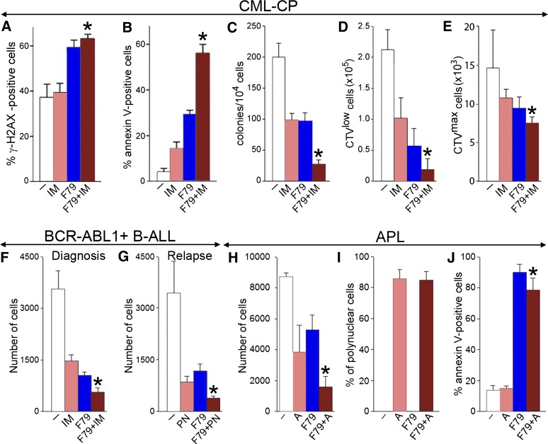
Figure 5.
F79 aptamer enhanced the effects of standard treatment in leukemia cells displaying genetic BRCA-deficient phenotype. (A-C) Lin−CD34+ CML-CP cells from 3 to 4 patients were untreated (−) (white) and treated with 1 μM imatinib (IM) (salmon), 5μM F79 (blue), and IM+F79 (brown) for 48 hours. (A) Percentage of cells with more than 20 γ-H2AX foci; *P < .01 in comparison with IM. (B) Percentage of annexin V–positive cells; *P < .001 in comparison with IM. (C) Number of colonies ± SD; *P < .01 in comparison with IM. (D,E) Lin−CD34+ CML-CP cells from 3 to 5 patients/group were labeled with CPD and incubated for 5 days with 1 μM IM (salmon), 5 μM F79 (blue), or IM+F79 (brown) or left untreated (white). (D) Mean number of Lin−CD34+CD38−CPDlow proliferating LSCs; *P = .02 in comparison with IM. (E) Mean number of Lin−CD34+CD38−CPDmax quiescent LSCs; *P = .01 in comparison with IM. (F) Mean number of xenograft cells from 3 freshly diagnosed BCR-ABL1 B-ALL patients treated for 5 days with 1 μM IM (salmon), 5 μM F79 (blue), IM+F79 (brown), or left untreated (white); *P < .01 in comparison with IM. (G) Mean number of xenograft cells from 3 relapsed B-ALL patients carrying BCR-ABL1(T315I) mutation treated for 5 days with 12.5 nM ponatinib (PN) (salmon), 5 μM F79 (blue), or PN+F79 (brown) or left untreated (white). Results represent mean number ± SD of living cells; *P < .05 in comparison with PN. (H-J) APL primary cells from 3 patients were incubated with 5 μM F79 aptamer (F79), 4 μM ATRA (A), or F79+ATRA or were left untreated (−). (H) Living cells were counted in Trypan blue 9 days later. (I) Polynuclear differentiated cells counted after staining in Giemsa. (J) Annexin V–positive cells assessed by a fluorescence-activated cell sorter; *P < .05 in comparison with group A.

