Abstract
Background
Baseball players are susceptible to a number of specific upper extremity injuries secondary to batting, pitching, or fielding. Fractures of the hook of hamate have been known to occur in batters. The purpose of this study is to present our experience with the surgical management of hook of hamate fractures and their short-term impact on the playing capability of competitive baseball players.
Methods
A retrospective chart review was performed on patients with hook of hamate fractures between the years 2000 and 2012. The inclusion criteria were (1) hook of hamate fracture, (2) competitive baseball players, and (3) surgical treatment of the injury. Patient demographics, mechanism of injury, surgical treatment, and outcome were collected from the medical records. Information on return to play was collected from the Internet when applicable.
Results
There were seven male patients that underwent eight procedures. The mechanism of injury was attributed to batting in six cases and rogue pitches in two cases. All surgeries consisted of hamate hook excision and ulnar tunnel decompression. One patient had concomitant carpal tunnel release. The median time between surgery and return to play was 5.7 weeks (range, 4.3 to 10.4 weeks).
Conclusions
The mechanism of hook of hamate fractures in baseball players is predictable, most often developing secondary to repetitive swinging. This injury may occur at all levels of competition. Ulnar tunnel decompression with hook of hamate excision provides good outcomes, with minimal complications and early return to play.
Keywords: Baseball, Fracture, Hook of hamate
Introduction
Fractures of the hook of the hamate comprise 2 to 4 % of all carpal fractures and occur most frequently among individuals who play golf, racquet sports, or baseball [24]. Baseball batters are exposed to either acute fractures or chronic stress injuries secondary to the impingement of the bat against the hook of the hamate during a batting swing [23]. These injuries often present as deep, ill-defined ulnar-sided wrist pain and routine radiographs may not identify the fracture.
Although a number of studies have reported the diagnostic and therapeutic management of hook of hamate fractures in athletes, and in baseball players specifically, many accounts were published over 20 years ago [3, 5, 16, 23, 27, 28]. Over the past few decades however, athletic performance across the board has improved due to improved training techniques, nutrition, technology, and equipment [6, 8, 11, 21, 29]. Current competitive athletes therefore are not entirely representative of their predecessors a few decades ago. The purpose of this study was to present a revised account of the etiology and management of hook of hamate fractures and their short-term impact on the playing capabilities of competitive baseball players.
Materials and Methods
This study was approved by our institutional review board and was conducted accordingly under its protocol and guidelines. A retrospective chart review of competitive baseball players treated for fractures of the hook of hamate between the years 2000 and 2012 was performed. The inclusion criteria were (1) hook of hamate fracture, (2) competitive baseball players, and (3) surgical treatment of the injury. Players were considered competitive if they participated at the high school, college, or professional level and were active members of their team at the time of injury. Individuals under the age of 18 were excluded from the study. Variables recorded from the medical records included basic patient demographics, the mechanism of injury, clinical findings, preoperative imaging, surgical diagnosis, surgical treatment, and complications. Information regarding the athlete’s return to play was collected from the Internet when not available in the medical record.
Eight hook of hamate fractures in seven competitive baseball players were identified. All patients were males and included three professional players (one major league, two minor league), three collegiate players, and one high school player. The subjects had a mean age of 21.7 years (range 18–26 years) at the time of surgery. The mechanism of injury was attributed to batting in six cases and a direct hit by a pitch in two cases. The dominant hand was involved in four cases. Out of the players that were injured during batting, all right-handed batters injured their left hand, while all left-handed batters injured their right hand. One of the two players who batted both right and left-handed presented with bilateral hamulus fractures. Background player information is summarized in Table 1.
Table 1.
Baseball player background information
| Patient | Competition level | Position | Hand dominance | Batting side | Side affected | Mechanism of injury | Age at surgery |
|---|---|---|---|---|---|---|---|
| 1 | NCAA division I | Outfielder | Right | Both | Bilateral | During batting | 21 |
| 2 | MLB | Outfielder | Left | Left | Right | During batting | 23 |
| 3 | MiLB (class AA) | 2nd base | Right | Both | Right | During batting | 24 |
| 4 | NCAA division I | Outfielder | Right | Right | Left | During batting | 20 |
| 5 | MiLB (class AA) | Outfielder | Right | Left | Right | During batting | 26 |
| 6 | NCAA division III | Outfielder | Right | Right | Left | Hit by pitch directly | 21 |
| 7 | High school | 1st base/pitcher | Left | Left | Right | Hit by pitch directly | 18 |
NCAA National Collegiate Athletic Association, MLB Major League Baseball, MiLB Minor League Baseball
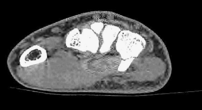
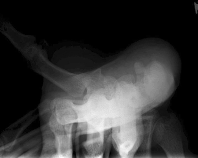
Of the eight fractures, two were considered nonunions. One patient had received conservative treatment that consisted of immobilization with an ulnar gutter splint for 6 weeks, several months prior to presentation (patient #1). The second patient, patient #7, had been symptomatic for 3 months prior to presentation and a CT scan demonstrated evidence of nonunion (Fig. 1). During clinical examination, patients had pain or tenderness over the hook of hamate on the volar surface of the hand. Only one patient reported paresthesias of the small and ring fingers. None of the patients demonstrated clawing or weakness or wasting of the intrinsic hand muscles. Three of the patients exhibited a positive Tinel’s sign at Guyon’s canal. The presence of a clinically suspected hook of hamate fracture was always confirmed with imaging studies. The modality of preoperative imaging varied. Posteroanterior and lateral plain films were initially obtained to rule out hook of hamate as well as other hand and wrist bony pathology. However, when the plain films were deemed inconclusive or negative, a CT scan or MRI was obtained. A summary of the physical findings for each patient is shown in Table 2 (Figs. 1 and 2).
Fig. 1.

CT scan illustrating a hook of hamate nonunion in an 18-year-old male
Table 2.
Physical findings
| Patient | Preoperative clinical exam |
|---|---|
| 1 | Negative Tinel’s sign at Guyon’s canal; pain in palm; no paresthesias or numbness in the ulnar nerve distribution; no intrinsic muscle weakness |
| Negative Tinel’s sign at Guyon’s canal; pain over hook of hamate; no paresthesias or numbness in ulnar nerve distribution; no intrinsic muscle weakness | |
| 2 | Positive Tinel’s sign at Guyon’s canal; point tenderness over hook of hamate; no paresthesias or numbness in ulnar nerve distribution; no intrinsic muscle weakness |
| 3 | Negative Tinel’s sign at Guyon’s canal; point tenderness over hook of hamate; no paresthesias or numbness in ulnar nerve distribution; no intrinsic muscle weakness |
| 4 | Positive Tinel’s sign at Guyon’s canal; point tenderness over hook of hamate; no paresthesias or numbness in ulnar nerve distribution; no intrinsic muscle weakness |
| 5 | Positive Tinel’s sign at Guyon’s canal; point tenderness over hook of hamate; paresthesias of ring and small fingers but no numbness; no intrinsic muscle weakness |
| 6 | Negative Tinel’s sign at Guyon’s canal; pain over hook of hamate; no paresthesias or numbness in ulnar nerve distribution; no intrinsic muscle weakness |
| 7 | Negative Tinel’s sign at Guyon’s canal; pain over hook of hamate; no paresthesias or numbness in ulnar nerve distribution; no intrinsic muscle weakness |
Fig. 2.

Carpal tunnel view revealing a fracture line at the mid-level of the hook of hamate
The surgery performed to treat this injury was excision of the hook of the hamate. This procedure was performed under either regional or general anesthesia, with tourniquet control. A Bruner-type incision was made starting just proximal to the wrist crease and extended over Guyon’s canal. Skin flaps were elevated and all bleeding was controlled with microcautery. The ulnar nerve and artery were identified proximally and followed distally, releasing Guyon’s canal. The ulnar nerve was followed to its deep branch and the deep arterial branch and mobilized. The sensory branches of the ulnar nerve were also mobilized. At all times, care was taken to avoid undue traction to the ulnar nerve and its branches. Ulnar nerve decompression was routinely performed in our patients as a preemptive step to avert postoperative ulnar nerve palsy, either due to compression within Guyon’s canal secondary to postoperative swelling or due to iatrogenic retraction [26], and to improve visualization of the neurovascular structures, as the deep motor branch of the ulnar nerve is at risk during both the dissection and excision of the hook of the hamate [14, 26]. The transverse carpal ligament was released from the hook of the hamate, but the attachment to the pisiform was preserved. The hook was observed, and subperiosteal dissection was carried out to facilitate removal of the hook, which was found to be loose. The base of the hook was then rongeured and rasps were employed to further smoothen the base of the fracture. The fingers were then passively moved to ensure that there was no impingement between the flexor tendons and the underlying resurfaced hamate. The wound was copiously irrigated and the tourniquet was deflated. Finally, the wound was closed in layers and a sterile dressing and a short arm splint were applied.
One patient had concomitant carpal tunnel release due to symptoms consistent with carpal tunnel syndrome. Postoperatively, a volar splint was applied for 3–4 weeks. Return to play was encouraged 4–6 weeks after surgery.
Results
With regard to the impact of these injuries on the playing capabilities of the athletes, three patients continued to play with the injury until the conclusion of the season (two collegiate, one minor league). Patient #1 was treated by cast immobilization for 6 weeks, followed by rehabilitation for 2 weeks at the initial time of injury. This resulted in asymptomatic fibrous nonunion, confirmed by CT scan. Although this collegiate player was able to play a whole season without further aggravation of his right hand injury, he was expecting to be drafted into a professional team. Surgery was therefore performed to prevent future aggravation of the nonunion. The time between injury and surgery was 9 months. Patient #4, also a collegiate player, continued to participate in games for almost 2 months after his injury and eventually had surgery at the end of the season. In the interval, he had received a steroid injection with minimal relief by a separate physician. Patient #5 played for 2 weeks with the injury until the end of the season. At the initial time of injury, the patient was immediately assessed; however, the diagnosis was missed and the patient was cleared to return to play. Due to persistent symptoms, the patient was reevaluated and the diagnosis was made. Two players (major league and collegiate) underwent surgery during the season and missed 28 and 25 games, respectively. One minor league player missed 36 games of a new season recovering from surgery. The median time between injury and surgical treatment was 33 days (range, 3 to 270 days). The median time between surgery and return to play was 5.7 weeks (range, 4.3 to 10.4 weeks). One minor complication was noted in one patient who experienced scar hypersensitivity. The short-term career impact of hook of hamate fractures on competitive baseball players is summarized in Table 3.
Table 3.
Short-term impact of hamate hook fractures on the career of competitive baseball players
| Patient | Time between injury and surgery (months) | Games missed secondary to hook of hamate fracture | Time from surgery until return to play (weeks) |
|---|---|---|---|
| 1 | 4 | – | 5.7 |
| 9 | 0—played until the end of the season; surgery performed at the end of the season | 5.7 | |
| 2 | 0.1 | 28 after surgery | 10.4 |
| 3 | 0.5 | 36 after surgery | 5.3 |
| 4 | 1.7 | 0—played until the end of the season; surgery performed at the end of the season | 5.6 |
| 5 | 0.5 | 0—played until the end of the season; surgery performed at the end of the season | – |
| 6 | 0.2 | 25 after surgery | 4.3 |
| 7 | 3 | – | 6.3 |
“–” unknown or missing data
aUnderwent surgery during the off-season and began a new season on the disabled list
Discussion
Baseball players are susceptible to a number of characteristic upper extremity injuries including rotator cuff impingement, osteochondritis dissecans of the capitellum, and medial collateral ligament injury of the elbow in pitchers [12, 22]; mallet finger injuries in fielders [18]; and hook of hamate fractures in batters [3, 7, 17, 23–25, 27, 28]. Initial accounts of hook of hamate fractures portrayed this injury as rare, in individuals who either fell on an outstretched hand or struck the ground while swinging a golf club [3–5, 23, 25, 28]. In a review of the literature conducted by Bishop and Beckenbaugh in 1987, hook of hamate fractures were believed to account for less than 2 % of carpal fractures [3]. More recently, the incidence of hamulus fractures among the general population has been quoted to be between 2 and 4 % of all carpal fractures [24]. However, the true incidence of this fracture may be higher than reported due to the possibility of missed diagnoses [17].
The body of the hamate is located dorsally and on the ulnar aspect of the carpus. Its hook (also known as the hamulus) is a long, thin structure that projects radially and volarly to the hypothenar eminence. The hamulus serves as an attachment site for the transverse carpal ligament, pisohamate ligament, flexor digiti minimi, and opponens digiti minimi [25, 28]. Owing to its anatomic proximity, an acute or chronically non-united displaced hamulus fracture may impinge on the adjacent branch of the ulnar nerve or tendons [16, 19, 27, 28]. These sequelae lend support to early operative treatment. In this study, three out of seven patients exhibited signs and symptoms of ulnar nerve neuropathy at the wrist, including a positive Tinel’s sign at Guyon’s canal, and were subsequently diagnosed with ulnar tunnel syndrome (UTS). The diagnosis of UTS based on Tinel’s sign alone may not be the most appropriate confirmatory study during the workup of ulnar neuropathy at the wrist. However, in the context of hook of hamate fractures and the proximity of Guyon’s canal to the hamate, Tinel’s sign was thought to be suggestive of UTS. The diagnosis of UTS is often made by a combination of the clinical impression and imaging findings, whereas electrodiagnostic tests are usually used to confirm the diagnosis [1]. Another potential consequence of a hook of hamate fracture is damage to the adjacent flexor tendons. A sharp bony fragment may irritate the adjacent flexor tendons, eventually fraying or completely rupturing either flexor digitorum profundus or superficialis [16, 19, 27, 28]. We did not see this complication in any of our patients, although at times, many months had passed between injury and surgery.
Compared to the base of the hamate, the hook of the hamate has fewer and smaller vascular foramina, and therefore the relatively poor blood supply to the hook is a contributing factor for fracture nonunion [13]. The results of this study are in agreement with the current literature, which suggests that surgical excision of a hook of hamate fracture produces favorable outcomes with minimal complications and a quick return to play in competitive athletes [23–25, 27, 28, 30]. Parker et al. presented a series of five patients with hook of hamate fractures. Four patients were professional baseball players. The players that were diagnosed with the fracture during the season and underwent surgical excision of the fragment were able to return to play within 6 weeks [23]. In our study, the two players that elected to have the surgical excision of the fragment mid-season missed 25 and 28 games spanning 4 to 6 weeks.
Demirkan et al. investigated the biomechanical implications of hook excision in a cadaveric study [9]. The results indicated that hamulus excision resulted in a decreased flexor digitorum profundus force across the long, ring, and small fingers. The magnitude of the force reduction varied depending on the position of the wrist and was enhanced with wrist extension and ulnar deviation. Furthermore, the excision produced increased excursion of the flexor digitorum profundus tendons as they became more lax and shifted to the ulnar aspect of the wrist [9]. Despite these cadaveric findings, none of our patients complained of any postoperative grip weakness or other clinically meaningful activity limitations, although it would have been useful if these objective findings were obtained in our patients.
The mechanism leading to fractures of the hook of hamate remains quite predictable in racquet sports and sports that involve clubs or bats, such as tennis, baseball, cricket, and golf [3, 17, 23–25, 27, 28]. While baseball players are the athletes of primary interest in this study, cricket players and golfers can also acquire this injury through a similar mechanism [2, 16, 20, 24]. Although in two instances injury was attributed to a rogue pitch, i.e., the batters were hit directly by a pitch on their hand, the results of this study support the conclusion that it is the hand that abuts against the end of the bat that is most vulnerable to injury [3, 24]. All of the right-handed batters injured their left hand while all of the left-handed batters injured their right hand. In one instance where the batter was a switch hitter, injury was sustained in both hands by switching back and forth between right- and left-handed batting stances. It is unclear, however, whether all fractures are the result of repeated microtrauma or an acute traumatic incident.
Other studies have proposed alternative surgical and nonsurgical treatments for these fractures such as open reduction and internal fixation (ORIF), splinting/casting, and ultrasound therapy [10, 15, 25, 31]. ORIF of hamulus fractures is aimed at restoring the native anatomy and function of the carpal bone. Scheufler et al. compared three patients that underwent ORIF to four patients that underwent fragment excision for hook of hamate fracture. The patients who underwent ORIF demonstrated significantly higher grip strength values postoperatively. These results however cannot be attributed to the surgical procedure alone due to the small sample size and marked age difference between the ORIF (44.5 years) and excision groups (20.3 years) [25]. Although ORIF may potentially yield improved postoperative grip strength for patients with these fractures, the recovery period is twice as long as fragment excision. The three patients treated with ORIF received a lower arm cast for 2 weeks postoperatively followed by physiotherapy and limited use of the hand for 6 weeks. Therefore, the utility of ORIF for hamulus fractures may be limited to the off-season in athletes, who generally desire quicker return to play.
Non-operative treatment should be discussed for those players who prefer to delay surgery. The risks associated with this decision however should be discussed in detail. At least three of our patients continued to play with the injury throughout the season (Table 3). Ultrasound therapy has some reported success. A case study of two baseball players with hook of hamate fracture nonunion 5 and 3.5 months post-injury were successfully treated with ultrasound therapy. The patients received therapy for 20 min a day for 4 months and were limited to daily activities during this time. Bone union was confirmed with a CT scan, and neither patient reported any pain or complications at a 1-year follow-up [15]. However, these athletes were 15 and 16 years of age and began treatment many months after their injury. Furthermore, it is possible that within the 4 months the fractures healed independently of the ultrasound therapy. Professional athletes may find it difficult to comply with strict and prolonged treatment guidelines due to time constraints and a hectic playing schedule, making this a less preferred treatment option.
The limitations of the study include the small sample size and a retrospective study design. In addition, patient rated outcomes such as a visual analog pain scale or QuickDASH were not available primarily because, in our experience, competitive athletes do not return for follow-up unless complications develop. Rather, we used game log records on the official team websites as surrogate markers of patient function. We believe that this method of tracking patient progress is sufficient given a primary aim of treatment, i.e., return to play. The surgical procedures performed for these athletes were not compared to other methods of treatment, such as short arm splint casts or ORIF. Also, the player’s hand positioning on the bat and the mechanics of their swing were not evaluated. Lack of data in this area raises the question about whether or not the injured baseball players were more susceptible to this injury due to unique anatomy or swinging techniques.
In conclusion, hook of hamate fractures remain a stubborn problem among competitive baseball players. The results of this study affirm the evidence that hook of hamate excision and ulnar tunnel decompression provide good outcomes, with minimal complications and relatively early return to play. Further research aimed at preventing this predictable injury is warranted.
Acknowledgments
Conflict of Interest
None of the authors have any conflicts of interest to declare.
References
- 1.Bachoura A, Jacoby SM. Ulnar tunnel syndrome. Orthop Clin North Am. 2012;43:467–474. doi: 10.1016/j.ocl.2012.07.016. [DOI] [PubMed] [Google Scholar]
- 2.Belliappa PP, Barton NJ. Hand injuries in cricketers. J Hand Surg. 1991;16B:212–214. doi: 10.1016/0266-7681(91)90180-v. [DOI] [PubMed] [Google Scholar]
- 3.Bishop AT, Beckenbaugh RD. Fracture of the hamate hook. J Hand Surg. 1987;13A:135–139. doi: 10.1016/0363-5023(88)90217-1. [DOI] [PubMed] [Google Scholar]
- 4.Carroll RE, Lakin JF. Fracture of the hook of hamate: radiographic visualization. Iowa Orthop J. 1993;13:178–182. [PMC free article] [PubMed] [Google Scholar]
- 5.Carter PR, Eaton RG, Littler JW. Ununited fracture of the hook of the hamate. J Bone Joint Surg. 1977;59A:583–588. [PubMed] [Google Scholar]
- 6.Carter AB, Kaminski TW, Douex AT, Jr, et al. Effects of high volume upper extremity plyometric training on throwing velocity and functional strength ratios of the shoulder rotators in collegiate baseball players. J Strength Cond Res. 2007;21:208–215. doi: 10.1519/00124278-200702000-00038. [DOI] [PubMed] [Google Scholar]
- 7.Collins CL, Comstock RD. Epidemiological features of high school baseball injuries in the United States, 2005–2007. Pediatrics. 2008;121:1181–1187. doi: 10.1542/peds.2007-2572. [DOI] [PubMed] [Google Scholar]
- 8.Crisco JJ, Greenwald RM, Blume JD, et al. Batting performance of wood and metal baseball bats. Med Sci Sports Exerc. 2002;34:1675–1684. doi: 10.1097/00005768-200210000-00021. [DOI] [PubMed] [Google Scholar]
- 9.Demirkan F, Calandruccio JH, Diangelo D. Biomechanical evaluation of flexor tendon function after hamate hook excision. J Hand Surg. 2003;28A:138–143. doi: 10.1053/jhsu.2003.50005. [DOI] [PubMed] [Google Scholar]
- 10.Dunn AW. Fractures and dislocations of the carpus. Surg Clin North Am. 1972;52:1513–1538. doi: 10.1016/s0039-6109(16)39895-4. [DOI] [PubMed] [Google Scholar]
- 11.Ebben WP, Hintz MJ, Simenz CJ. Strength and conditioning practices of Major League Baseball strength and conditioning coaches. J Strength Cond Res. 2005;19:538–546. doi: 10.1519/R-15464.1. [DOI] [PubMed] [Google Scholar]
- 12.Erne HC, Zouzias IC, Rosenwasser MP. Medial collateral ligament reconstruction in the baseball pitcher’s elbow. Hand Clin. 2009;25:339–346. doi: 10.1016/j.hcl.2009.05.006. [DOI] [PubMed] [Google Scholar]
- 13.Failla JM. Hook of hamate vascularity: vulnerability to osteonecrosis and nonunion. J Hand Surg. 1993;18A:1075–1079. doi: 10.1016/0363-5023(93)90405-R. [DOI] [PubMed] [Google Scholar]
- 14.Fredericson M, Kim BJ, Date ES, et al. Injury to the deep motor branch of the ulnar nerve during hook of hamate excision. Orthopedics. 2006;29:456–458. doi: 10.3928/01477447-20060501-08. [DOI] [PubMed] [Google Scholar]
- 15.Fujioka H, Tanaka J, Yoshiya S, et al. Ultrasound treatment of nonunion of the hook of hamate in sports activities. Knee Surg Sports Traumatol Arthrosc. 2004;12:162–164. doi: 10.1007/s00167-003-0425-0. [DOI] [PubMed] [Google Scholar]
- 16.Futami T, Aoki H, Tsukamoto Y. Fractures of the hook of hamate in athletes. 8 cases followed for 6 years. Acta Orthop Scand. 1993;64:469–471. doi: 10.3109/17453679308993670. [DOI] [PubMed] [Google Scholar]
- 17.Guha AR, Marynissen H. Stress fractures of the hook of hamate. Br J Sports Med. 2002;36:224–225. doi: 10.1136/bjsm.36.3.224. [DOI] [PMC free article] [PubMed] [Google Scholar]
- 18.Lubahn JD. Mallet finger fractures: a comparison of open and closed technique. J Hand Surg. 1989; 14 (2pt 2):394–6. [DOI] [PubMed]
- 19.Minami A, Ogino T, Usui M, et al. Finger tendon rupture secondary to fracture of the hamate. A case report. Acta Orthop Scand. 1985;56:96–97. doi: 10.3109/17453678508992992. [DOI] [PubMed] [Google Scholar]
- 20.Murray PM, Cooney WP. Golf-induced injuries of the wrist. Clin Sports Med. 1996;15:85–109. [PubMed] [Google Scholar]
- 21.Nicholls RL, Elliott BC, Miller K. Impact injuries in baseball: prevalence, aetiology and the role of equipment performance. Sports Med. 2004;34:17–25. doi: 10.2165/00007256-200434010-00003. [DOI] [PubMed] [Google Scholar]
- 22.Ouellette H, Labis J, Bredella M, et al. Spectrum of shoulder injuries in the baseball pitcher. Skeletal Radiol. 2008;37:491–498. doi: 10.1007/s00256-007-0389-0. [DOI] [PubMed] [Google Scholar]
- 23.Parker RD, Berkowitz MS, Brahms MA, et al. Hook of the hamate fractures in athletes. Am J Sports Med. 1986;14:517–523. doi: 10.1177/036354658601400617. [DOI] [PubMed] [Google Scholar]
- 24.Rettig AC. Athletic injuries of the wrist and hand. Part 1: traumatic injuries of the wrist. Am J Sports Med. 2003;31:1038–1048. doi: 10.1177/03635465030310060801. [DOI] [PubMed] [Google Scholar]
- 25.Scheufler O, Radmer S, Erdmann D, et al. Therapeutic alternatives in nonunion hook of hamate fractures: personal experience in 8 patients and review of literature. Ann Plast Surg. 2005;55:149–154. doi: 10.1097/01.sap.0000168697.05149.75. [DOI] [PubMed] [Google Scholar]
- 26.Smith P, 3rd, Wright TW, Wallace PF, et al. Excision of the hook of the hamate: a retrospective survey and review of the literature. J Hand Surg. 1988;13A:612–615. doi: 10.1016/s0363-5023(88)80107-2. [DOI] [PubMed] [Google Scholar]
- 27.Stark HH, Chao EK, Zemel NP, et al. Fracture of the hook of hamate. J Bone Joint Surg. 1989;71A:1202–1207. [PubMed] [Google Scholar]
- 28.Stark HH, Jobe FW, Boyes JH, et al. Fracture of the hook of hamate in athletes. J Bone Joint Surg. 1977;59A:575–582. [PubMed] [Google Scholar]
- 29.Szymanski DJ, McIntyre JS, Szymanski JM, et al. Effect of wrist and forearm training on linear bat-end, center of percussion, and hand velocities and on time to ball contact of high school baseball players. J Strength Cond Res. 2006;20:231–240. doi: 10.1519/R-17134.1. [DOI] [PubMed] [Google Scholar]
- 30.Watson HK, Rogers WD. Nonunion of the hook of the hamate: an argument for bone grafting the nonunion. J Hand Surg. 1989;14A:486–490. doi: 10.1016/s0363-5023(89)80008-5. [DOI] [PubMed] [Google Scholar]
- 31.Whalen JL, Bishop AT, Linscheid RL. Nonoperative treatment of acute hamate hook fractures. J Hand Surg. 1992;17A:507–511. doi: 10.1016/0363-5023(92)90363-t. [DOI] [PubMed] [Google Scholar]


