Fig. 1.
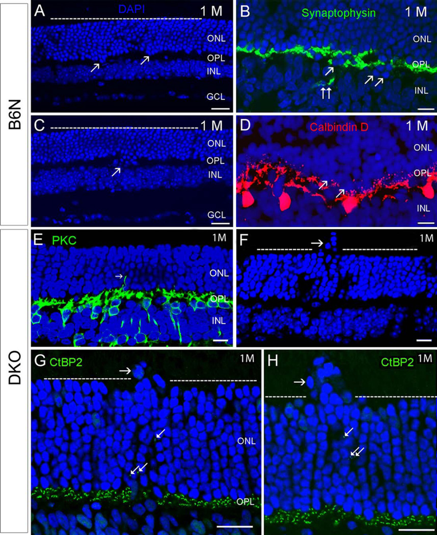
Comparison of retinal degeneration between C57BL/6N and DKO rd8 mice (abbreviates them as B6N and DKO, respectively, in all figures and legends) at one month old. A–D: In B6N mice, most of the retinal architecture is unremarkable (A, C). Occasionally, a few or subpopulation of photoreceptor nuclei visualized by DAPI staining, protruded toward the INL in focal regions of retina (arrows in A, C). Correspondingly, a few synaptophysin-labeled photoreceptor terminals (B, double arrows) and Calbindin D-labeled HC (D) extend into the INL. E–H: In DKO mice, some photoreceptor nuclei migrate into the retinal inner and outer segments in local regions (horizontal arrows in F, G, and H), resulting in an irregular outer margin of the ONL (dashed lines in F, G, H). Correspondingly, PKC-labeled RBC dendrites (E, arrow) and CtBP2-labeled synaptic ribbons (arrows in G and H) stretch into the INL. Scale bar: 20 µm for A, C, 10 µm for B, D, E–H. ONL, outer nuclear layer. OPL, outer plexiform layer. INL, inner nuclear layer. IPL, inner plexiform layer. HC, horizontal cell. GCL, ganglion cell layer).

