Abstract
Background
The foundation for successful arrhythmia ablation is the mapping of electric propagation to identify underlying mechanisms. In atrial fibrillation (AF), however, mapping is difficult so that ablation has often targeted electrogram features, with mixed results. We hypothesized that wide field-of-view (panoramic) mapping of both atria would identify causal mechanisms for AF and allow interpretation of local electrogram features, including complex fractionated atrial electrograms (CFAE).
Methods and Results
Contact mapping was performed using biatrial multipolar catheters in 36 AF subjects (29 persistent). Stable AF rotors (spiral waves) or focal sources were seen in 35 of 36 cases and targeted for ablation (focal impulse and rotor modulation) before pulmonary vein isolation. In 31 of 36 subjects (86.1%), AF acutely terminated (n=20; 16 to sinus rhythm) or organized (n=11; 19±8% slowing) with 2.5 minutes focal impulse and rotor modulation (interquartile range, 1.0–3.1) at one source, defined as the primary source. Subjects exhibited 2.1±1.0 concurrent AF sources of which the primary, by phase mapping, precessed in limited areas (persistent 2.5±1.7 versus paroxysmal 1.7±0.5 cm2; P=0.30). Notably, source regions showed mixed electrogram amplitudes and CFAE grades that did not differ from surrounding atrium (P=NS). AF sources were not consistently surrounded by CFAE (P=0.67).
Conclusions
Stable rotors and focal sources for human AF were revealed by contact panoramic mapping (focal impulse and rotor modulation mapping), but not by electrogram footprints. AF sources precessed within areas of ≈2 cm2, with diverse voltage characteristics poorly correlated with CFAE. Most CFAE sites lie remote from AF sources and are not suitable targets for catheter ablation of AF.
Keywords: atrium, contact panoramic mapping, electrophysiology, fibrillation, focal sources, FIRM ablation, human, rotors
In seminal work, Haïssaguerre et al1 reported that human atrial fibrillation (AF) may be triggered by pulmonary vein (PV) ectopy whose ablation may prevent AF initiation. However, the mechanisms that sustain human AF, once triggered,2 are undefined. Classically, mapping of electric propagation was the foundation to identify causal mechanisms for supraventricular3–5 and ventricular6 arrhythmias for ablation. However, this is difficult in AF because spatial nonuniformity7–9 mandates wide field-of-view mapping to identify drivers, and because AF mapping studies often have not used interventions to separate causal from bystander sites.10,11
In the absence of proven sustaining mechanisms, AF ablation has empirically2 targeted low voltage and complex fractionated atrial electrograms (CFAE)12 independent of their relation to AF propagation, with mixed outcomes.13,14 We recently demonstrated for the first time,15 using panoramic mapping of both atria with contact electrodes, that human AF is sustained by stable spiral waves (rotors) or focal sources, as shown in some animal models.16 Prospective brief ablation at these sources (focal impulse and rotor modulation, FIRM) acutely terminated or organized AF before PV isolation and improved long-term freedom from AF.
We hypothesized that human AF rotors and focal sources would be identified by biatrial propagation mapping but not by electrograms showing CFAE or low voltage. Our rationale was as follows. First, the causal mechanisms for common arrhythmia circuits defined by classical mapping exhibit variable electrogram features.3–6 Second, rotors in animal models of AF precess in limited areas such that electrograms at any one site may vary.17 Third, although AF sources in high-resolution animal and numeric studies16 are surrounded by fractionated electrograms,18 clinically measured CFAE reflect diverse mechanisms, including far-field artifact.19 Fourth, ablation of CFAE sites has often led to disappointing results.13,14
We tested our hypothesis by using biatrial mapping of AF propagation to identify primary AF rotors and focal sources, where targeted ablation can acutely terminate or substantially organize AF without PV isolation, relative to CFAE grade and electrogram voltage in patients with persistent and paroxysmal AF.
Methods
Patient Enrollment
We prospectively enrolled 36 patients with standard indications for AF ablation at the Veterans Affairs and University of California Medical Centers in San Diego; these patients were included in the FIRM-guided limb of the recently reported Conventional Ablation for Atrial Fibrillation With or Without Focal Impulse and Rotor Modulation (CONFIRM) trial.20 The protocol was approved by our joint institutional review board (University of California/Veterans Affairs Medical Centers, San Diego, CA), and all patients provided written informed consent. The only exclusion was an inability or refusal to provide consent.
Electrophysiological Study
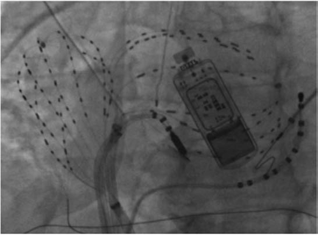
Electrophysiology study was performed >5 half-lives after discontinuing antiarrhythmic medications (>60 days after amiodarone; Table 1). Via femoral venous access, 64 pole basket catheters (Constellation, Boston Scientific, MA) were advanced to the right atrium and transseptally to the left atrium for 128 biatrial electrodes (Figure 1). Baskets were manipulated to ensure optimal atrial contact judged fluoroscopically, by electrograms and intracardiac echocardiography.20 Unfractionated heparin was infused to maintain activated clotting time >350 seconds, with no adverse events from the basket catheters.15
Table 1.
Clinical Characteristics
| Characteristics | |
|---|---|
| Persistent/paroxysmal AF | 29/7 |
| Age, y | 63±9 |
| History of AF, mo | 52 (38–110) |
| Left atrial diameter, mm | 48±7 |
| LVEF, % | 53±15 |
| CHADS2 score | |
| 0 or 1 | 13 (36%) |
| ≥2 | 23 (64%) |
| NYHA class | |
| 0–I | 29 (81%) |
| II–III | 7 (19%) |
| Comorbid conditions | |
| Hypertension | 31 (86%) |
| Diabetes mellitus | 12 (33%) |
| Prior stroke/TIA | 6 (17%) |
| Coronary disease | 18 (50%) |
| Hypercholesterolemia | 30 (86%) |
| Prior conventional ablation | 15 (42%) |
| Number failing amiodarone | 22 (61%) |
| Days since amio discontinued | 365 (69–730) |
| Concomitant drug therapy | |
| ACEI/ARB | 21 (58%) |
| β-Adrenoceptor antagonists | 24 (67%) |
| Statins | 19 (53%) |
Normally distributed variables are listed as mean±SD; non-normally distributed values are listed as median (interquartile range). AF indicates atrial fibrillation; ACEI/ARB, angiotensin-converting enzyme inhibitors/angiotensin receptor blockers; LVEF, left ventricular ejection fraction; NYHA, New York Heart Association; and TIA, transient ischemic attack.
Figure 1.
Contact mapping (focal impulse and rotor modulation mapping) of atrial fibrillation (AF) in both atria. Fluoroscopy shows 64 pole basket catheters used to record AF in the right and left atria, as well as a coronary sinus catheter, an ablation catheter and an esophageal temperature probe. A subcutaneous ECG monitor, implanted preprocedurally to record baseline AF burden, then used postprocedurally to stringently document AF recurrence, is also shown.
When spontaneous AF was not observed (n=5 patients), AF was induced by burst pacing at cycle lengths 500, 450, 400, 350, and 300 ms, then in 10 ms steps to AF. AF was then analyzed after ≥10 minutes. Unipolar and bipolar intracardiac signals were filtered at 0.05 to 500 Hz then digitized at 1 kHz to 16-bit resolution (Bard Pro, Billerica, MA).
Panoramic Mapping of AF Sources
Contact mapping of AF was performed with a novel system (RhythmView, Topera, Inc, San Diego, CA), as recently described.15,20,21 Briefly, electrograms were filtered to exclude noise and far-field signals using rate-dynamics of human left and right atrial action potential duration22–25 to estimate minimum activation time, and conduction velocity24,26 to identify physiological propagation paths. Electrograms were analyzed to construct isopotential movies of successive AF cycles (Movies I–III in the online-only Data Supplement).
From AF isopotential movies, rotors were identified by rotational activity and focal impulses were identified by centrifugal activation from a point of origin. Rotors and focal sources were diagnosed only if stable for thousands of cycles, mapped in time-lapse fashion (multiple epochs) for >10 minutes (≥3000 cycles) before ablation, to exclude transient pivot points of passive fibrillatory activity.11,27,28 Isochronal snapshots of individual cycles of rotors and focal sources are illustrated in Figures 2A, 3A, and 4A.
Figure 2.
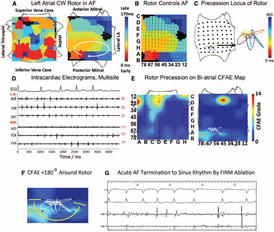
Stable rotor source for human atrial fibrillation (AF), adjacent to moderate complex fractionated atrial electrograms (CFAE). A, Left atrial rotor during AF. B, Activation emanates from rotor to surrounding atrium during AF. C, Rotor precesses over time along the color-coded time line (Movie I in the online-only Data Supplement). D, Electrograms at various sites, referenced to E. E, Panoramic CFAE/AF source map showing AF rotor adjacent to moderate CFAE (grades 8–10), but with higher grade CFAE at distant sites. F, CFAE surrounds AF rotor precession by <180°. G, Focal impulse and rotor modulation (FIRM) ablation at the rotor terminates AF to sinus rhythm hyperacutely in <1 minute before any other ablation. CFAE on the ablation catheter extinguishes as AF terminates. Orientation: the left atrium is opened horizontally across the mitral value with the valve halves folded up and down. The right atrium is opened vertically through the tricuspid value with the valve halves folded laterally and septally.
Figure 3.
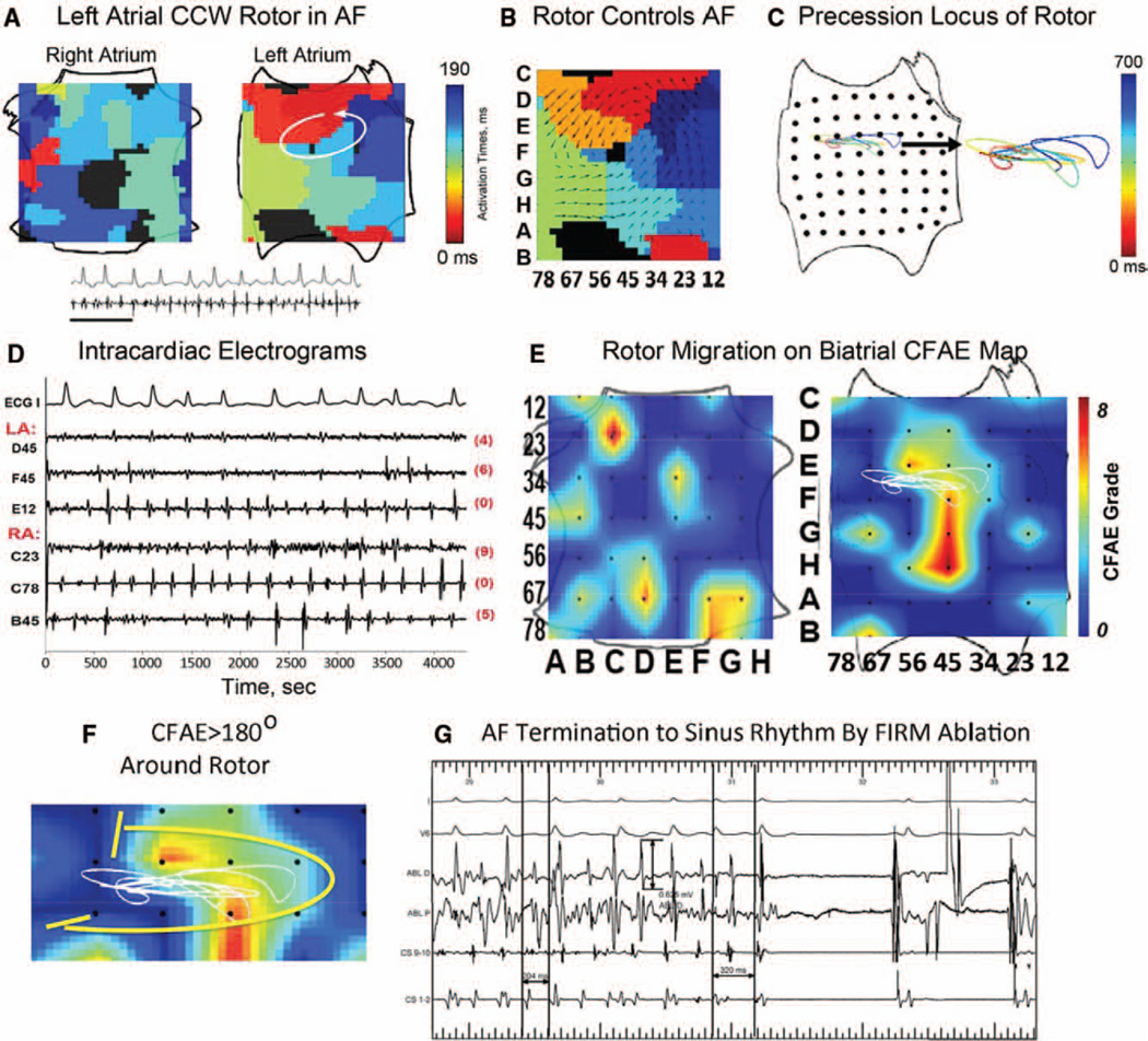
Stable left atrial rotor for atrial fibrillation (AF) overlies minimal/low grade complex fractionated atrial electrograms (CFAE). A, Left atrial rotor during AF (time bar = 1 second; Movie II in the online-only Data Supplement); that (B) controls activation in surrounding atrium. C, Rotor precession (color-coded time line). D, Electrograms showing low CFAE score at the rotor (=6), referenced to E. E, Panoramic rotor/CFAE map. AF rotor precession overlies CFAE, but of low grade (yellow, 4–6). F, CFAE surrounds rotor by >180°, (G) focal impulse and rotor modulation (FIRM) ablation at the rotor terminated AF hyperacutely to sinus rhythm in <1 minute. Note the absence of CFAE on ablation distal electrode (0.625 mV). The panel also shows contact artifact on the ablation catheter and AF CL slowing just before termination. Left and right atrial orientations as in Figure 2A. CCW indicates counterclockwis.
Figure 4.
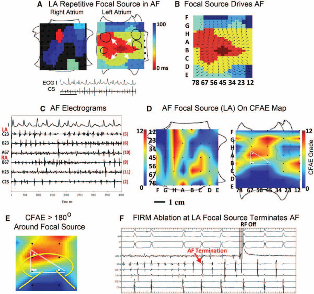
Stable repetitive atrial fibrillation (AF) focal source, inconsistent relation to complex fractionated atrial electrograms (CFAE). A, Left atrial focal source during AF (time bar = 1 second; Movie III in the online-only Data Supplement); B, activation emanates from focal source to atrium; C, AF electrograms in both atria, referenced to D; D, Panoramic CFAE map showing AF focal source precession (white dots bounded by lines) overlying CFAE (grades 7–10), but also with high CFAE grade remotely; E, CFAE surrounds AF source >180°. F, Focal impulse and rotor modulation (FIRM) ablation at focal source terminated AF toward end of application (RF Off =RF, radiofrequency ablation, OFF), before pulmonary vein isolation. Left and right atrial orientations as in Figure 2A.
To define whether activation emanated from AF rotors or focal impulses (supporting their role as sources), we analyzed vectorial direction.18 Isochronal snapshots of AF cycles (Figures 2A–4A) were analyzed in 10 ms bins. Vectors were calculated from activation times at the locations forming the spatial boundaries of each 10, 20, 30… ms bin.
We defined the location of stable human AF sources with the knowledge that fibrillatory rotors are predicted to precess (wobble).17,29 The Hilbert transform30 was applied to unipolar electro-grams as in seminal animal models of AF18 and human ventricular fibrillation.31 The rotor singularity was defined at the crossing point of real and imaginary components,30 and tracked every 20 to 40 ms. Points were joined using third-order Bézier curve interpolation such that first and second derivatives were continuous at the precession locus, and excluding outlying points from interpolation error. Rotor precession loci are illustrated in Figures 2C, 3B, 6, and Movies I to II in the online-only Data Supplement. Focal beat origins in AF were identified at the earliest site of activation, and successive origins were tracked (Figure 4C; Movie III in the online-only Data Supplement). We computed precession areas for each stable AF source.
Figure 6.
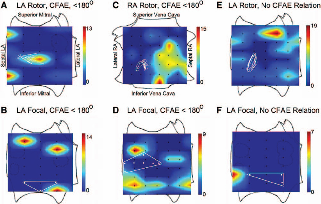
Human atrial fibrillation (AF) sources showing no overlap and minimal surrounding electrogram fractionation. A, Rotor and (B) focal sources with adjacent but not surrounding (<180°) complex fractionated atrial electrograms (CFAE) in left atria; C, rotor in right atrium and (D) focal source in left atrium with adjacent but not surrounding (<180°) CFAE; E, rotor and (F) focal sources with no clear relation to CFAE. Color scale: CFAE grade, higher numbers (warmer colors) indicate more fractionation. Left and right atrial orientations provided in A and C.
Focal Impulse and Rotor Map (FIRM)–guided Ablation
FIRM-guided ablation15 at AF sources was performed before conventional ablation, using a Thermocool catheter (Biosense-Webster, Inc, Diamond Bar, CA) at 25 to 35 W or, in heart failure patients, an EPT Blazer catheter (Boston Scientific, San Jose, CA) at 40 to 50 W target 52°C. FIRM ablation was applied at each stable source for the end point of AF termination or ≤10 minutes, whichever came first. Whenever AF terminated we vigorously attempted reinduction. If AF was reinduced, then FIRM-guided ablation was repeated at ≤3 sources (per institutional review board protocol),15 if necessary, for ≤30 minutes of total FIRM ablation. The composite acute end point from FIRM included AF termination with nonreinducibility, or ≥10% AF slowing that represents elimination of AF sources in computer simulations32 and clinical studies (that used a >3%–4% cut point).33
Because patients may have multiple AF sources, we focused on the primary AF source defined as that source where FIRM ablation first terminated AF or, if AF did not terminate, first organized AF (slowed AF by ≥10%).
Electrogram Quantification
We quantified CFAE and electrogram voltage at each biatrial bipole for 4 seconds. Bipolar signals were recorded directly or created in software from unipolar signals, filtered at 30 to 400 Hz.34
CFAE were graded as described by Scherr et al34 for the Carto XP method (Biosense-Webster Inc). This method identifies electrograms with voltage 0.05 to 0.15 mV, then counts the number of intervals between electrograms that fall within 70 to 120 ms (interval confidence level, ICL). Previous reports show that this and other automated algorithms closely correlate with visual CFAE.35 Because ICL=2/s (ICL 5/2.5 seconds)34 has been considered significant CFAE, we used ICL ≥7 in 4 seconds to avoid overdetection. Biatrial maps of CFAE ICL grade were created using symmetrical median filtering at points of poor electrode signal and linearly interpolating grade across electrodes in 2 dimensions.
Areas of confluent CFAE were calculated within absolute CFAE grade ≥7.34 Overlap areas between AF sources and CFAE (grade ≥7) were calculated using graphing software (Photoshop, Adobe Systems, CA). We determined whether sources were surrounded by CFAE (grade ≥7) for ≥180°, as in animal models.18 Electrogram voltages were calculated at each electrode as peak-to-peak amplitude. We averaged CFAE grade and electrogram voltages (1) within AF source precession loci and (2) for the ipsilateral atrium outside the source (surrounding regions) because it would be useful to know whether AF sources have a unique voltage fingerprint in their ipsilateral atrium.
Statistical Analysis
Continuous data are represented as mean±SD or median and interquartile range as appropriate. Normality was evaluated using the Kolmogorov–Smirnov test. Comparisons between 2 groups were made with Student t tests and summarized with mean±SD for independent samples if normally distributed or, if not normally distributed, evaluated with the Mann–Whitney U test and summarized with medians and quartiles. Nominal values are expressed as n (%) and compared with χ2 tests or the Fisher exact test for comparisons when expected cell frequency was <5. A value of P<0.05 was considered statistically significant.
Results
Patient characteristics are summarized in Table 1. Stable AF-sustaining sources were detected in 97% (35/36) of our patients (Table 2). This study focuses on the 31 patients in whom FIRM ablation met the acute end point. These individuals exhibited 2.1±1.0 concurrent sources, of which 71.8% were rotors and 28.2% focal impulses, 77.5% were left atrial and 22.5% were right atrial. Before conventional ablation, FIRM ablation at the primary source acutely terminated AF with nonreinducibility in n=20 (16 to sinus rhythm, 2 to typical atrial flutter, and 2 to other atrial tachycardias) with 2.5 minutes of ablation (interquartile range, 1.0–3.1) at the primary source, or slowed AF in n=11 (19±8% slowing) (Table II in the online-only Data Supplement).
Table 2.
Spatial Dynamics of Atrial Fibrillation Sources
| Characteristic | Persistent AF | Paroxysmal AF | P |
|---|---|---|---|
| Baseline AF cycle length, ms | 170±21 | 189±13 | 0.03 |
| Patients with detected sources | 28/29 (97%) | 7/7 (100%) | |
| Patients in whom FIRM was completed | 26/29 (89.7%) | 6/7 (85.7%) | |
| Patients reaching acute end point | 24/29 (82.8%) | 7/7 (100%) | |
| No. of concurrent AF sources | 2.4±1.1 | 1.9±0.7 | 0.23 |
| No. of rotors, %* (LA/RA) | 74.1% (29/14) | 61.5% (6/2) | |
| No. of focal impulses, %* (LA/RA) | 25.9% (15/0) | 38.5% (5/0) | |
| Primary source | |||
| No. of rotors (LA, RA) | 18 (14/4) | 4 (3/1) | |
| No. of focal impulses (LA, RA) | 6 (6/0) | 3 (3/0) | |
| Precession area, cm2 | 2.5±1.7 | 1.7±0.5 | 0.3 |
| FIRM-ablation time to AF termination | 2.5 (IQR, 1–3) | 3.0 (IQR, 1–4.1) | 0.49 |
| Primary source/min¶ | (4.5±7.3) | (3.9±4.1) | |
| Total FIRM-ablation time | 19.9 (IQR, 3.1–27.5) | 8.9 (IQR, 3–10.5) | 0.01 |
| All sources/min‡ | 17.5±10.5 | 7.7±4.2 |
AF indicates atrial fibrillation; FIRM, focal impulse and rotor modulation; LA, left atrial; and RA, right atrial.
Percentages are within each patient group;
duration of FIRM ablation at the rotor/focal source where AF terminated then was nonreinducible; and
total duration of FIRM ablation for all sources (before conventional ablation).
Achievement of the acute end point did not differ for patients studied at first ablation or with prior ablation (18/21 versus 13/15; P=0.37, Fisher exact test; Table II in the online-only Data Supplement). Patients undergoing first or prior ablations did not differ in left atrial diameter (P=0.46), age (P=0.43), or left ventricular ejection fraction (P=0.19) (Table I in the online-only Data Supplement).
Localized Sources in Human AF Precess in Limited Spatial Areas
Stable AF rotors and focal sources precessed in nonrepeating limited atrial areas over time for each patient (Table 2).
Figure 2 shows a stable rotor in persistent AF, summarized by the isochronal snapshot of clockwise activation coded red-to-blue (early to late) in the inferior left atrium (Figure 2A). Movie I in the online-only Data Supplement shows multiple cycles. On directionality analysis, activation emanated from this rotor to surrounding atrium (Figure 2B) with peripheral collision of wavefronts from prior cycles. Rotor precession contributed to altered surrounding atrial activation over multiple AF revolutions (Figure 2C; Movie I in the online-only Data Supplement).
Figure 3A shows a stable counterclockwise rotor during persistent AF, with activation again emanating peripherally (Figure 3B) from its precession locus in the high posterior left atrium (Figure 3C; Movie II in the online-only Data Supplement). Figure 4A shows a repetitive focal source on the posterior left atrium outside the PVs during paroxysmal AF, with centrifugal emanation from its origin, confirmed by directionality analysis (Figure 4B). Notably, the focal origin also precessed within a limited area over successive cycles (Figure 4C).
Table 2 summarizes that rotors and focal sources for human AF precessed within circumscribed spatial areas that were similar between persistent and paroxysmal AF.
AF Rotors and Focal Impulses Exhibited Diverse Electrogram Characteristics
AF sources exhibited electrograms of similar voltage and CFAE grade with surrounding ipsilateral atrium, as shown in Figures 2–5 and summarized in Table III in the online-only Data Supplement.
Figure 5.
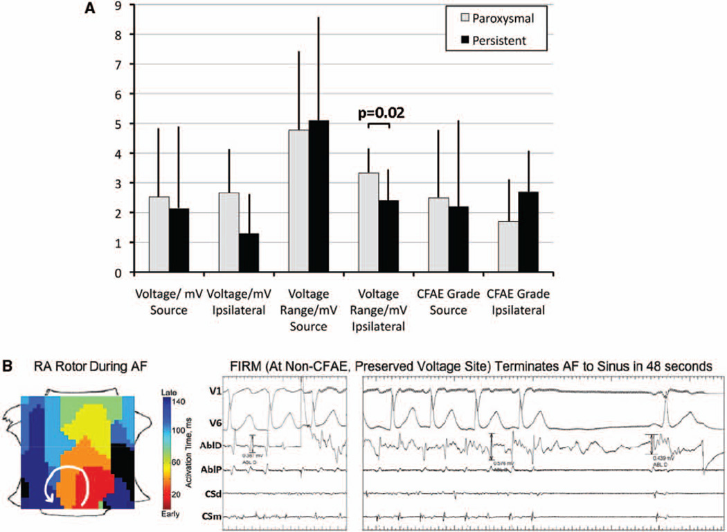
A, Comparison of complex fractionated atrial electrograms (CFAE) grade and electrogram voltage between atrial fibrillation (AF) sources and surrounding ipsilateral atrium for paroxysmal and persistent AF patients (B) preserved electrogram amplitude (≈0.4–0.5 mV) without CFAE at right atrial rotor in persistent AF, where focal impulse and rotor modulation (FIRM) hyperacutely terminated AF to sinus rhythm in 48 seconds before any other ablation. The patient remains AF-free after 15 months on implantable ECG monitor. Right atrial orientation as in Figure 2A.
Figure 2D presents electrograms at sites within and remote from the rotor precession area, with CFAE grade indicated. Repeating this for all biatrial bipoles resulted in a panoramic biatrial CFAE map (Figure 2E), in which the rotor showed minimal overlap with CFAE and <180° surrounding CFAE (Figure 2F). The highest CFAE grades lay remote from the AF rotor, in the anterior left atrium and right atrium.
Figure 3 shows similar analyses during persistent AF in which the rotor did overlap with CFAE (Figure 3D) with >180° surrounding CFAE (Figure 3E). Nevertheless, the highest CFAE grades lay remote from the rotor in the midposterior left atrium and right atrium. In Figure 4, an AF focal source partially overlapped CFAE (grade ≥7), with ϡ80° surrounding CFAE (Figure 4E), but again the highest CFAE grade sites lay remote from the source.
Figure 5A illustrates that AF sources and surrounding regions did not differ in electrogram voltage, voltage range, or CFAE grade. AF sources in persistent and paroxysmal AF patients exhibited inconsistent voltages, indicated by tall SD bars, that did not differ from ipsilateral voltages. In a secondary analysis, voltage range in ipsilateral atrium was higher for paroxysmal than persistent AF (P=0.02, Mann–Whitney U test). For illustration, Figure 5B shows a right atrial rotor where electrograms had preserved voltage (≈0.4–0.5 mV) and no CFAE (zero overlap), yet where FIRM terminated AF to sinus rhythm in 48 seconds.
Figure 6A through 6F further illustrates the inconsistent and diverse relationship of stable AF sources to CFAE. None of these AF sources overlapped CFAE regions, and few were surrounded by CFAE. Even when AF sources related spatially to CFAE, high CFAE grades were always observed remote from the source. These results are summarized in Table III in the online-only Data Supplement.
Discussion
Wide-area contact mapping of AF (FIRM mapping) in both atria revealed stable rotors and focal sources that sustained human AF, yet that did not exhibit consistent or characteristic electrogram features. AF sources precessed within patient-specific areas of ≈2 cm2 were sustained for thousands of cycles during mapping and showed activation that emanated to surrounding atrial tissue. The causal role of sources in sustaining AF was also proven by the acute response of AF to FIRM ablation. Notably, however, rotors did not show an electrogram fingerprint. These data explain the mixed clinical experience of CFAE ablation.
Stable Sources for Human AF
This study and the CONFIRM trial15 show that human AF is often perpetuated by stable rotors and focal sources, as recently validated by additional centers.36 FIRM ablation in the CONFIRM trial substantially improved freedom from AF at up to 2 years to 82.4% using continuous subcutaneous ECG monitors versus 44.9% in patients receiving conventional ablation alone.15
These mechanistic insights build on reports from animal models of AF,29,30,37 and, by demonstrating stable AF drivers, explain reports in which human AF shows consistent spatial activation vectors,7 consistent gradients in intra-atrial rate and frequency,8,9,38 and consistent ECG patterns39 over time. Stable sources at patient-specific locations also explain why AF may terminate unpredictably at any stage of conventional ablation, including early before the PVs are isolated,40 or abruptly after lengthy ablation with little preceding slowing40 or organization.41
Mechanistically, it remains unclear how multiple sources interact to cause AF. If asynchronous, sources may create AF through interference and wave collision.32 However, we15 and others36 have also mapped human AF from a single source where FIRM terminated AF, rendered it nonreinducible and eliminated AF on follow-up (for instance, cases in Figures 2 and 3). In these cases, it remains to be determined whether AF may result from the spatial precession of sources, with dynamically varying impingement of wavefronts on complex fiber architecture, or gradients in rate or repolarization near the source.18 Precession of focal sources in our work supports studies that focal beats may represent transmural breakthrough from epicardial or transmural reentry.10,42
The area of AF source precession ≈2 cm2 provides a rationale for brief FIRM ablation because 6 to 10 lesions of ≈0.25 to 0.5 cm2 would cover this area (3–5 minutes). Although improved spatial electrode resolution may refine area analyses, from a purely clinical perspective, the 0.5 to 0.7 cm diameter of ablation lesions coupled with clinical catheter motion may limit the need for substantially higher mapping resolution.
Relationship of AF Sources to Specific Electrogram Features
Although AF rotors and focal sources were spatially related to CFAE or regions of low amplitude in some patients, many showed no relationship.
Patients in whom AF sources were surrounded by CFAE for >180° (Figures 3 and 4) agree with animal models18 in which rotors are surrounded by fractionated electrograms representing fibrillatory conduction. In other patients, <180° surrounding CFAE may conceivably reflect preferred fibrillatory conduction from anisotropic propagation,26,43 scar,44 and tissue dynamics.24,25 However, the fact that CFAE were frequently observed remote from AF sources (Figures 2–6) supports data that CFAE reflect diverse mechanisms, including autonomic stimulation downstream from ganglionated plexi,45 as well as far-field signal detection.19
Clinical Implications
The complexity of AF has previously limited real-time mapping during ablation, causing an empirical search for surrogates of extra-PV mechanisms, such as CFAE. AF propagation maps from real (contact) electrograms reduces inaccuracies from virtual (noncontact) electrograms in AF46 and enables the identification of AF sources whose precession loci may serve as targets for FIRM ablation. Notably, source areas did not exhibit an electrogram footprint, such as disorganized or organized (CFAE) electrograms, low or high voltage. These results explain why extensive CFAE ablation produces sporadic acute and chronic impact on AF.13,14
These results emphasize a back to basics approach for AF, in which arrhythmia mechanisms should be proven before interpreting electrograms. For instance, in patients with atrioventricular AV node reentry, high-frequency potentials have unclear significance unless mapped near Koch’s triangle,4 sharp spikes recorded from the AV annulus indicate accessory pathway potentials only once the existence of the pathway has been proven,3 fractionated ventricular electrograms may be unrelated to continuation of ventricular tachycardia unless concealed entrainment is identified,6 and so on. In AF, local electrogram interpretation is further complicated by undefined, multiple, and varying activation waves impinging on the mapping antenna.
Limitations
We chose to analyze only primary AF rotors and focal sources. Analyzing secondary sources would complicate interpretation of any electrogram relationship. Because the stability of CFAE is debated,12,47 we mapped CFAE simultaneously at all basket electrodes. We did not perform other CFAE analyses, such NavX (St. Jude Medical, Minneapolis, MN) because automated algorithms and visual grading of CFAE may be similar35 and because this would not alter the inconsistent relationship of AF sources to CFAE (eg, source sites in Figures 3 and 5B would not be fractionated under any criteria). Basket splines may be subjected to tangential distortion that increases electrode spacing that may overestimate CFAE near AF sources48 and alter precise source areas estimates, but this should not materially impact analyses between AF sources, CFAE overlap, or surrounding CFAE. Directionality analysis used interpolation, but AF isochrones also show activity that emanates from sources. We analyzed the area relationship with AF sources favorably with respect to CFAE by comparing only the ipsilateral atrium, although this knowledge would not be available without mapping AF sources. Finally, our patient population was mostly men, although recent studies from other centers36 show similar FIRM-guided mapping and ablation results in women.
Conclusions
Wide field-of-view contact mapping (FIRM-mapping) revealed stable rotors and focal sources that sustain human AF but do not exhibit electrogram footprints. Rotors and focal sources for human AF precessed within patient-specific areas of ≈2 cm2 that were identified neither by CFAE nor low voltage. The vast majority of CFAE lay remote from AF sources and are not suitable targets for ablation.
Supplementary Material
CLINICAL PERSPECTIVE.
Recent clinical trials show that ablation for atrial fibrillation (AF) remains suboptimal, even in paroxysmal AF, largely because the mechanisms that sustain AF (after it has been triggered by pulmonary vein [PV], ectopy) are undefined. To address this problem, physicians may empirically ablate sites exhibiting complex fractionated atrial electrograms (CFAE). Some studies propose that CFAE sites sustain clinical AF, but this has not been proven mechanistically and CFAE ablation has been disappointing. We recently demonstrated in the Conventional Ablation with or without Focal Impulse and Rotor Modulation (CONFIRM) trial that paroxysmal AF and persistent AF are typically driven by stable rotors (spiral waves) or focal sources where targeted ablation (FIRM) can rapidly terminate AF and greatly improve outcome from PV isolation. In the present study, we analyzed whether AF sustaining sources (rotors/focal sources) could be identified by CFAE or other signal characteristics in n=36 patients. Contact mapping of both atria in AF revealed sources that precessed in small stable areas (≈2 cm2) for prolonged periods. Notably, sources exhibited inconsistent signals with or without CFAE, with low or preserved amplitudes. Moreover, although rotors or focal sources are localized to small atrial regions, CFAE were widespread and often remote from sources. In summary, stable rotors and focal sources for clinical AF were universally revealed by wide-area mapping of both atria but not by a signal footprint. Most CFAE sites lay remote from AF sources and thus are not ideal ablation targets.
Acknowledgments
We are grateful to Judith Hildreth, RN; Cherie Jaynes, RN; Elizabeth Greer, RN; Stephanie Yoakum, NP; Donna Cooper, RN; Anthony Moyeda, CVT; and Kenneth Hopper CVT for their great technical assistance, and Kathleen Mills, BA for coordinating and administering the study. Finally, we would like to express our gratitude to Dr Jose Jalife for his extremely helpful comments on this manuscript.
Sources of Funding
This work was supported in part by grants from the American Heart Association to David E. Krummen, and from the National Institutes of Health (HL83359, HL83559-S1, HL103800) and Doris Duke Charitable Foundation to Sanjiv M. Narayan.
Disclosures
Dr Narayan reports being coinventor on intellectual property owned by the University of California and licensed to Topera, Inc. Dr Narayan holds equity in Topera. Topera has not sponsored any research, including that presented here. Dr Narayan also reports having received honoraria from Medtronic, St. Jude Medical and Biotronik Corporations and grant support from Biosense-Webster. His Institution has received fellowship support from Medtronic, Boston Scientific, St. Jude Medical and Biotronik. Dr Shivkumar is a scientific advisor to Topera (uncompensated). His institution has received fellowship support from Medtronic, Boston Scientific, Biosense-Webster and St Jude Medical. Dr Rappel reports being coinventor on intellectual property owned by the University of California and licensed to Topera, Inc. Dr Rappel holds equity in Topera. Topera has not sponsored any research, including that presented here. Dr. Miller reports having received honoraria from Medtronic, St. Jude Medical and Biotronik Corporations, Boston Scientific Corporation and Biosense-Webster. He is a scientific advisor to Stereotaxis and Topera (modest compensation). His institution has received fellowship support from Medtronic, Boston Scientific, Biosense-Webster and Biotronik. Dr Krummen’s institution has received fellowship support from Medtronic, Boston Scientific, St. Jude Medical and Biotronik.
Footnotes
The online-only Data Supplement is available at http://circep.ahajournals.org/lookup/suppl/doi:10.1161/CIRCEP.111.977264/-/DC1.
References
- 1.Haïssaguerre M, Jaïs P, Shah DC, Takahashi A, Hocini M, Quiniou G, Garrigue S, Le Mouroux A, Le Métayer P, Clémenty J. Spontaneous initiation of atrial fibrillation by ectopic beats originating in the pulmonary veins. N Engl J Med. 1998;339:659–666. doi: 10.1056/NEJM199809033391003. [DOI] [PubMed] [Google Scholar]
- 2.Calkins H, Kuck KH, Cappato R, Brugada J, Camm AJ, Chen SA, Crijns HJ, Damiano RJ, Jr, Davies DW, DiMarco J, Edgerton J, Ellenbogen K, Ezekowitz MD, Haines DE, Haissaguerre M, Hindricks G, Iesaka Y, Jackman W, Jalife J, Jais P, Kalman J, Keane D, Kim YH, Kirchhof P, Klein G, Kottkamp H, Kumagai K, Lindsay BD, Mansour M, Marchlinski FE, McCarthy PM, Mont JL, Morady F, Nademanee K, Nakagawa H, Natale A, Nattel S, Packer DL, Pappone C, Prystowsky E, Raviele A, Reddy V, Ruskin JN, Shemin RJ, Tsao HM, Wilber D. Heart Rhythm Society Task Force on Catheter and Surgical Ablation of Atrial Fibrillation. 2012 HRS/EHRA/ECAS expert consensus statement on catheter and surgical ablation of atrial fibrillation: recommendations for patient selection, procedural techniques, patient management and follow-up, definitions, endpoints, and research trial design: a report of the Heart Rhythm Society (HRS) Task Force on Catheter and Surgical Ablation of Atrial Fibrillation. Developed in partnership with the European Heart Rhythm Association (EHRA), a registered branch of the European Society of Cardiology (ESC) and the European Cardiac Arrhythmia Society (ECAS); and in collaboration with the American College of Cardiology (ACC), American Heart Association (AHA), the Asia Pacific Heart Rhythm Society (APHRS), and the Society of Thoracic Surgeons (STS). Endorsed by the governing bodies of the American College of Cardiology Foundation, the American Heart Association, the European Cardiac Arrhythmia Society, the European Heart Rhythm Association, the Society of Thoracic Surgeons, the Asia Pacific Heart Rhythm Society, and the Heart Rhythm Society. Heart Rhythm. 2012;9:632–696. e21. doi: 10.1016/j.hrthm.2011.12.016. [DOI] [PubMed] [Google Scholar]
- 3.Jackman WM, Wang XZ, Friday KJ, Roman CA, Moulton KP, Beckman KJ, McClelland JH, Twidale N, Hazlitt HA, Prior MI. Catheter ablation of accessory atrioventricular pathways (Wolff-Parkinson-White syndrome) by radiofrequency current. N Engl J Med. 1991;324:1605–1611. doi: 10.1056/NEJM199106063242301. [DOI] [PubMed] [Google Scholar]
- 4.Jackman WM, Beckman KJ, McClelland JH, Wang X, Friday KJ, Roman CA, Moulton KP, Twidale N, Hazlitt HA, Prior MI. Treatment of supraventricular tachycardia due to atrioventricular nodal reentry, by radiofrequency catheter ablation of slow-pathway conduction. N Engl J Med. 1992;327:313–318. doi: 10.1056/NEJM199207303270504. [DOI] [PubMed] [Google Scholar]
- 5.Feld GK, Fleck RP, Chen PS, Boyce K, Bahnson TD, Stein JB, Calisi CM, Ibarra M. Radiofrequency catheter ablation for the treatment of human type 1 atrial flutter. Identification of a critical zone in the reentrant circuit by endocardial mapping techniques. Circulation. 1992;86:1233–1240. doi: 10.1161/01.cir.86.4.1233. [DOI] [PubMed] [Google Scholar]
- 6.Stevenson WG, Khan H, Sager P, Saxon LA, Middlekauff HR, Natterson PD, Wiener I. Identification of reentry circuit sites during catheter mapping and radiofrequency ablation of ventricular tachycardia late after myocardial infarction. Circulation. 1993;88(4 Pt 1):1647–1670. doi: 10.1161/01.cir.88.4.1647. [DOI] [PubMed] [Google Scholar]
- 7.Gerstenfeld EP, Sahakian AV, Swiryn S. Evidence for transient linking of atrial excitation during atrial fibrillation in humans. Circulation. 1992;86:375–382. doi: 10.1161/01.cir.86.2.375. [DOI] [PubMed] [Google Scholar]
- 8.Lazar S, Dixit S, Marchlinski FE, Callans DJ, Gerstenfeld EP. Presence of left-to-right atrial frequency gradient in paroxysmal but not persistent atrial fibrillation in humans. Circulation. 2004;110:3181–3186. doi: 10.1161/01.CIR.0000147279.91094.5E. [DOI] [PubMed] [Google Scholar]
- 9.Sahadevan J, Ryu K, Peltz L, Khrestian CM, Stewart RW, Markowitz AH, Waldo AL. Epicardial mapping of chronic atrial fibrillation in patients: preliminary observations. Circulation. 2004;110:3293–3299. doi: 10.1161/01.CIR.0000147781.02738.13. [DOI] [PubMed] [Google Scholar]
- 10.Allessie MA, de Groot NM, Houben RP, Schotten U, Boersma E, Smeets JL, Crijns HJ. The electropathological substrate of longstanding persistent atrial fibrillation in patients with structural heart disease: longitudinal dissociation. Circ Arrhythm Electrophysiol. 2010;122:1674–1682. doi: 10.1161/CIRCEP.109.910125. [DOI] [PubMed] [Google Scholar]
- 11.Cuculich PS, Wang Y, Lindsay BD, Faddis MN, Schuessler RB, Damiano RJ, Jr, Li L, Rudy Y. Noninvasive characterization of epicardial activation in humans with diverse atrial fibrillation patterns. Circulation. 2010;122:1364–1372. doi: 10.1161/CIRCULATIONAHA.110.945709. [DOI] [PMC free article] [PubMed] [Google Scholar]
- 12.Nademanee K, McKenzie J, Kosar E, Schwab M, Sunsaneewitayakul B, Vasavakul T, Khunnawat C, Ngarmukos T. A new approach for catheter ablation of atrial fibrillation: mapping of the electrophysiologic substrate. J Am Coll Cardiol. 2004;43:2044–2053. doi: 10.1016/j.jacc.2003.12.054. [DOI] [PubMed] [Google Scholar]
- 13.Oral H, Chugh A, Good E, Wimmer A, Dey S, Gadeela N, Sankaran S, Crawford T, Sarrazin JF, Kuhne M, Chalfoun N, Wells D, Frederick M, Fortino J, Benloucif-Moore S, Jongnarangsin K, Pelosi F, Jr, Bogun F, Morady F. Radiofrequency catheter ablation of chronic atrial fibrillation guided by complex electrograms. Circulation. 2007;115:2606–2612. doi: 10.1161/CIRCULATIONAHA.107.691386. [DOI] [PubMed] [Google Scholar]
- 14.Oral H, Chugh A, Yoshida K, Sarrazin JF, Kuhne M, Crawford T, Chalfoun N, Wells D, Boonyapisit W, Veerareddy S, Billakanty S, Wong WS, Good E, Jongnarangsin K, Pelosi F, Jr, Bogun F, Morady F. A randomized assessment of the incremental role of ablation of complex fractionated atrial electrograms after antral pulmonary vein isolation for long-lasting persistent atrial fibrillation. J Am Coll Cardiol. 2009;53:782–789. doi: 10.1016/j.jacc.2008.10.054. [DOI] [PubMed] [Google Scholar]
- 15.Narayan SM, Krummen DE, Shivkumar K, Clopton P, Rappel WJ, Miller JM. Treatment of atrial fibrillation by the ablation of localized sources: CONFIRM (Conventional Ablation for Atrial Fibrillation With or Without Focal Impulse and Rotor Modulation) trial. J Am Coll Cardiol. 2012;60:628–636. doi: 10.1016/j.jacc.2012.05.022. [DOI] [PMC free article] [PubMed] [Google Scholar]
- 16.Skanes AC, Mandapati R, Berenfeld O, Davidenko JM, Jalife J. Spatiotemporal periodicity during atrial fibrillation in the isolated sheep heart. Circulation. 1998;98:1236–1248. doi: 10.1161/01.cir.98.12.1236. [DOI] [PubMed] [Google Scholar]
- 17.Zlochiver S, Yamazaki M, Kalifa J, Berenfeld O. Rotor meandering contributes to irregularity in electrograms during atrial fibrillation. Heart Rhythm. 2008;5:846–854. doi: 10.1016/j.hrthm.2008.03.010. [DOI] [PMC free article] [PubMed] [Google Scholar]
- 18.Kalifa J, Tanaka K, Zaitsev AV, Warren M, Vaidyanathan R, Auerbach D, Pandit S, Vikstrom KL, Ploutz-Snyder R, Talkachou A, Atienza F, Guiraudon G, Jalife J, Berenfeld O. Mechanisms of wave fractionation at boundaries of high-frequency excitation in the posterior left atrium of the isolated sheep heart during atrial fibrillation. Circulation. 2006;113:626–633. doi: 10.1161/CIRCULATIONAHA.105.575340. [DOI] [PubMed] [Google Scholar]
- 19.Narayan SM, Wright M, Derval N, Jadidi A, Forclaz A, Nault I, Miyazaki S, Sacher F, Bordachar P, Clémenty J, Jaïs P, Haïssaguerre M, Hocini M. Classifying fractionated electrograms in human atrial fibrillation using monophasic action potentials and activation mapping: evidence for localized drivers, rate acceleration, and nonlocal signal etiologies. Heart Rhythm. 2011;8:244–253. doi: 10.1016/j.hrthm.2010.10.020. [DOI] [PMC free article] [PubMed] [Google Scholar]
- 20.Narayan SM, Krummen DE, Rappel WJ. Clinical mapping approach to diagnose electrical rotors and focal impulse sources for human atrial fibrillation. J Cardiovasc Electrophysiol. 2012;23:447–454. doi: 10.1111/j.1540-8167.2012.02332.x. [DOI] [PMC free article] [PubMed] [Google Scholar]
- 21.Narayan SM, Patel J, Mulpuru S, Krummen DE. Focal impulse and rotor modulation ablation of sustaining rotors abruptly terminates persistent atrial fibrillation to sinus rhythm with elimination on follow-up: a video case study. Heart Rhythm. 2012;9:1436–1439. doi: 10.1016/j.hrthm.2012.03.055. [DOI] [PMC free article] [PubMed] [Google Scholar]
- 22.Narayan SM, Bode F, Karasik PL, Franz MR. Alternans of atrial action potentials during atrial flutter as a precursor to atrial fibrillation. Circulation. 2002;106:1968–1973. doi: 10.1161/01.cir.0000037062.35762.b4. [DOI] [PubMed] [Google Scholar]
- 23.Narayan SM, Franz MR. Quantifying fractionation and rate in human atrial fibrillation using monophasic action potentials: implications for substrate mapping. Europace. 2007;9(Suppl 6):vi89–vi95. doi: 10.1093/europace/eum212. [DOI] [PubMed] [Google Scholar]
- 24.Narayan SM, Kazi D, Krummen DE, Rappel WJ. Repolarization and activation restitution near human pulmonary veins and atrial fibrillation initiation: a mechanism for the initiation of atrial fibrillation by premature beats. J Am Coll Cardiol. 2008;52:1222–1230. doi: 10.1016/j.jacc.2008.07.012. [DOI] [PMC free article] [PubMed] [Google Scholar]
- 25.Narayan SM, Franz MR, Clopton P, Pruvot EJ, Krummen DE. Repolarization alternans reveals vulnerability to human atrial fibrillation. Circulation. 2011;123:2922–2930. doi: 10.1161/CIRCULATIONAHA.110.977827. [DOI] [PMC free article] [PubMed] [Google Scholar]
- 26.Lalani GG, Schricker A, Gibson M, Rostamian A, Krummen DE, Narayan SM. Atrial conduction slows immediately before the onset of human atrial fibrillation: a bi-atrial contact mapping study of transitions to atrial fibrillation. J Am Coll Cardiol. 2012;59:595–606. doi: 10.1016/j.jacc.2011.10.879. [DOI] [PMC free article] [PubMed] [Google Scholar]
- 27.Konings KT, Smeets JL, Penn OC, Wellens HJ, Allessie MA. Configuration of unipolar atrial electrograms during electrically induced atrial fibrillation in humans. Circulation. 1997;95:1231–1241. doi: 10.1161/01.cir.95.5.1231. [DOI] [PubMed] [Google Scholar]
- 28.Schilling RJ, Kadish AH, Peters NS, Goldberger J, Davies DW. Endocardial mapping of atrial fibrillation in the human right atrium using a noncontact catheter. Eur Heart J. 2000;21:550–564. doi: 10.1053/euhj.1999.1851. [DOI] [PubMed] [Google Scholar]
- 29.Davidenko JM, Pertsov AV, Salomonsz R, Baxter W, Jalife J. Stationary and drifting spiral waves of excitation in isolated cardiac muscle. Nature. 1992;355:349–351. doi: 10.1038/355349a0. [DOI] [PubMed] [Google Scholar]
- 30.Gray RA, Pertsov AM, Jalife J. Spatial and temporal organization during cardiac fibrillation. Nature. 1998;392:75–78. doi: 10.1038/32164. [DOI] [PubMed] [Google Scholar]
- 31.Nash MP, Mourad A, Clayton RH, Sutton PM, Bradley CP, Hayward M, Paterson DJ, Taggart P. Evidence for multiple mechanisms in human ventricular fibrillation. Circulation. 2006;114:536–542. doi: 10.1161/CIRCULATIONAHA.105.602870. [DOI] [PubMed] [Google Scholar]
- 32.Haissaguerre M, Lim KT, Jacquemet V, Rotter M, Dang L, Hocini M, Matsuo S, Knecht S, Jais P, Virag N. Atrial fibrillatory cycle length: Computer simulation and potential clinical importance. Europace. 2007;9(Suppl 6):vi64–vi70. doi: 10.1093/europace/eum208. [DOI] [PubMed] [Google Scholar]
- 33.Takahashi Y, O’Neill MD, Hocini M, Dubois R, Matsuo S, Knecht S, Mahapatra S, Lim KT, Jaïs P, Jonsson A, Sacher F, Sanders P, Rostock T, Bordachar P, Clémenty J, Klein GJ, Haïssaguerre M. Characterization of electrograms associated with termination of chronic atrial fibrillation by catheter ablation. J Am Coll Cardiol. 2008;51:1003–1010. doi: 10.1016/j.jacc.2007.10.056. [DOI] [PubMed] [Google Scholar]
- 34.Scherr D, Dalal D, Cheema A, Cheng A, Henrikson CA, Spragg D, Marine JE, Berger RD, Calkins H, Dong J. Automated detection and characterization of complex fractionated atrial electrograms in human left atrium during atrial fibrillation. Heart Rhythm. 2007;4:1013–1020. doi: 10.1016/j.hrthm.2007.04.021. [DOI] [PubMed] [Google Scholar]
- 35.Hunter RJ, Diab I, Thomas G, Duncan E, Abrams D, Dhinoja M, Sporton S, Earley MJ, Schilling RJ. Validation of a classification system to grade fractionation in atrial fibrillation and correlation with automated detection systems. Europace. 2009;11:1587–1596. doi: 10.1093/europace/eup351. [DOI] [PubMed] [Google Scholar]
- 36.Shivkumar K, Ellenbogen KA, Hummel JD, Miller JM, Steinberg JS. Acute termination of human atrial fibrillation by identification and catheter ablation of localized rotors and sources: first multicenter experience of focal impulse and rotor modulation (FIRM) ablation. J Cardiovasc Electrophysiol. 2012;23:1277–1285. doi: 10.1111/jce.12000. [DOI] [PMC free article] [PubMed] [Google Scholar]
- 37.Ryu K, Shroff SC, Sahadevan J, Martovitz NL, Khrestian CM, Stambler BS. Mapping of atrial activation during sustained atrial fibrillation in dogs with rapid ventricular pacing induced heart failure: evidence for a role of driver regions. J Cardiovasc Electrophysiol. 2005;16:1348–1358. doi: 10.1111/j.1540-8167.2005.00266.x. [DOI] [PubMed] [Google Scholar]
- 38.Lemola K, Ting M, Gupta P, Anker JN, Chugh A, Good E, Reich S, Tschopp D, Igic P, Elmouchi D, Jongnarangsin K, Bogun F, Pelosi F, Jr, Morady F, Oral H. Effects of two different catheter ablation techniques on spectral characteristics of atrial fibrillation. J Am Coll Cardiol. 2006;48:340–348. doi: 10.1016/j.jacc.2006.04.053. [DOI] [PubMed] [Google Scholar]
- 39.Xi Q, Sahakian AV, Ng J, Swiryn S. Atrial fibrillatory wave characteristics on surface electrogram: ECG to ECG repeatability over twenty-four hours in clinically stable patients. J Cardiovasc Electrophysiol. 2004;15:911–917. doi: 10.1046/j.1540-8167.2004.03577.x. [DOI] [PubMed] [Google Scholar]
- 40.Haïssaguerre M, Sanders P, Hocini M, Takahashi Y, Rotter M, Sacher F, Rostock T, Hsu LF, Bordachar P, Reuter S, Roudaut R, Clémenty J, Jaïs P. Catheter ablation of long-lasting persistent atrial fibrillation: critical structures for termination. J Cardiovasc Electrophysiol. 2005;16:1125–1137. doi: 10.1111/j.1540-8167.2005.00307.x. [DOI] [PubMed] [Google Scholar]
- 41.Forclaz A, Narayan SM, Scherr D, Linton N, Jadidi AS, Nault I, Rivard L, Miyazaki S, Uldry L, Wright M, Shah AJ, Liu X, Xhaet O, Derval N, Knecht S, Sacher F, Jaïs P, Hocini M, Haïssaguerre M. Early temporal and spatial regularization of persistent atrial fibrillation predicts termination and arrhythmia-free outcome. Heart Rhythm. 2011;8:1374–1382. doi: 10.1016/j.hrthm.2011.05.008. [DOI] [PMC free article] [PubMed] [Google Scholar]
- 42.Baxter WT, Mironov SF, Zaitsev AV, Jalife J, Pertsov AM. Visualizing excitation waves inside cardiac muscle using transillumination. Biophys J. 2001;80:516–530. doi: 10.1016/S0006-3495(01)76034-1. [DOI] [PMC free article] [PubMed] [Google Scholar]
- 43.Klos M, Calvo D, Yamazaki M, Zlochiver S, Mironov S, Cabrera JA, Sanchez-Quintana D, Jalife J, Berenfeld O, Kalifa J. Atrial septopulmonary bundle of the posterior left atrium provides a substrate for atrial fibrillation initiation in a model of vagally mediated pulmonary vein tachycardia of the structurally normal heart. Circ Arrhythm Electrophysiol. 2008;1:175–183. doi: 10.1161/CIRCEP.107.760447. [DOI] [PMC free article] [PubMed] [Google Scholar]
- 44.Oakes RS, Badger TJ, Kholmovski EG, Akoum N, Burgon NS, Fish EN, Blauer JJ, Rao SN, DiBella EV, Segerson NM, Daccarett M, Windfelder J, McGann CJ, Parker D, MacLeod RS, Marrouche NF. Detection and quantification of left atrial structural remodeling with delayed-enhancement magnetic resonance imaging in patients with atrial fibrillation. Circulation. 2009;119:1758–1767. doi: 10.1161/CIRCULATIONAHA.108.811877. [DOI] [PMC free article] [PubMed] [Google Scholar]
- 45.Lin J, Scherlag BJ, Zhou J, Lu Z, Patterson E, Jackman WM, Lazzara R, Po SS. Autonomic mechanism to explain complex fractionated atrial electrograms (CFAE) J Cardiovasc Electrophysiol. 2007;18:1197–1205. doi: 10.1111/j.1540-8167.2007.00976.x. [DOI] [PubMed] [Google Scholar]
- 46.Earley MJ, Abrams DJ, Sporton SC, Schilling RJ. Validation of the non-contact mapping system in the left atrium during permanent atrial fibrillation and sinus rhythm. J Am Coll Cardiol. 2006;48:485–491. doi: 10.1016/j.jacc.2006.04.069. [DOI] [PubMed] [Google Scholar]
- 47.Habel N, Znojkiewicz P, Thompson N, Müller JG, Mason B, Calame J, Calame S, Sharma S, Mirchandani G, Janks D, Bates J, Noori A, Karnbach A, Lustgarten DL, Sobel BE, Spector P. The temporal variability of dominant frequency and complex fractionated atrial electrograms constrains the validity of sequential mapping in human atrial fibrillation. Heart Rhythm. 2010;7:586–593. doi: 10.1016/j.hrthm.2010.01.010. [DOI] [PubMed] [Google Scholar]
- 48.Correa de Sa DD, Thompson N, Stinnett-Donnelly J, Znojkiewicz P, Habel N, Muller JG, Bates JH, Buzas JS, Spector PS. Electrogram fractionation: the relationship between spatiotemporal variation of tissue excitation and electrode spatial resolution. Circ Arrhythm Electrophysiol. 2011;4:909–916. doi: 10.1161/CIRCEP.111.965145. [DOI] [PubMed] [Google Scholar]
Associated Data
This section collects any data citations, data availability statements, or supplementary materials included in this article.


