Abstract
BACKGROUND
Atherosclerosis is the main cause of cardiovascular disease which is caused by a high-fat diet. Many of these patients use boiled quince leaves for their treatment. However, the supporting scientific information is limit. The aim of this study was to evaluate the effect of quince leaf on the progression of atherosclerosis and whether it can be an appropriate alternative to statins.
METHODS
24 male rabbits were randomly divided into two groups: normal diet (6 n) and high-cholesterol diet (2% cholesterol, 18 n) for 8 weeks. At the end of the 8 weeks, both groups underwent blood sampling and their biochemical markers were measured. Then, all animals in the normal-diet group and three of the high-cholesterol diet group were killed to investigate atheromic plaque in their coronary artery. The 15 remaining rabbits of the high-cholesterol diet group were randomly divided into 3 groups (5 n) after discontinuation of the fatty diet. The first group was not given any treatment, the second received atorvastatin (0.5 mg/kg) orally, and the third received quince leaf extract (50 mg/kg) orally for 12 weeks. At the end of this period, after blood sampling, biopsy of coronary artery was performed for histological study.
RESULTS
The results showed that atorvastatin and quince leaf significantly decreased total cholesterol, triglyceride, LDL, AST, ALT, AP, BUN, and Cr levels compared with the first group of the high-cholesterol diet group (P < 0.05). No significant difference was found between atorvastatin and quince leaf extract groups in biochemical markers and atherosclerotic plaque in coronary artery.
CONCLUSION
Atorvastatin and quince leaf extract can effectively prevent the progression of atherosclerosis in coronary arteries. According to the results of this study and also lower toxic effects of herbal medication compared to synthetic medication, leaf extract can be a substitute for statins in treatment and prevention of cardiovascular disease. The anti-atherosclerotic effect of quince leaf is most likely related to its antioxidant components.
Keywords: Hyperlipidemia, Atherosclerosis, Coronary Arteries, Biochemical Markers, Atheromic Plaque, Atorvastatin, Quince Leaf Extract
Introduction
Studies have illustrated that a high-fat diet causes severe oxidative stress in the vascular tissue.1 Additionally, it is the cause of mortality and morbidity.1,2 Many believed that it can be induced from a simple dysfunction of endothelial lining.1,3-5 Moreover, of the factors causing heart failure life style, fatty regimen, hypertension, and a fatty diet, particularly LDL cholesterol, are mainly responsible for hypercholesterolemia that is related to increased damage in vascular tissue by free radical oxidative stress.3,6,7 Although cholesterol-lowering drugs such as statins were used for several decades and are effective in preventing cardiovascular disorders, their usage is often limited because of their adverse effects, such as rhabdomyolysis. These effects are very pronounced when a statin is taken with another type of cholesterol-lowering drug, in particular fibrate. This resulted in the introduction of plants as a source of natural antioxidants.8-10 Due to the adverse effects associated with synthetic lipid-lowering drugs, the quest for natural products with lipid-lowering potential and without or minimal side effects is necessary. It has been found that many plants are useful as antioxidant and antimicrobial agents and can be used as forms of herbal medicine.11-16Among these plants, quince is a suitable source of antioxidant. It contains appreciable amounts of vitamin E and phenolic compounds (catechins, phenolic acids, and kaempferol-3-o-rutinoside), which also have protective effects. Given the high levels of phenolic compounds in the quince leaves (such as kaempferol-phosphate), they are more effective than the fruit and seeds in the promoting of health and are a useful and inexpensive source of bioactive elements.11,13,15,16 The quince leaves are widely used to treat diarrhea, heart palpitations, and eye disease.17 Phenolic compounds from dietary antioxidants are the most natural antioxidants.18 This study evaluated the effect of the quince leaves on biochemical markers such as lipid profiles, liver enzymes, and kidney function, and compared them to a standard medication called atorvastatin.
Materials and Methods
Drugs and chemicals
All drugs and biochemicals used in this study were purchased from Sigma Co (St, Louis, Mo, USA) and from Merck Company (Darmstadt, Germany). All other chemicals an reagents were of analytical grade.
Atherogenic Food
Pure cholesterol powder was purchased from Farzan Teb Co. (Merck, Germany). Daily food dosage was measured for 3 consecutive days. Intake rate was obtained for calculating the atherogenic diet formula, and the amount of 54 gr was considered for all animals. A high-fat diet with 2% cholesterol was prepared and stored at 4°C.
Preparation of the extract
The quince green dried leaves were purchased from Herbs Chemist (Tabriz, Iran). Methanolic 70% extract was prepared by maceration method. The extracts were filtered three times and concentrated to dryness under vacuum. Then, the percentage of the obtained dry extract (7%) was determined and it was kept at subzero degrees centigrade until administration time. The extract was dissolved in water in order to be given orally by gavage needle.
Methods
24 male New Zealand white rabbits (the least number of animals in each group = 6, with 2500 ± 200 weight, and mean age of 8 weeks) were purchased from the Pasteur Institute of Iran. They were kept at 22°C, and given free access to food (from Sahand Niroo Co., Tabriz, Iran) and water at least 7 days before the experimental study. After adaptation, rabbits were randomly divided into two groups of normal diet (n = 6), and high-cholesterol diet (n = 18). The rabbits with the high-cholesterol diet were fed an atherogenic diet containing 2% cholesterol (54 g/day) for 8 weeks. At the end of week 8, groups underwent blood sampling from marginal ear vein. For histopathological studies, all rabbits of the normal diet group plus three rabbits selected randomly from the high-cholesterol diet group were killed and biopsy was performed from left coronary artery. Then, the remaining rabbits of the high-cholesterol diet group were randomly divided into three groups of five rabbits each after stopping atherogenic diet for 12 weeks. The first group was fed with a normal diet without any treatment and served as control group. The second group was given the same diet as the control group plus atorvastatin at a dose of 0.5 mg/kg once a day orally (atorvastatin group). The third group was fed the same diet as the previous groups plus a supplement of quince leaf extract (5 mg/kg once a day orally). At the end of week 12, the blood samples were taken again, and coronary biopsy was performed on all rabbits after thoracotomy. All animal care and experimental protocols were confirmed by the Medical Ethics Committee of Tabriz and complied with the guidelines of the National Institute of Health (NIH publication 86-23 revised 1985).
Sample collection
Blood samples were collected from all rabbits in two steps that have been mentioned previously. Plasma was prepared by centrifugation at 3000 g for 15 minutes. Serum levels of lipid profiles such as total cholesterol (TC), triglyceride (TG), low density lipoprotein (LDL) cholesterol, high density lipoprotein (HDL) cholesterol, and liver enzymes including aspartate transaminase (AST), alanine transaminase (ALT), and alkaline phosphatase (AP), and other biomarkers for kidney functionality (BUN, Cr) were recorded by an Auto Analyzer.
Histopathological study
For sampling, animals were anesthetized by appropriate doses of ketamine and xylazine by intramuscular injection. Fixation was perfused by saline buffer formaldehyde-cold PBS 4%, PH: 7.2 into heart ventricle. Histological biopsy of the coronary artery was performed after thoracotomy.
After thoracotomy, coronary specimens were immersed in formalin 10% for 48-72 hour. Then, they were embedded in paraffin and cut into thick sections (5µm), subsequently deparaffinized by graded concentrations of ethanol and xylene, and then were stained by specific Weigert’s Iron hematoxylin staining. Finally, surfaces of the whole plaques were measured and compared to surfaces of total coronary arteries by Motic Software. The plaque thickness was confirmed by three observers.
Statistical analysis
Results were analyzed by SPSS for Windows (version 16; SPSS Inc., Chicago, IL., USA) and Student’s t-test (assuming equal variances), Mann-Whitney test, and Kruskal-wallis test (P < 0.05 was considered significant) were performed to determine the statistical significance of data obtained from the two groups and compare them to each other). The diagrams were depicted by Microsoft Excel.
Results
Determination of biochemical markers
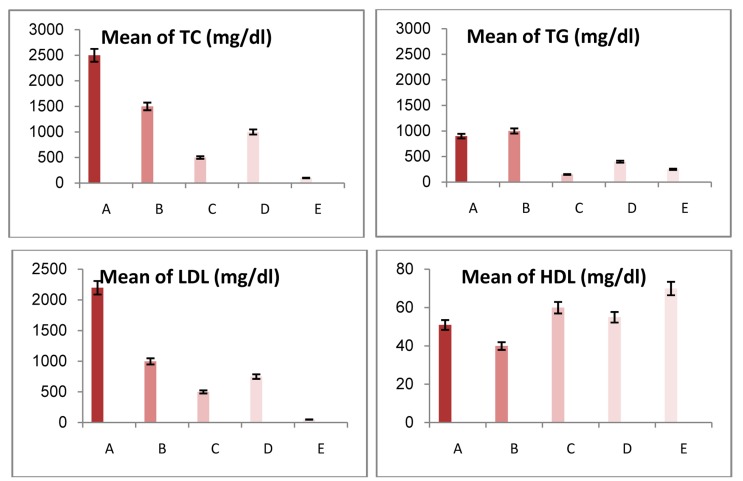
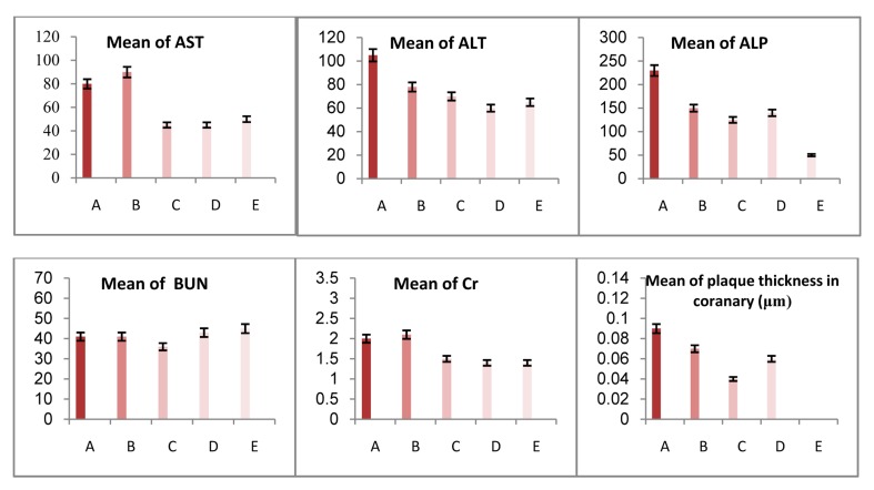
Lipid profile levels in the blood plasma of different groups are shown in table 1 and figure 1. In the high-cholesterol diet group TC, TG, and LDL levels increased significantly compared with the normal diet group, and HDL decreased (P < 0.05). Moreover, the biomarkers AST, ALT, AP, BUN, and Cr increased significantly in all groups during the two months of using the high-cholesterol diet (Table 1 and Figure 2) (P < 0.05). After three months of discontinuing the high-cholesterol diet, all biochemical markers were measured. This showed that the cholesterol level of the control group of the high-cholesterol diet group was lower compared with before stopping. However, it was still significantly high compared with the normal diet group (P < 0.05). The 0.5 mg/kg dose of atorvastatin and 50 mg/kg of quince leaf extract significantly decreased TC, TG, LDL, AST, ALT, and Cr levels compared to the control group of the high-cholesterol diet group (P < 0.05). However, in both of these groups TC, LDL, and AP levels were significantly higher compared with the normal diet group(P < 0.05), but no significant difference was found in TG, HDL, and BUN levels. In both atorvastatin and quince leaf extract groups, the levels of AST, and ALT showed no significant difference to the normal diet group.
Table 1.
Comparison of lipid profile, liver enzymes, and plaque thickness in normal and high-cholesterol diet animals (end of 2 months) and after stopping highcholesterol diet (end of 3 months)
| Groups |
Lipid profile (mg/dl) |
Liver enzymes |
Plaque thickness (µm) |
|||||||
|---|---|---|---|---|---|---|---|---|---|---|
| TC | TG | LDL | HDL | AST | ALT | AP | BUN | Cr | Aorta artery | |
| Normal-diet | 76.7 ± 23.2 | 179.3 ± 14.8 | 22.8 ± 6.4 | 68.7 ± 8.5 | 50.3 ± 6.4 | 68.0 ± 10.1 | 56.3 ± 1.9 | 5.0 ± 2.65 | 1.46 ± 0.20 | - |
| High-cholesterol diet | 467.6 ± 1002.1 | 1925.7 ± 2008.2* | 2232.8 ± 914.6* | 50.6 ± 2.7* | 83.0 ± 7.4* | 104.6 ± 7.5* | 230.0 ± 31.9* | 41.14 ± 1.03 | 2.02 ± 0.31* | 0.09 ± 0.04* |
| Control | 1406.0 ± 343.1* | 1037.3 ± 228.9* | 1073.3 ± 56.8* | 42.3 ± 4.8* | 86.3 ± 10.9 | 78.0 ± 2.0 | 145.7 ± 26.0 | 41.67 ± 0.88 | 2.03 ± 0.20 | 0.06 ± 0.01* |
| Atorvastatin | 813.7 ± 427.7*, ** | 386.7 ± 185.1* | 682.3 ± 368.2 | 54.0 ± 8.1 | 45.0 ± 4.9** | 57.7 ± 6.0** | 134.0 ± 15.1* | 42.67 ± 1.45 | 1.44 ± 0.43** | 0.06 ± 0.03* |
| Quince leaf extract | 511.7 ± 174.4*,** | 138.3 ± 68.3** | 534.0 ± 52.3*,** | 60.0 ± 6.1 | 45.0 ± 7.0** | 68.7 ± 2.7** | 121.5 ± 39.5* | 41.00±2.08 | 1.54±0.22** | 0.04± 0.01 |
TC: total cholesterol; TG: triglyceride; LDL: low density lipoprotein; HDL: high density lipoprotein; AST: aspartate transaminase; ALT: alanine transaminase; AP: alkaline phosphatase
compared with normal diet group
compared with control group
Figure 1.
Lipid profile changes in the blood plasma in different groups A: High-cholesterol diet group; B: Control group after stopping high-cholesterol diet; C: Quince group; D: Atorvastatin group; E: Normal diet group TC: Total cholesterol, TG: Triglyceride, LDL: Low density lipoprotein, HDL: High density lipoprotein
Figure 2.
The alternations in biomarkers and coronary plaque thickness in all groups A: High-cholesterol diet group; B: Control group after stopping high-cholesterol diet; C: Quince group; D: Atorvastatin group; E: Normal diet group AST: Aspartate transaminase; ALT: Alanine transaminase; AP: Alkaline phosphatase; BUN: Blood urea nitrogen, Cr: Creatinine
Histopathologic findings of coronary arteries
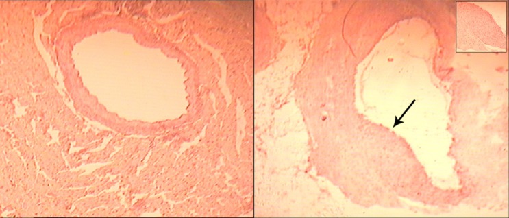
Histological sections of stained coronary arteries and the alternation in coronary plaque thickness from all the groups of rabbits are shown in figures 2, 3, and 4. Atherosclerotic changes were not observed in the normal diet group (Figure 3a). However, in the intimal surface of the coronary arteries from the high-cholesterol diet group, hyperplasia was seen in media layer along with high infiltration of cells to plaque formation site (Figure 3b). The thickness of the plaque had extended in all experimental groups after stopping the high-cholesterol diet. The thickness of plaques in the control group had extended compared with the high-cholesterol diet group. No considerable difference was seen in the extent of atherosclerotic thickness between the atorvastatin and quince leaf extract groups, and the control group (Figure 4a-c).
Figure 3.
Photomicrograph of a section of normal coronary artery (control group) (a) and hyperplasia were seen in media layer along with high infiltration of cells to plaque formation site (b) X40
Figure 4.
Photomicrograph of a coronary artery section of the control group after stopping high-cholesterol diet (a), atorvastatin (b), and quince leaf extract (c) groups No considerable difference was seen in the extent of atherosclerotic thickness between the atorvastatin and quince leaf extract groups, and control group
Discussion
Beyond the shadow of a doubt, hyperlipidemia is the most important risk factor for atherosclerosis.1,19 Using a daily high-cholesterol diet leads to its accumulation in the plasma membrane that causes membrane damage. This as an atherogenic stimulus increases the production of growth factors and proliferation of smooth muscle cells (SMC).20Therefore, any factors that reduce cholesterol levels can affect this process.
In this study, high-cholesterol level in plasma was shown by hypercholesterolemic regimen. Our results were similar to the study by Adaramoye et al. in which rats received high-cholesterol diets.21 Additionally, we demonstrated that lipid profiles (TC, TG, LDL, HDL) were returned to nearly normal levels by a standard diet for 12 weeks after stopping the hypercholesterolemic diet in the control group. Salazar et al. also reported that the biochemical parameters of rabbits were returned to normal by returning them to a standard diet. However, the histological parameters were not fully recovered.22
In The present study, atorvastatin has been used as a standard drug. This drug is one of the statin family of drugs that reduce cholesterol by inhibiting the HMG-CoAreductase. Furthermore, since atorvastatin was used in this study (oral administration) the results were quite predictable.23Until now, this drug has been used with the high-cholesterol diet by the prophylactic method. However, we have used this drug after accumulation of plaque, in order to demonstrate the reduction or returning of plaque and biochemical marker changes. The obtained results indicated that the drug’s ability to return the patient to normal condition (normal lipid profiles and biomarkers) has been effective, but it was less than the group receiving quince leaf extract. The early effects of statin therapy have been to lower LDL by 24-63%.24
Several studies have indicated that herbal or synthetic medicine decrease mortality of cardiovascular disease (CVD) by cholesterol regulation.25Accordingly, many efforts have been made to reduce CVD risk through cholesterol regulation. The health advantages of plant foods have been noted by some studies.26,27 Plants contain a variety of flavonoids such as flavonols, flavones, anthocyanidins, and quercetin.28 Moreover, we evaluated the efficacy of quince leaf extract on atheromic plaque. Hayek et al. in their study, showed the reduction in oxidation by flavonoids.29In another study, two flavonoids in the form of glucuronide and sulfate compounds were administered orally to rats and their antioxidant capacity increased.
Researches have illustrated that phenolic compounds, typically flavonol derivatives such as kaempferol glycoside and 0-3 kaempferol retinoid, are found inquince leaf and act as a filter protection against UV radiation.12,13,15
Flavonoids cause vascular expansion by increasing nitric oxide; this is an antioxidant property against LDL.30,31 Epidemiological studies have reported the beneficial effect of red wine and foods rich in flavonoids in reducing the risk of high cholesterol.31-33 Another study compared the protective effect of methanolic quince leaf extract and green tea on erythrocyte hemolysis that is caused by oxidative damage from free radicals.21 The anti-hemolytic effect in humans was demonstrated by both leaves. This study also revealed that the antioxidant capacity of leaf extract was lower than green tea extract. Moreover, there was no linear correlation between antioxidant activity and free radical reduction. In fact, the antioxidant activity of the leaves could be caused by the antagonistic or synergistic activity of bioactive compounds that are still unknown.34,35
In addition, the current study revealed a significant reduction in lipid profile (TC, TG, and LDL) and increase in HDL cholesterol in all experimental groups after stopping the atherogenic diet. These results illustrated that the group receiving quince leaf extract was more similar to the normal diet group, and the atorvastatin group had a better status than the group which was not taking medication. On the other hand, Suk et al. showed that serum levels of rats receiving a high-cholesterol diet were higher compared with rats receiving a standard diet. After receiving a standard diet for 6 weeks, they were able to return to their normal serum cholesterol levels. However, no change was observed in their triglyceride level.36 In our study, both levels of TC, and TG reduced significantly; the group receiving quince leaf extract was more similar to the group receiving standard regimen in this respect. To confirm, the same results were observed in rabbits fed on apple juice and the group that received high-cholesterol. This result indicates that apple juice has been effective in the adjustment of dyslipidemia caused by a high-cholesterol diet.37 On the other hand, consumption has been associated with a high-cholesterol diet.
The aqueous quince leaf extract was adminestrated in the three doses of 50, 100, and 200 mg/kg with isoproterenol (ISO) by Rajadurai et al. They administrated them orally, injected, and compared the results to alpha-tocopherol. Our study revealed that quince leaf extract in the dose of 200 mg/kg could regulate lipid profile levels, and reduce CK and LDH enzymes elevated by ISO. The effect of aqueous quince leaf extract 200 mg/kg was found to be equal to the effect of alpha-tocopherolin 60 mg/kg.17 The investigation method and objective of this study were different to our study; an atherogenic diet was used with quince extract to evaluate the preventive effect of the extract. Furthermore, we investigated the influence of the development or regression process of quince extract. In this study, we used total extract while previous studies did not mention the component of the leaf extract used.
Our study also showed the reduction of biochemical markers including AST, ALT, ALP, BUN, and Cr by a high-cholesterol diet regimen in the control, atorvastatin, and quince leaf extract groups. These results were consistent with the study by Singanan et al.35 (they demonstrated the hepatoprotective effect of quince leaf extract), and Suk et al.36 (they studied on rat for 6 weeks). However, in the histological results, liver damage caused by a high-fat diet did not improve with a standard diet.36 As expected, an atherogenic diet, as a progression of atheroma catalyst, resulted in an increase of this complication in the aortic and coronary artery in the control group. Increase plaque, in this group, showed that damage to the endothelium caused by a high-cholesterol diet provided a context for further stenosis in blood vessels. In both atorvastatin and leaf extract receiving groups plague formation was considerably higher. In this regard our findings were consistent with the results of other studies.35,36 Our study indicated that plaque formation in the coronary artery in rabbits receiving atherogenic diet. However, after three months follow-up with quince leaf extract 50mg/kg and atorvastatin 0.5 mg/kg, neither drug were able to inhibit the increase in plaque in the coronary; plaque thickness was higher in the control group, which had not been taking any medication, than the quince leaf extract group. This difference was not significant. Decorde et al. showed that the phenols of grape, black grapes, apple juice, and apple decreased atherosclerotic plaque in hamsters by 93%, 78%, 60%, and 48%, respectively.38 A survey on red wine showed a significant reduction in plaque in arteries.39
Inflammation has an important role in the development of atherosclerosis. The abatement of inflammation in the coronary plaque in the group receiving atorvastatin is due to the anti-inflammatory effects of statins that has already been proven.40
In some researches, the activity of some enzymes during the inflammatory process were inhibited.41-43 Plaque stabilizing effects of statins may also be due to the reduction of some inflammatory cytokine after plasma lipid lowering. The reduced anti-inflammatory cytokine levels, such as CRP, with the lowering in TC and LDL prevented the development of atherosclerosis.37
Conclusion
The results of the present study confirmed the correlation between hypercholesterolemia and atherosclerosis, and also that quince leaf extract, like atorvastatin, can effectively reduce a high-fat diet-induced atherosclerosis.
However, according to histological determination, atorvastatin and quince leaf extract were not able to prevent plaque increasing in the coronary after plaque formation for 12 weeks. Both treatments have been able to reduce serum levels to nearly normal level after plaque formation and accumulation; the quince leaf extract was more effective than atorvastatin which may be due to its phenolic components. It seems that the decrease in plaque must be studied for a longer time at least for the dose used in our study.
Further studies are recommended in order to find the exact mechanism of the quince leaf extract in endothelial function improvement. More effective clinical applications of these natural compounds will be determined.
Footnotes
Conflicts of Interest
Authors have no conflict of interests.
REFERENCES
- 1.Crowther MA. Pathogenesis of atherosclerosis. Hematology Am Soc Hematol Educ Program. 2005:436–41. doi: 10.1182/asheducation-2005.1.436. [DOI] [PubMed] [Google Scholar]
- 2.Asgary S, JafariDinani N, Madani H, Mahzoni P, Naderi GH. Effect of Glycyrrhiza glabra extract on aorta wall atherosclerotic lesion in hyper-cholestero-lemic rabbits. Pak J Nutr. 2007;6(4):313–7. [Google Scholar]
- 3.Wang Y, Tuomilehto J, Jousilahti P, Antikaimnen R, Mahonen M, Katzmarzyk PT, et al. Lifestyle factors in relation to heart failure among Finnish men and women. Circ Heart Fail. 2011;4(5):607–12. doi: 10.1161/CIRCHEARTFAILURE.111.962589. [DOI] [PubMed] [Google Scholar]
- 4.Jialal I, Devaraj S, Kaul N. The Effect of α-Tocopherol on Monocyte Proatherogenic Activity. J Nutr. 2001;131(2):389S–94S. doi: 10.1093/jn/131.2.389S. [DOI] [PubMed] [Google Scholar]
- 5.Campbell JH, Efendy JL, Smith NJ, Campbell GR. Molecular basis by which garlic suppresses atherosclerosis. J Nutr. 2001;131(3s):1006S–9S. doi: 10.1093/jn/131.3.1006S. [DOI] [PubMed] [Google Scholar]
- 6.Hansson GK. Inflammation, atherosclerosis, and coronary artery disease. N Engl J Med. 2005;352(16):1685–95. doi: 10.1056/NEJMra043430. [DOI] [PubMed] [Google Scholar]
- 7.Poredos P, Jezovnik MK. Dyslipidemia, statins, and venous thromboembolism. Semin Thromb Hemost. 2011;37(8):897–902. doi: 10.1055/s-0031-1297368. [DOI] [PubMed] [Google Scholar]
- 8.Reddy R, Chahoud G, Mehta JL. Modulation of cardiovascular remodeling with statins: fact or fiction? Curr Vasc Pharmacol. 2005;3(1):69–79. doi: 10.2174/1570161052773915. [DOI] [PubMed] [Google Scholar]
- 9.Ali MS, Pervez MK. Marmenol: a 7-geranyloxycoumarin from the leaves of Aegle marmelos Corr. Nat Prod Res . 2004;18(2):141–6. doi: 10.1080/14786410310001608037. [DOI] [PubMed] [Google Scholar]
- 10.du Toit R, Volsteedt Y, Apostolides Z. Comparison of the antioxidant content of fruits, vegetables and teas measured as vitamin C equivalents. Toxicology. 2001;166(1-2):63–9. doi: 10.1016/s0300-483x(01)00446-2. [DOI] [PubMed] [Google Scholar]
- 11.Silva BM, Andrade PB, Valentao P, Ferreres F, Seabra RM, Ferreira MA. Quince (Cydoniaoblonga Miller) fruit (pulp, peel, and seed) and Jam: antioxidant activity. J Agric Food Chem. 2004;52(15):4705–12. doi: 10.1021/jf040057v. [DOI] [PubMed] [Google Scholar]
- 12.Silva BM, Valentao P, Seabra RM, Andrade PB. Quince (Cydoniaoblonga Miller): an interesting dietary source of bioactive compounds. In: Papadopoulos K, editor. Food Chemistry Research Developments. New York, NY: Nova Publishers; 2008. pp. 243–66. [Google Scholar]
- 13.Oliveira AP, Pereira JA, Andrade PB, Valentao P, Seabra RM, Silva BM. Phenolic profile of Cydoniaoblonga Miller leaves. J Agric Food Chem. 2007;55(19):7926–30. doi: 10.1021/jf0711237. [DOI] [PubMed] [Google Scholar]
- 14.Chockalingam V, Kadali SS, Gnanasambantham P. Antiproliferative and antioxidant activity of Aeglemarmelos (Linn) leaves in Dalton's Lymphoma Ascites transplanted mice. Indian J Pharmacol. 2012;44(2):225–9. doi: 10.4103/0253-7613.93854. [DOI] [PMC free article] [PubMed] [Google Scholar]
- 15.Oliveira AP, Pereira J, Andrade PB, Valentao P, Seabra RM, Silva BM. Organic acids composition of Cydoniaoblonga Miller leaf. Food Chemistry. 2008;111(2):393–9. doi: 10.1016/j.foodchem.2008.04.004. [DOI] [PubMed] [Google Scholar]
- 16.Costa RM, Magalhaes AS, Pereira JA, Andrade PB, Valentao P, Carvalho M, et al. Evaluation of free radical-scavenging and antihemolytic activities of quince (Cydoniaoblonga) leaf: a comparative study with green tea (Camellia sinensis). Food ChemToxicol. 2009;47(4):860–5. doi: 10.1016/j.fct.2009.01.019. [DOI] [PubMed] [Google Scholar]
- 17.Rajadurai M, Prince PS. Comparative effects of Aeglemarmelos extract and alpha-tocopherol on serum lipids, lipid peroxides and cardiac enzyme levels in rats with isoproterenol-induced myocardial infarction. Singapore Med J. 2005;46(2):78–81. [PubMed] [Google Scholar]
- 18.Fiorentino A, D'Abrosca B, Pacifico S, Mastellone C, Piscopo V, Caputo R, et al. Isolation and structure elucidation of antioxidant polyphenols from quince (Cydonia vulgaris) peels. J Agric Food Chem. 2008;56(8):2660–7. doi: 10.1021/jf800059r. [DOI] [PubMed] [Google Scholar]
- 19.Lusis AJ. Atherosclerosis. Nature. 2000;407(6801):233–41. doi: 10.1038/35025203. [DOI] [PMC free article] [PubMed] [Google Scholar]
- 20.Mcmurry HF, Chahwala SB. Amlodipine exerts a potent antimigrational effect on aortic smooth muscle cells in culture. J Cardiovasc Pharmacol. 1999;20:54–6. [Google Scholar]
- 21.Adaramoye OA, Akintayo O, Achem J, Fafunso MA. Lipid-lowering effects of methanolic extract of Vernonia amygdalina leaves in rats fed on high cholesterol diet. Vasc Health Risk Manag. 2008;4(1):235–41. doi: 10.2147/vhrm.2008.04.01.235. [DOI] [PMC free article] [PubMed] [Google Scholar]
- 22.Salazar JJ, Ramirez AI, de Hoz R, Rojas B, Ruiz E, Tejerina T, et al. Alterations in the choroid in hypercholesterolemic rabbits: reversibility after normalization of cholesterol levels. Exp Eye Res. 2007;84(3):412–22. doi: 10.1016/j.exer.2006.10.012. [DOI] [PubMed] [Google Scholar]
- 23.Fuhrman B, Aviram M. Preservation of paraoxonase activity by wine flavonoids: possible role in protection of LDL from lipid peroxidation. Ann N Y AcadSci. 2002;957:321–4. doi: 10.1111/j.1749-6632.2002.tb02933.x. [DOI] [PubMed] [Google Scholar]
- 24.Serdar Z, Aslan K, Dirican M, Sarandol E, Yesilbursa D, Serdar A. Lipid and protein oxidation and antioxidant status in patients with angiographically proven coronary artery disease. ClinBiochem. 2006;39(8):794–803. doi: 10.1016/j.clinbiochem.2006.02.004. [DOI] [PubMed] [Google Scholar]
- 25.Kwiterovich PO. The effect of dietary fat, antioxidants, and pro-oxidants on blood lipids, lipoproteins, and atherosclerosis. J Am Diet Assoc. 1997;97(7 Suppl):S31–S41. doi: 10.1016/s0002-8223(97)00727-x. [DOI] [PubMed] [Google Scholar]
- 26.Yokozawa T, Cho EJ, Sasaki S, Satoh A, Okamoto T, Sei Y. The protective role of Chinese prescription Kangen-karyu extract on diet-induced hypercholesterolemia in rats. Biol Pharm Bull. 2006;29(4):760–5. doi: 10.1248/bpb.29.760. [DOI] [PubMed] [Google Scholar]
- 27.Zhang HW, Zhang YH, Lu MJ, Tong WJ, Cao GW. Comparison of hypertension, dyslipidaemia and hyperglycaemia between buckwheat seed-consuming and non-consuming Mongolian-Chinese populations in Inner Mongolia, China. Clin Exp Pharmacol Physiol. 2007;34(9):838–44. doi: 10.1111/j.1440-1681.2007.04614.x. [DOI] [PubMed] [Google Scholar]
- 28.Terao J, Kawai Y, Murota K. Vegetable flavonoids and cardiovascular disease. Asia Pac J Clin Nutr. 2008;17(Suppl 1):291–3. [PubMed] [Google Scholar]
- 29.Hayek T, Fuhrman B, Vaya J, Rosenblat M, Belinky P, Coleman R, et al. Reduced progression of atherosclerosis in apolipoprotein E-deficient mice following consumption of red wine, or its polyphenols quercetin or catechin, is associated with reduced susceptibility of LDL to oxidation and aggregation. Arterioscler Thromb Vasc Biol. 1997;17(11):2744–52. doi: 10.1161/01.atv.17.11.2744. [DOI] [PubMed] [Google Scholar]
- 30.Hogg N, Struck A, Goss SP, Santanam N, Joseph J, Parthasarathy S, et al. Inhibition of macrophage-dependent low density lipoprotein oxidation by nitric-oxide donors. J Lipid Res. 1995;36(8):1756–62. [PubMed] [Google Scholar]
- 31.Benito S, Buxaderas S, Mitjavila MT. Flavonoid metabolites and susceptibility of rat lipoproteins to oxidation. Am J Physiol Heart Circ Physiol. 2004;287(6):H2819–H2824. doi: 10.1152/ajpheart.00471.2004. [DOI] [PubMed] [Google Scholar]
- 32.Hertog MG, Kromhout D, Aravanis C, Blackburn H, Buzina R, Fidanza F, et al. Flavonoid intake and long-term risk of coronary heart disease and cancer in the seven countries study. Arch Intern Med. 1995;155(4):381–6. [PubMed] [Google Scholar]
- 33.Leikert JF, Rathel TR, Wohlfart P, Cheynier V, Vollmar AM, Dirsch VM. Red wine polyphenols enhance endothelial nitric oxide synthase expression and subsequent nitric oxide release from endothelial cells. Circulation. 2002;106(13):1614–7. doi: 10.1161/01.cir.0000034445.31543.43. [DOI] [PubMed] [Google Scholar]
- 34.Silva BM, Andrade PB, Martins RC, Valentao P, Ferreres F, Seabra RM, et al. Quince (Cydoniaoblonga miller) fruit characterization using principal component analysis. J Agric Food Chem. 2005;53(1):111–22. doi: 10.1021/jf040321k. [DOI] [PubMed] [Google Scholar]
- 35.Singanan V, Singanan M, Begum H. The Hepatoprotective Effect of Bael Leaves (Aegle Marmelos) in alcohol Induced Liver Injury in Albino Rats. International Journal of Science & Technology. 2007;2(2):83–92. [Google Scholar]
- 36.Suk FM, Lin SY, Chen CH, Yen SJ, Su CH, Liu DZ, et al. Taiwanofungus camphoratus activates peroxisome proliferator-activated receptors and induces hypotriglyceride in hypercholesterolemic rats. Biosci Biotechnol Biochem. 2008;72(7):1704–13. doi: 10.1271/bbb.70810. [DOI] [PubMed] [Google Scholar]
- 37.Setorki M, Asgary S, Eidi A, Rohani AH, Esmaeil N. Effects of apple juice on risk factors of lipid profile, inflammation and coagulation, endothelial markers and atherosclerotic lesions in high cholesterolemic rabbits. Lipids Health Dis. 2009; 8:39. doi: 10.1186/1476-511X-8-39. [DOI] [PMC free article] [PubMed] [Google Scholar]
- 38.Decorde K, Teissedre PL, Auger C, Cristol JP, Rouanet JM. Phenolics from purple grape, apple, purple grape juice and apple juice prevent early atherosclerosis induced by an atherogenic diet in hamsters. Mol Nutr Food Res. 2008;52(4):400–7. doi: 10.1002/mnfr.200700141. [DOI] [PubMed] [Google Scholar]
- 39.da Luz PL, Serrano Junior CV, Chacra AP, Monteiro HP, Yoshida VM, Furtado M, et al. The effect of red wine on experimental atherosclerosis: lipid-independent protection. Exp Mol Pathol. 1999;65(3):150–9. doi: 10.1016/s0014-4800(99)80004-5. [DOI] [PubMed] [Google Scholar]
- 40.Liu Y, Yan F, Liu Y, Zhang C, Yu H, Zhang Y, et al. Aqueous extract of rhubarb stabilizes vulnerable atherosclerotic plaques due to depression of inflammation and lipid accumulation. Phytother Res. 2008;22(7):935–42. doi: 10.1002/ptr.2429. [DOI] [PubMed] [Google Scholar]
- 41.Nair MP, Mahajan S, Reynolds JL, Aalinkeel R, Nair H, Schwartz SA, et al. The flavonoid quercetin inhibits proinflammatory cytokine (tumor necrosis factor alpha) gene expression in normal peripheral blood mononuclear cells via modulation of the NF-kappa beta system. Clin Vaccine Immunol. 2006;13(3):319–28. doi: 10.1128/CVI.13.3.319-328.2006. [DOI] [PMC free article] [PubMed] [Google Scholar]
- 42.Kwon KH, Murakami A, Tanaka T, Ohigashi H. Dietary rutin, but not its aglyconequercetin, ameliorates dextran sulfate sodium-induced experimental colitis in mice: attenuation of pro-inflammatory gene expression. Biochem Pharmacol. 2005;69(3):395–406. doi: 10.1016/j.bcp.2004.10.015. [DOI] [PubMed] [Google Scholar]
- 43.Huang MT, Liu Y, Ramji D, Lo CY, Ghai G, Dushenkov S, et al. Inhibitory effects of black tea theaflavin derivatives on 12-O-tetradecanoylphorbol-13-acetate-induced inflammation and arachidonic acid metabolism in mouse ears. Mol Nutr Food Res. 2006;50(2):115–22. doi: 10.1002/mnfr.200500101. [DOI] [PubMed] [Google Scholar]


