
Fig. 10.
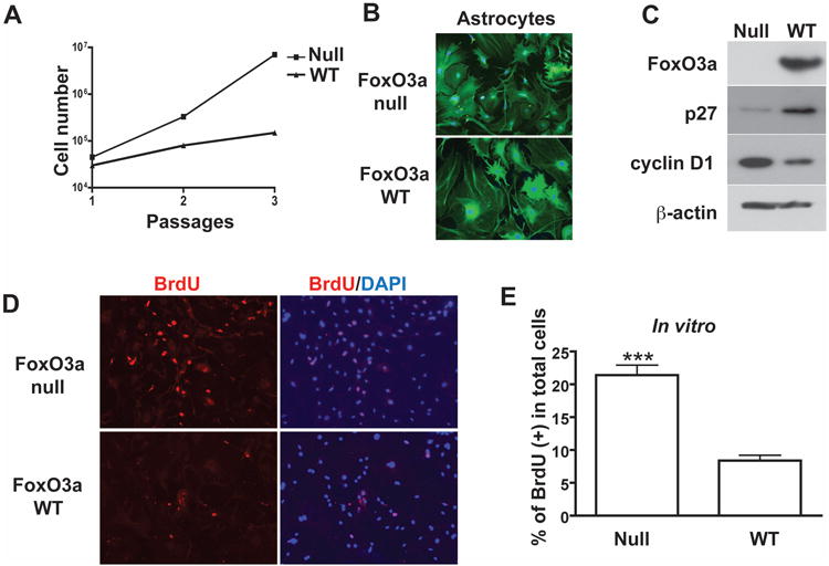
Astrocytes from Foxo3a-null mice exhibit higher proliferative potential in vitro. A, astrocytes were isolated from 4 week old of Foxo3a-null or Foxo3a-WT mouse brain and cultured in vitro. Total cell number was counted in each passage. B, after 3 passages, purity of astrocyte cultures was determined by GFAP and DAPI staining. C, astrocyte lysates were collected and subjected to Western blotting for Foxo3a, cyclin D1, and p27Kip1. β-actin was used as a loading control. D and E, mouse astrocytes were stained with BrdU and DAPI. Proliferating cells were counted as the percent of BrdU+ in DAPI+ nuclei. A total of 30 pictures from 4 mice were calculated. ***, p < 0.001 in comparison to control.