

Abstract
Sub-100 nm hollow gold nanoparticle superstructures were prepared in a direct one-pot reaction. A gold-binding peptide conjugate, C6-AA-PEPAu (PEPAu = AYSSGAPPMPPF), was constructed and used to direct the simultaneous synthesis and assembly of gold nanoparticles. Transmission electron microscopy and electron tomography revealed that the superstructures are uniform and consist of monodisperse gold nanoparticles arranged into a spherical monolayer shell.
Individual nanoparticles have size-, shape-, and composition-dependent properties. Nanoparticle assemblies can exhibit unique ensemble properties that depend on the organization of the nanoparticles.1 Therefore, to control the ensemble properties of a nanoparticle assembly, methods to precisely control the placement of nanoparticles within the assembly must be developed. Achieving rational control of nanoparticle assembly from the “bottom up” is a significant synthetic challenge and represents one of the most important hurdles in modern nanoscience.1a, b Practical methods that address this challenge would ideally (1) allow for expeditious construction of arbitrarily complex nanoparticle superstructures, (2) enable assembly of nanoparticles of any composition, (3) permit precise control over structural attributes, including nanoparticle size and position, and (4) require few preparative steps.
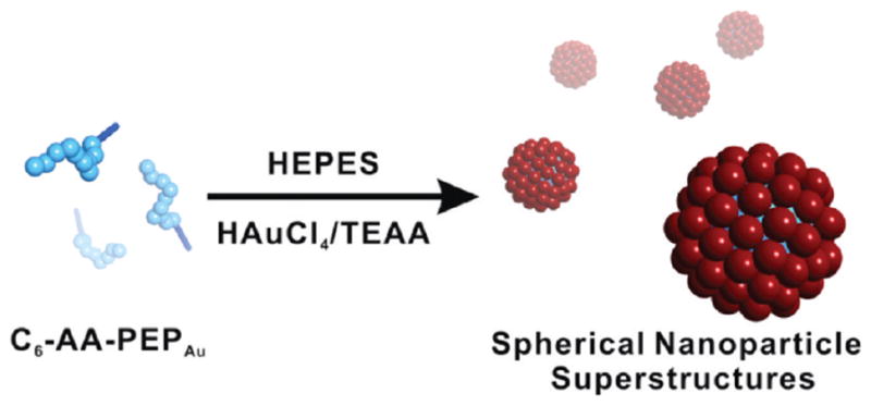
We recently introduced a new methodology for designing and assembling nanoparticle superstructures that begins to address these important criteria.2 This methodology relies on unique peptide conjugate molecules comprising peptides with sequences of amino acids that recognize and bind specific inorganic compositions and organic tethers designed to influence the self-assembly of these peptides.3 In the presence of inorganic salts and reducing agents, properly designed peptide conjugate molecules can orchestrate a one-pot reaction in which the synthesis and assembly of nanopar-ticles into complex superstructures occurs simultaneously. From the outset of our studies in this area, we targeted hollow spherical nanoparticle superstructures4 because of their potential practical application as capsules or delivery agents5 and because of the inherent challenge of expeditiously generating such structures in a directed one-pot synthesis. Here we introduce a new peptide conjugate that directs the construction of such structures (Scheme 1). Importantly, we demonstrate that when this methodology is used, small modifications of the peptide conjugate can dramatically impact the topology of the final nanoparticle superstructure.
Scheme 1.
Direct Preparation of Hollow Spherical Gold
Nanoparticle Superstructures (Red Spheres = Gold Nanoparticles)
In our previous work, we showed that C12-PEPAu conjugates (PEPAu6 = AYSSGAPPMPPF), when mixed with a gold salt and a reducing agent, direct the synthesis and assembly of 1D gold nanoparticle double helices exhibiting tailorable metrics and structures.2 These results prompted us to systematically investigate how shortening the length of the aliphatic tether would impact the synthesis and assembly process and the ultimate structure of the product nanoparticle superstructure. Accordingly, we prepared C10-PEPAu, C8-PEPAu, and C6-PEPAu using established methods.2a Each conjugate was individually dissolved in 0.1 M HEPES buffer [pH 7.3 ± 0.1; HEPES = 4-(2-hydroxyethyl)-1-piperazineethanesulfonic acid], and an aliquot of chloroauric acid/triethylammonium acetate (HAuCl4/TEAA) buffer solution (0.1 M HAuCl4 in 1.0 M TEAA, pH 7.0) was added to each solution. In this system, HEPES served as the primary reducing agent.7 C10-PEPAu reactions yielded 1D nanoparticle superstructures, while C6-PEPAu and C8-PEPAu reactions yielded only dispersed individual nanoparticles or nanoparticle aggregates (Figure S3 in the Supporting Information). On the basis of these observations, we concluded that the C6 and C8 tethers were not sufficient for directing either the assembly of the peptide conjugate or the organization of the nanoparticles.
We note that both the amino acid sequence and the organic tether can influence the self-assembly of the peptide conjugate. Therefore, we prepared C6-AA-PEPAu, reasoning that the alanine residues attached to the N-terminus of PEPAu could impact the self-assembly of the conjugate.8 Interestingly, C6-AA-PEPAu self-assembled into spherical structures (diameter = 136.5 ± 2.6 nm) upon incubation in water (24 h), as evidenced by transmission electron microscopy (TEM) (Figure S4) and atomic force microscopy (AFM) (Figures S5 and S6). However, no self-assembly was observed in HEPES (Figure S7). AFM height images suggested that the spheres spread onto the MICA surface, resulting in broader and flatter structures (height = 29.3 ± 1.0 nm and diameter = 525.2 ± 8.9 nm; Figure S5). AFM phase images of the structures collected after TEM imaging and consequent exposure to high vacuum revealed that the structures had collapsed in the middle (Figure S6). Collectively, these AFM data suggest that the self-assembled C6-AA-PEPAu spherical structures are hollow.9
Encouraged by these observations, we proceeded to evaluate whether C6-AA-PEPAu could direct the self-assembly of gold nanoparticles into spherical superstructures (Scheme 1). HAuCl4/ TEAA was added to a solution of C6-AA-PEPAu in HEPES buffer to yield a colorless solution. After incubation at room temperature for 24 h, the solution became light-purple, suggesting the formation of gold nanoparticle assemblies. TEM analysis of samples of this solution revealed the exclusive formation of spherical structures comprising gold nanoparticles (8.3 ± 0.2 nm) (Figure 1a, b and Figure S8). The UV–vis spectrum of the spherical superstructures exhibited a maximum absorbance at 540 nm, which, as expected, is red-shifted compared to that of colloidal solutions of 9 nm gold nanoparticles (~517 nm).10 Close examination of the TEM images indicated that, with dark edges and light interiors, the structures likely consist of self-assembled C6-AA-PEPAu cores (35.3 ± 1.0 nm) surrounded by gold nanoparticle monolayer shells (51.6 ± 0.8 nm) (Figure 1a, b and Figure S8). Electron tomography was used to probe the three-dimensional structure of the nanoparticle assemblies. Tilted images collected at 1° tilt intervals from −70 to 70° were combined computationally to generate a three-dimensional electron density map (tomogram). Sample tomographic slices (Figure 1c) and the surface-rendered 3D tomogram (Figure 1d) definitively revealed that the structures are spherical capsules comprising a single layer of gold nanoparticles.
Figure 1.

(a, b) TEM images of spherical gold nanoparticle superstructures. (c) X–Y computational slices (i–viii) of the 3D tomographic volume containing the nanoparticle assembly (scale bar = 20 nm). (d) 3D surface rendering of the tomographic volume.
At this stage of our investigation, it was clear that C6-AA-PEPAu directs the assembly of gold nanoparticles into spherical super-structures and that hollow self-assembled C6-AA-PEPAu structures serve as the underlying structure-directing entities. In fact, when we performed the same reaction using AA-PEPAu instead of C6-AA-PEPAu, random gold nanoparticle aggregates were observed as the product (Figure S10). We note that the average diameter of the nanoparticle superstructures is smaller than what one might expect on the basis of the average diameter of the C6-AA-PEPAu nano-spheres that self-assemble in water. This suggests that the assembly of C6-AA-PEPAu and gold nanoparticles in HEPES buffer may in fact be coupled and that both the peptide conjugate and nanoparticle components could influence the size, shape, and self-assembly of the resulting superstructure.
To more completely understand the mechanism of superstructure formation, we studied samples of the reaction mixture at increasing time points using both TEM and AFM. After ~20 min, we observed islands of nanoparticles containing nanoparticles of uniform diameter (4.7 ± 0.1 nm) (Figure 2a and Figure S11). Over the course of several hours, the nanoparticles within the islands grew (5.4 ± 0.2 nm at 2.5 h, Figure 2b; 6.2 ± 0.1 nm at 4.5 h, Figure 2c). We hypothesize that as small nanoparticles form in solution, they provide a surface onto which C6-AA-PEPAu can attach and aggregate. Aggregates of C6-AA-PEPAu then attract more nanoparticles, leading to the formation of island structures. At a certain critical point, the island structures accumulate and concentrate enough C6-AA-PEPAu in one area that it becomes favorable for C6-AA-PEPAu to self-assemble into spherical structures, thus driving the spherical assembly of the gold nanoparticles (Figure 2d). We note that this assembly process also results in a small number of incomplete structures (see arrow in Figure 2d) that need additional nanoparticles to complete their spherical nanoparticle monolayer shells.
Figure 2.

Progress of superstructure formation: TEM images and particle size distributions after (a) 20 min, (b) 2.5 h, (c) 4.5 h, and (d) 38 h of reaction. The arrow indicates an incomplete nanoparticle superstructure.
In summary, we have discovered that C6-AA-PEPAu conjugates direct the formation of well-defined hollow spherical nanoparticle superstructures. Remarkably, C6-AA-PEPAu and C12-PEPAu,2a which have slightly different compositions, direct the formation of dramatically different nanoparticle superstructures (hollow spheres vs double helices2a). This result points toward the versatility of this methodology and the ability to program the shape of the ultimate nanoparticle superstructure simply by programming specific information into the composition of the peptide conjugate.
Supplementary Material
Acknowledgments
Funding for this work was provided by the University of Pittsburgh and the NSF (DMR-0954380). The authors thank the MEMS Department and PINSE for access to electron microscopy and AFM instrumentation, respectively.
Footnotes
Supporting Information Available: Experimental procedures and additional supporting data. This material is available free of charge via the Internet at http://pubs.acs.org.
References
- 1.(a) Kotov NA, Stellacci F. Adv Mater. 2008;20:4221. [Google Scholar]; (b) Nie ZH, Petukhova A, Kumacheva E. Nat Nanotechnol. 2010;5:15. doi: 10.1038/nnano.2009.453. [DOI] [PubMed] [Google Scholar]; (c) Chang WS, Slaughter LS, Khanal BP, Manna P, Zubarev ER, Link S. Nano Lett. 2009;9:1152. doi: 10.1021/nl803796d. [DOI] [PubMed] [Google Scholar]; (d) Hentschel M, Saliba M, Vogelgesang R, Giessen H, Alivisatos AP, Liu N. Nano Lett. 2010;10:2721. doi: 10.1021/nl101938p. [DOI] [PubMed] [Google Scholar]
- 2.(a) Chen CL, Zhang PJ, Rosi NL. J Am Chem Soc. 2008;130:13555. doi: 10.1021/ja805683r. [DOI] [PMC free article] [PubMed] [Google Scholar]; (b) Chen CL, Rosi NL. J Am Chem Soc. 2010;132:6902. doi: 10.1021/ja102000g. [DOI] [PubMed] [Google Scholar]
- 3.Chen CL, Rosi NL. Angew Chem, Int Ed. 2010;49:1924. doi: 10.1002/anie.200903572. and references therein. [DOI] [PubMed] [Google Scholar]
- 4.(a) Caruso F, Caruso RA, Mohwald H. Science. 1998;282:1111. doi: 10.1126/science.282.5391.1111. [DOI] [PubMed] [Google Scholar]; (b) Wong MS, Cha JN, Choi KS, Deming TJ, Stucky GD. Nano Lett. 2002;2:583. [Google Scholar]; (c) Park S, Lim JH, Chung SW, Mirkin CA. Science. 2004;303:348. doi: 10.1126/science.1093276. [DOI] [PubMed] [Google Scholar]; (d) Rana RK, Murthy VS, Yu J, Wong MS. Adv Mater. 2005;17:1145. [Google Scholar]; (e) Liu B, Zeng HC. J Am Chem Soc. 2004;126:8124. doi: 10.1021/ja048195o. [DOI] [PubMed] [Google Scholar]; (f) Vasquez Y, Sra AK, Schaak RE. J Am Chem Soc. 2005;127:12504. doi: 10.1021/ja054442s. [DOI] [PubMed] [Google Scholar]
- 5.(a) Jin YD, Gao XH. J Am Chem Soc. 2009;131:17774. doi: 10.1021/ja9076765. [DOI] [PMC free article] [PubMed] [Google Scholar]; (b) Wu GH, Mikhailovsky A, Khant HA, Fu C, Chiu W, Zasadzinski JA. J Am Chem Soc. 2008;130:8175. doi: 10.1021/ja802656d. [DOI] [PMC free article] [PubMed] [Google Scholar]
- 6.Slocik JM, Stone MO, Naik RR. Small. 2005;1:1048. doi: 10.1002/smll.200500172. [DOI] [PubMed] [Google Scholar]
- 7.(a) Habib A, Tabata M, Wu YG. Bull Chem Soc Jpn. 2005;78:262. [Google Scholar]; (b) Xie JP, Lee JY, Wang DIC. Chem Mater. 2007;19:2823. [Google Scholar]
- 8.Jahnke E, Lieberwirth I, Severin N, Rabe JP, Frauenrath H. Angew Chem, Int Ed. 2006;45:5383. doi: 10.1002/anie.200600610. [DOI] [PubMed] [Google Scholar]
- 9.Seo SH, Chang JY, Tew GN. Angew Chem, Int Ed. 2006;45:7526. doi: 10.1002/anie.200600688. [DOI] [PubMed] [Google Scholar]
- 10.Link S, El-Sayed MA. J Phys Chem B. 1999;103:8410. JA106833G. [Google Scholar]
Associated Data
This section collects any data citations, data availability statements, or supplementary materials included in this article.