Abstract
In 2007 to 2008, a fruit rot of Melon (Cucumis melo L.) caused by Sclerotium rolfsii occurred sporadically in a farmer's vinyl house in Jinju City. The symptoms started with watersoaking lesion and progressed into the rotting of the surface of fruit. White mycelial mats appeared on the lesion at the surface of the fruit and a number of sclerotia formed on the fruit near the soil line. The sclerotia were globoid in shape, 1~3 mm in size, and white to brown in color. The hyphal width was measured 3 to 8 µm. The optimum temperature for mycelial growth and sclerotia formation was 30 on PDA. Typical clamp connections were observed in hyphae of grown for 4 days on PDA. On the basis of symptoms, mycological characteristics and pathogenicity to the host plant, this fungus was identified as Sclerotium rolfsii Saccardo. This is the first report of the fruit rot of Melon caused by S. rolfsii in Korea.
Keywords: Cucumis melo, Fruit rot, Melon, Sclerotium rolfsii
A fruit rot of Melon (Cucumis melo L.) caused by Sclerotium rolfsii was observed in a farmer's vinyl house in Jinju City in 2007 and 2008. This disease mainly occurs on fruit in vinyl houses. It has not been recorded in Korea (The Korean Society of Plant Pathology, 2004).
The sclerotial diseases occur primarily in warm climates and, especially in the hot and humid conditions in vinyl houses. Sclerotial diseases affect a wide variety of plants, including many vegetables, flowers, legumes, cereals, forage plants and weeds (Agrios, 2005). The pathogen causes damping-off of seedlings, stem canker, crown blight, and rot on root, crown, bulb, tuber and fruit (Gobayashi et al., 1992).
Recently, S. rolfsii has become a serious pathogen in Korea and is one of the most important soil borne pathogens in Gyeongnam province. Some sclerotial diseases, such as Disporum smilacinum (Kwon and Jee, 2007), Allium victorialis (Kwon et al., 2008), Elsholtzia splendens (Kwon and Park, 2008) and Petunia hybrida (Kwon, 2008), were recently reported by authors in Korea.
Symptoms
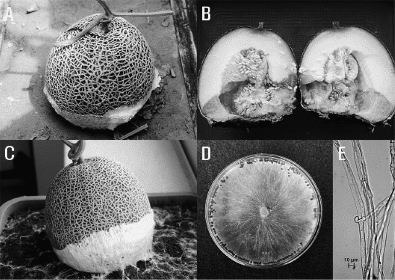
White mycelial mats appeared on the surface of basal fruits (Fig. 1A). The white mycelia were readily visible nearby soil surface fruit and grew inside of fruit tissues. The symptoms started with watersoaking lesion and progressed into the surface of the fruit. The fungus produced numerous small globoid sclerotia of uniform size which were initially white but turned brown. This was observed both on PDA and on the host plant in the field. The heavily infected fruits became watery and eventually rotted (Fig. 1B).
Fig. 1.
Symptoms of fruit rot of Melon (Cucumis melo) and mycological characteristics of the pathogenic fungus, Sclerotium rolfsii. A, Abandoned fruit became a secondary inoculum source in the field; B, Longitudinal section of a severely infected fruit; C, Symptoms after artificial inoculation; D, Mycelial mat and sclerotia grown on PDA after 20 days; E, Clamp connection.
Disease occurrence
In Gyeongsangnam-do, the Melon is cultivated as a cash crop and its cultivation increases annually. These cultivation practices favored an outbreak of the disease. The infection rate reached 2% between May 2007 and May 2008. Abundant sclerotia were often produced on the surfaces of infected fruits and near the soilline in vinyl houses, which play an important role of secondary inoculum as soil born diseases in the fields.
Mycological characteristics
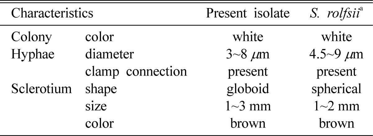
Infected fruits were collected from the fields and isolated to globoid dark brown sclerotia on surface fruit. The sclerotia were disinfected in a 1% NaOCl solution for 60 seconds then washed in distilled water 3 times. The fungus was isolated on potato dextrose agar (PDA). After incubated for 4 days at 25℃, the mycelial tips were cut and transferred to fresh PDA for further study. The growth pattern of the fungus was examined after incubation for 20 days at 25 to 30℃. The fungus grew between 10℃ and 35℃ and optimal temperature for the growth was 30℃ on PDA. The white mycelium usually formed many narrow hyphal strands in the aerial mycelium which were 3 to 8 µm in width. After 4 days, this mycelium had typical clamp connection structure (Fig. 1E). After 20 days, sclerotia were examined for the characteristics. Small, uniformly sized, globoid sclerotia were produced in great numbers.The sclerotia were initially white then turned to dark brown at maturation. The maximum numbers of sclerotia were produced at 25 to 30℃. The size of sclerotia were 1 to 3 mm (Fig. 1D, Table 1).
Table 1.
Comparison of mycological characteristics between the isolate obtained from Melon (Cucumis melo L.) and described previously Sclerotium rolfsii

aDescribed by Mordue (1974)
Pathogenicity test
The pathogenicity of the fungus on Melon (Cucumis melo) was tested in a greenhouse at Gyeongsangnamdo Agricultural Research and Extension Services in May 2008. Field soil was sterilized in an autoclave 3 times for 5 days. Inoculum was prepared from mycelial mats grown for 7 days on PDA. The fruits were inoculated with 100 g of the inoculum source near the basal end of fruit. The first symptom appeared 4 days after inoculation. It developed to severe fruit rot (Fig. 1C). The pathogen was reisolated from the lesions to prove Koch's postulate.
On the basis of symptoms, mycological characteristics and pathogenicity to the host plant, this fungus was identified as Sclerotium rolfsii Saccardo.
The fungus is stored at the Korean Agricultural Culture Collection (KACC No. 43067), National Institute of Agricultural Biotechnology, Rural Development Administration, Suwon.
References
- 1.Agrios GN. Plant pathology. 5th ed. London: Academic press; 2005. p. 922. [Google Scholar]
- 2.Gobayashi T, Katumoto K, Abiko K, Abe Y, Kakishima M. Illustrated genera of plant pathogenic fungi in Japan. The Whole Farming Educational Association; 1992. p. 685. (in Japanese) [Google Scholar]
- 3.Kwon JH. Occurrence of stem rot of Petunia hybrida caused by Sclerotium rolfsii. Korean J Mycol. 2008;36:203–205. (in Korean) [Google Scholar]
- 4.Kwon JH, Jee HJ. Occurrence of stem rot of Disporum smilacinum caused by Sclerotium rolfsii in Korea. Plant Pathol J. 2007;23:212–214. [Google Scholar]
- 5.Kwon JH, Jee HJ, Park CS. Occurrence of bulb rot of Allium victorialis var. platyphyllum caused by Sclerotium rolfsii in Korea. Plant Pathol J. 2008;24:90–92. [Google Scholar]
- 6.Kwon JH, Park CS. Occurrence of stem rot of Elsholtzia Splendens caused by Sclerotium rolfsii. Res Plant Dis. 2008;14:131–133. (in Korean) [Google Scholar]
- 7.Mordue JEM. Sclerotium rolfsii. CMI descriptions of pathogenic fungi and bacteria. No. 410. Kew, Surrey, England: Commonwealth Mycological Institute; 1974. [Google Scholar]
- 8.The Korean Society of Plant Pathology. List of plant diseases in Korea. 4th ed. 2004. p. 779. (in Korean) [Google Scholar]


