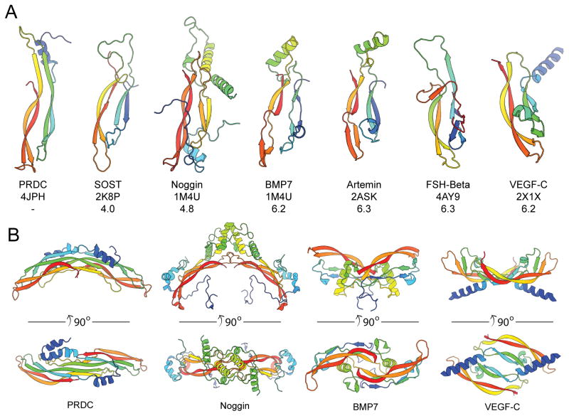
Figure 8. Structural comparison of cystine-knot monomers and dimers.
(A) Ribbon representations of a single chain for several cystine-knot proteins, colored as rainbow from the N- (blue) to C-terminus. Each structure was superimposed with the PRDC monomer (Chain B). PDB identifiers are indicated along with the Z-score from a pair-wise alignment using the Dali-lite server. (B) Comparison of the dimerization mechanisms of PRDC to other cystine-knot proteins. The PRDC dimer exhibits a growth factor-like architecture with a head-to-tail assembly, similar to BMP7 and VEGF-C, whereas Noggin assembles in a head-to-head fashion.

