Introduction
I'll play it, and tell you what it is later.—Miles Davis
“Listen, how would you like to come on tour with my new band?” Benny Wilson asked me in the summer of 1965 during my final year of graduate school. A few days later, I received a letter from the National Institutes of Health (NIH) with the news that I had been awarded a three-year fellowship to do postdoctoral research at the Medical Research Council (MRC) Laboratory of Molecular Biology in Cambridge. Benny was an exciting young jazz pianist and blues organist, the son of a Baptist minister in Eugene who transported his Hammond organ to gigs in the back of his elegant blue Mercedes 220S Cabriolet. It was a great temptation to join Benny on the road, but there would be scant opportunity to do biochemistry. On the other hand, I guessed that I would find musicians in Cambridge. That fall, in our final weekend in the Bay Area, my wife and I found ourselves at Ken Kesey's home in La Honda, where his famous bus “Furthur” was parked in the driveway. We watched hours and hours of raw 16-mm movie footage taken by the Merry Pranksters on their infamous trip, surrounded by hippies and Hells Angels. In the morning, we raced up La Honda Road to Skyline, where we had breakfast with Neal Cassady, at the counter of Alice's Restaurant in Woodside. Cassady was as vivid as Kerouac had portrayed him as Dean Moriarty in On the Road. At breakfast, he maintained a rapid stream of incomprehensible parallel conversations: with me, the person sitting on the other side of him, the waitress, and someone who was not there. Over the next few days, we reluctantly said goodbye to our native culture, crossed the country to New York by train, and boarded a cut-rate Icelandic Airlines propeller flight to London via Keflavik.
My postdoctoral adviser, the Welsh protein chemist Ieuan Harris, had booked us a room at the Prince Regent, a nice pub near the middle of Cambridge. On our first morning, I rolled out of bed, took the bus to the MRC, and was greeted by the receptionist with “Oh, Dr. Noller. Let me go find Dr. Perutz!” I was stunned. Did she mean Nobel laureate Max Perutz, director of the MRC Laboratory of Molecular Biology and one of the gods of molecular biology? I followed her down the hall. She said, “Dr. Perutz is out of his office. Let's see if he's in his laboratory.”
Just then, the door of the cold room flew open, and a small bald man in a white coat stepped out, carrying an Erlenmeyer flask containing a red liquid. Max Perutz, founder of the science of protein crystallography, was doing a hemoglobin prep with his own hands. He stopped, smiled modestly, extended a free hand, and welcomed me warmly to the MRC. I was struck by his utter lack of pretention and his generosity in taking time to talk with an obscure postdoctoral fellow on his first day, even as condensation formed on the surface of his Erlenmeyer. I felt very out of place, in way over my head, and very lucky.
Wat is de Bedoeling van het Leven?—Jack Moore
I was born in Oakland and grew up in the East Bay. Neither of my parents had been to college, but they and my grandparents stressed the importance of education. They encouraged and pushed me every step of the way as best they could. My father, Harry F. Noller, Sr., the first-generation son of an Englishman and a Swede, was a self-taught mechanical engineer with a high school education. During his career, he made the unlikely transition from a job as draftsman to leading the team of engineers that developed the first mechanical calculator with a memory at Marchant Calculating Machine Co. in Emeryville. My mother, Charlotte Silva Noller, whose family were Portuguese from the Azores, had gone to business school and became a full-time homemaker after I was born. After World War II, we lived in a one-bedroom apartment in Berkeley on lower Dwight Way, surrounded by a vast industrial slum. When the wind came from the south, we inhaled pungent aromas emitted by the tall Cutter Laboratories smokestack across the street. What was going on in those “laboratories,” I wondered? I was one of the few Caucasian kids at Columbus School, where I had many friends and supportive teachers. In a well intended but misguided moment, my teachers advised my parents that I should skip a grade. I was interested in everything, particularly things to do with “science,” including the Lone Ranger Atom Bomb Ring that I sent away for with some Kix breakfast cereal box tops. On it was mounted a tiny telescope-like viewer, inside of which you could watch the tracks of subatomic particles as they were emitted from some unspecified radioisotope.
Following the war, the atomic bomb was always in the news. From the sidewalk in front of our apartment, my dad pointed out a white circular structure that was just visible over the eucalyptus trees, high in the Berkeley Hills. The structure housed the cyclotron, he explained, where scientists had “split the atom” and helped to develop the atomic bomb. Little could we imagine that fifty years later, I would be working in that very building collecting x-ray data using synchrotron radiation. Our neighbor Dick Lukens was a biochemist. He listened patiently to my questions, even loaning me a sophisticated college paleontology textbook when I asked about dinosaurs. My dad took me to Chabot Observatory in the Oakland Hills, where the public could look at the planets and stellar objects through a powerful telescope. Sometimes he took me with him to Marchant, where everyone greeted him with a smile, from the screw-machine operators and secretaries to the engineers. I was fascinated by the enormous complexity of the insides of the calculators, with over 2000 rapidly moving parts that interacted precisely with one another to add, subtract, multiply, and divide, whirring and clattering as they processed their mechanical arithmetic. My dad told stories about a man he knew named Dr. Cornish, a sort of independent medical researcher who was in the headlines for bringing a dog called Lazarus “back to life” in his lab in West Berkeley. Many interesting things were happening right around me. At the age of 6 or 7, I began to wonder, how do you make a living as a scientist? It was not obvious.
My parents built a modest home in Orinda, just to the east of the Berkeley Hills, where we moved in 1948. Without a doubt, we were the least affluent family in that upscale community. In the sixth grade at the Orinda Union School, I was lucky to have, as my teacher, the wonderful Verna Givens, who sacrificed a major chunk of her salary to buy a high-quality microscope for our class. She limited its use to only a few selected students, and I was lucky to be one of them. With that microscope, I watched protozoa swim around in pond water for the first time. In the eighth grade, much to the detriment of our academic performance, my friends and I discovered the intriguing and weird world of science fiction magazines, where we were introduced to many far-out concepts, some real and some imaginary, by the pioneering writings of Robert Heinlein, Philip K. Dick, Arthur C. Clarke, Ray Bradbury, and others. We speculated about “the fourth dimension,” time travel, and relativity. We had no idea what we were talking about, but we were getting very high on an avalanche of strange new ideas.
In 1952, I entered Acalanes High School, an institution blessed with an extraordinary faculty, many of whom had been educated at the University of California, Berkeley, bringing its high standards along with them. Some, like Josephine Ochoa, our intimidating Latin teacher, instilled in us academic discipline and fear that brought the top students in our class to their knees. Our exceptional physics teacher, John Annis, introduced us to the concepts of experimental controls and intellectual rigor. However, I was most intrigued by biology and chemistry. Berkeley scientists were again making the headlines, but this time they were not the Manhattan Project physicists. Now, the headlines of the Oakland Tribune announced “Life in the Test Tube” and “Secret of Life” in stories about the reconstitution of tobacco mosaic virus (TMV) from its purified protein and RNA in Wendell Stanley's lab and the demonstration that the purified TMV RNA by itself could cause tobacco plants to produce the virus. When the list of majors offered at Berkeley was passed around our high school class, recalling our family friend Dick Lukens, the word “biochemistry” popped out at me. Did this mean what I thought it did? Could it be possible that you might be able to do both chemistry and biology? For an entire career? One day, I decided to drive into Berkeley to find out for myself. The biochemistry department was housed in a bland four-story concrete building that stood across the road from the Cal football stadium. Over the entrance were the words “Biochemistry and Virus Laboratory.” I was thrilled. “Virus Laboratory!” Magical words. On the walls of the lobby were huge blowups of beautiful electron micrographs of viruses: bacteriophage T2, poliovirus, and right out of the headlines, the famous TMV, taken by the pioneering electron microscopist Robley Williams. Struggling to contain my excitement, I told the receptionist that I wanted to find out about biochemistry. She picked up the phone and called Professor Donald McDonald, a carbohydrate chemist. He welcomed me into the office next to his laboratory and spent about an hour patiently telling me about biochemistry, what his research was about, and what the biochemistry major at Berkeley was like. Professor McDonald's generosity that day, in taking time to talk to a high school kid who walked in off the street unannounced, had a huge impact on me.
My parents could not afford to send me away to college, and because Berkeley was a fifteen-minute drive from our house, it was a foregone conclusion that I would go to college at Cal. My mother's father, M. R. Silva, who had himself quit school in the fifth grade to take a job packing bullets at the Hercules Powder Co. to help support his family, stepped in and sprung for the $54 per semester fees, plus the cost of my textbooks. I began commuting from Orinda to Berkeley, drifting through the corners of the twisty Wildcat Canyon Road in my MG TD. After the rigor of Acalanes, the classes at Berkeley did not seem all that difficult. The exception was calculus, which was taught completely and incomprehensibly by teaching assistants; I nearly ended my career, along with my sanity, in trying to understand the formal definition of a limit as presented in our impenetrable calculus textbook. To fulfill the biochemistry major requirements, which included nearly all of the classes required for both the chemistry and biology majors, I was often in class from 8 to 5.
On top of this, I was playing the saxophone with various bands in the evenings. On the weekends, I worked with friends who were building and racing sports cars and motorcycles. I rode my 500cc BSA Gold Star on the trails in the Oakland Hills. A girlfriend studying in the theater department recruited me to play in the pit band for the Mask and Dagger Review and on stage for a production of Georg Büchner's Woyzeck. On Thursday nights, there were the legendary jam sessions at George's Northgate Café on Euclid Avenue near the campus, where I sat in with some of the best musicians in the Bay Area while puzzling over physical chemistry problem sets at a back table between tunes. Many of those players at Northgate have since earned their own bins at record stores, including the saxophonist Pharoah Sanders, the bassist Barre Phillips, and the pianist-composer Clare Fischer. Our usual drummer was the (now) film and Broadway actor Larry Bryggman. Along with the music scene, I discovered the Berkeley hipster underground, where I met many of the characters who had migrated to Telegraph Avenue from North Beach: people with names like “Barney Google,” “Hube the Cube,” and “Leonard the Locomotive.” I was getting spread dangerously thin.
After graduating from Berkeley in 1960, my first research opportunity was a year working as a lab technician in the Laboratory of Medical Entomology at the Kaiser Foundation Research Institute under Eliezer Benjamini, an entomologist studying flea-bite hypersensitivity. I was first stationed on the grounds of the United States government plague lab at the Presidio in San Francisco, where we collected and analyzed flea saliva. At lunch, I got my first close-up exposure to seasoned scientists, their contests of wry wit interlaced with spellbinding anecdotes: the suave Leo Kartman's stories about tracking down the causative agents of tropical diseases, which led to his knighthood, and Bruce Hudson's accounts of trapping plague-infested rodents in the High Sierras. On one memorable excursion, we visited an abandoned house in the Fillmore District, where police had discovered a corpse along with an infestation of human fleas, with which we hoped to replenish the lab's dwindling Pulex irritans colony. We wore white pants fastened at the ankles with rubber bands and entered the house armed with hand-held aspirators. Minutes into our safari, my pants seemed to have become spray-painted gray around the ankles, a motif that slowly moved up my pant legs as we maneuvered around the blood-stained bed. We worked quickly, recovering a large jar full of P. irritans. Even after showering thoroughly at the lab and decontaminating our clothes in a creosote-laced tub, a few dozen fleas managed to survive the trip home, much to my mother's profound anguish.
In early 1961, I was moved to a lab at the Kaiser Foundation Hospital in Richmond, across the Bay, where I began working with the biochemist Janis Young, who had earned her Ph.D. degree at Berkeley working on the amino acid sequence of the coat protein of none other than TMV with Heinz Fraenkel-Conrat. Janis was a talented biochemist and a patient teacher, generous enough to give me a free hand in much of our work. I was soon joined by a new co-worker at the Richmond lab who had just received a master's degree in entomology at Berkeley. He had spellbinding stories to tell: escaping from Russia to Germany from the Soviet Union during the war, joining the Hitler Jugend, jumping from planes with the U.S. 82nd Airborne Division, doing hard time in the Maryland State Penitentiary for armed assault, and expulsion from the doctoral program at Berkeley after brandishing a loaded pistol on a departmental field trip. My education continued to expand in many dimensions.
One evening, I even got to work in the legendary Fraenkel-Conrat lab, using their rotary evaporator to concentrate a flaskfull of flea extract. The only other person working late was the distinguished Japanese protein chemist Akira Tsugita, a major player on the TMV project. He was surprisingly friendly and showed me how to use the evaporator, asked questions, and cracked jokes in his barely comprehensible English, smiling through gold teeth. The lab was small and cramped, equipment jammed in its narrow aisles. It reeked of pyridine, the characteristic smell of protein chemistry in that era. It was hard to believe that from these cramped and cluttered spaces came the magic that had led to the discoveries of the “secret of life” and “life in a test tube.” That evening, the untrained observer might even have mistaken me in my white lab coat for one of those very researchers. My chest swelled with pride as the flea extract slowly reduced in volume under my steady, expert gaze.
The end of my rookie year in a real lab was signaled by a telegram with the message that I had been accepted to graduate school in the chemistry department at the University of Oregon in Eugene with a teaching assistantship to cover my tuition and living expenses. My immediate reaction was that someone must have made a huge mistake. I was pleasantly stunned. This was another happy accident, the outcome of which was not at all obvious to me at that moment.
After my first year of graduate school in Eugene, which was packed with courses, exams, and playing gigs with a local group called The Jazz Prophets, I was accepted into the laboratory of Sidney Bernhard at the Institute of Molecular Biology. He had recently arrived from the NIH, where he had made his reputation in the field of enzyme mechanisms, in particular, the serine proteases. Sidney had done his graduate work at Columbia with Louis Hammett, the father of physical organic chemistry. The Institute of Molecular Biology's atmosphere was much more like that of an artist's studio than a factory or hospital. Faculty and students clustered around the blackboard in the hallway in open-ended discussions. On nice days, the whole institute would go for potluck picnics. Sidney himself played some jazz piano with me at parties. The sculptor Louis Durchanek was our facilities manager. There was probably more scientific discussion over coffee (and cigarettes) than actual pipetting. It was a culture that nurtured (and respected) creativity of all kinds, including the scientific kind. My project was to map the active site of the serine protease subtilisin using a novel substrate analog called furylacryloylimidazole. I made a stable furylacryloyl-subtilisin adduct, which we characterized kinetically and spectroscopically, and then used it to identify the sequence around the active-site serine. While I was writing up the sequence paper, I received a wake-up call in the form of a report in Nature from Fred Sanger, who had scooped me. Nevertheless, I had enough results to write my Ph.D. thesis. Sidney suggested that I go to Cambridge for my postdoctoral research to work with Ieuan Harris, who was studying a more challenging metabolic enzyme, a step or two up in complexity from serine proteases.
Who are you? What are you working on?—Sydney Brenner
My postdoctoral project in Ieuan Harris's lab was to determine the amino acid sequence of the enzyme glyceraldehyde-3-phosphate dehydrogenase. At that time, the enzyme was the largest protein to be sequenced by using traditional direct protein sequencing methods. I collaborated with another postdoctoral fellow, the very talented and funny Australian Barrie Davidson. Besides us, there were two “research students” working on their doctoral projects. One of them was Graham Jones, a red-headed Welshman who resonated with Barrie's humor; the other was Jo Butler, a more traditional Cambridge student. Barrie and I got on well together and soon had a strong start on the sequence. Our use of ion-exchange columns to separate the tryptic and chymotryptic peptides, à la Stanford Moore and William Stein, was not received well by Ieuan. He tried to persuade us to use just a Sephadex sizing column followed by paper electrophoresis, a simpler approach that he himself had developed. Our use of high concentrations of urea on columns, in an attempt to recover some of the poorly soluble peptides, came under more serious fire when Ieuan discovered a cluster of urea crystals clogging the flow cell of his precious UV detector and spilling over onto the floor. While cleaning up the mess and with Ieuan safely out of earshot, Barrie and Graham burst into song, set to the melody of Leonard Bernstein's “Maria” from West Side Story.
 |
Across the hall, Shyam Dube, a postdoctoral fellow working with Brian Clarke and Kjeld Marcker, was sequencing methionyl-tRNA using the RNA oligonucleotide sequencing methods that Fred Sanger had developed just down the hall from us. Shyam was a dapper guy, with slicked-back wavy black hair, a spotless white lab coat, stylish glasses, and immaculately polished shoes. Shyam chain-smoked as he monitored the movement of the blazingly radioactive 32P-labeled tRNA through his column by the buzzing of his Geiger counter, without gloves or radiation shield, his cigarettes burning a series of parallel brown grooves in the wooden bench top. Shyam was known to disappear in the middle of the afternoon, driving his large American sedan into the middle of Cambridge to go “tea dancing,” where he refined his skills at picking up attractive widows and divorcees. At an adjacent bench, César Milstein, an Argentine postdoctoral fellow who would later become known for his discovery of monoclonal antibodies, was trying to sequence an immunoglobulin light chain using myeloma cell lines that overproduced single antibodies. Downstairs in the basement, John Abelson, Howard Goodman, and Art Landy, three more American postdoc fellows, worked night and day isolating and sequencing the su3 suppressor tRNA using the Sanger oligonucleotide method. Further into the basement, Paul Sigler, Brian Matthews, and David Blow were hunched over a small table wedged into a hallway connecting the MRC lab to Addenbrooke's Hospital, building the first molecular model of chymotrypsin, adjusting the metal Kendrew skeletal models of the amino acids with Allen wrenches and rulers, and measuring their positions from an electron density map displayed in a stack of transparent plastic sheets. With little warning, I had been dropped into the very center of the rapidly exploding new field of molecular biology.
In Berkeley, I had met Tony Tanner, who was a British hipster on leave from Cambridge University and was to become Head of English Studies at King's College. Tony urged me to contact him when I arrived in Cambridge. I found him in his “rooms,” a sort of spacious, high-ceilinged, chilly apartment in the ancient stone buildings where fellows of King's resided. He invited us to a “sherry party” that he was giving the following week. We arrived to find Tony's rooms filled with tweedy Cambridge intellectuals: professors, dons, fellows, research students, and undergraduates. I felt very uncomfortable, partly because I did not recognize a soul in the room apart from Tony and partly because they all seemed at least an order of magnitude brighter and more articulate than anyone I had ever met. I quickly found a glass of sherry and tried to avoid exposing my 200-word Californian vocabulary by staying as inconspicuous as possible. As my anxiety peaked, I was approached by a man of compact dimensions, whose presence dominated the room. I immediately recognized him from his distinctive South African accent and from his visit to Eugene a few years before. “Hello. I'm Sydney Brenner. Who are you?” he asked. When he realized I was working at the MRC, he asked, “What are you working on?” When I replied, he announced, “That's stupid! If you're a protein chemist, why don't you work on something interesting, like ribosomes?” I felt catapulted into my nightmare of nightmares. Perhaps the most brilliant of all of the Cambridge scientists, the intellect most feared by the lunch table of Nobel laureates at the MRC, whose landmark research papers were the most subtle, elegant, and impenetrable, had singled me out in the crowd, exposing my utter scientific cluelessness to the Cambridge academic society and thus to the world.
In the following days and weeks, as I tried to retrieve the fragments of my shattered ego, I found myself unable to get the word “ribosome” out of my head. What Sydney had done, as he had done for many other young scientists, was to try to make me realize that I have only one life to live and one scientific career. You can spend your life and career working on something boring or something exciting. What I had failed to realize was that the choice was up to me, and Sydney had forcefully pointed this out. I went to the library at the MRC and began reading everything I could find about ribosomes. I asked the other researchers what they knew about ribosomes and who was working on them. Someone said that Alfred Tissières was forming a group in Geneva to study ribosomes. My next lucky break was when Ieuan told me that the Hungarian chemist Michael Sajgò would be coming to his lab and that he would be replacing me. I wrote to Alfred to ask if he could use a protein chemist to work on ribosomes. He wrote back immediately, wanting to know when I could start.
In the spring of 1966, my wife and I sold our MG YB saloon (for £30), bought a Bedford van (for £54), installed a mattress in the back of the van, and prepared to head for Geneva to meet Alfred. As we were packing, our landlady advised us about survival on the continent. “Be sure to pack a tinned ham,” she warned, in all seriousness. We stopped to visit friends in Paris, where we slept in the van in a cushy neighborhood on the Île Saint-Louis, and then drove to Geneva via the Burgundian city of Dijon, where we were served a truly consciousness-altering coq au vin at a roadside restaurant. Alfred turned out to be utterly delightful, with a civilized, almost aristocratic demeanor and a wry sense of humor. He was tall and gaunt, with wispy gray hair, deep-set blue eyes, and hollow cheeks. It was difficult to tell how old he was. He was from the canton of Valais in the Swiss Alps and an accomplished alpiniste, who had participated in one of the first major British expeditions to the Himalayas. He had been a graduate student in the biochemistry department at Cambridge, where he had driven a classic Bentley. He and the Bentley next moved to Jim Watson's lab at Harvard University to work on ribosomes. In Jim's lab, Alfred had discovered that ribosomes separated into two subunits when the magnesium ion concentration was lowered. Alfred's own lab was now attempting to characterize the protein composition of the ribosome, so he wanted a protein chemist to come join in the effort. I accepted on the spot. We returned to Cambridge via Monte Carlo. We watched the Grand Prix of Monaco as John Frankenheimer was filming the movie Grand Prix, starring James Garner, with appearances by the actual Formula 1 drivers. Most impressive was the camera car, a powerful Lola T70 sports-racing car driven by former Formula 1 champion Phil Hill, who drove the circuit at speed, surrounded by the race cars, with an enormous 70-mm Mitchell Panavision Reflex camera mounted just behind his head.
Alfred's lab was populated by several of Jim Watson's students from Harvard, including Peter Moore, Ray Gesteland, and Gary Gussin. The founding postdoctoral fellow in his group was another American, Rob Traut, who had been introduced to protein synthesis as a graduate student in Fritz Lipmann's lab at The Rockefeller Institute. Rob had then gone to the MRC, where he had done landmark experiments on peptidyl transferase with Robin Monro. Rob was running polyacrylamide tube gels of 30S and 50S subunits, trying to figure out just how many proteins there were. The gels he showed me were frightening: there were literally dozens of different protein bands. Worse yet, these proteins were impossible to work with because they were insoluble! To deal with this, Peter Moore was developing a column chromatography procedure, which was based on an earlier attempt by Jean-Pierre Waller and which used a carboxymethylcellulose column run in 6 m urea with a linear salt gradient to separate the proteins of the 30S subunit. The flatter he ran the gradient, the more peaks appeared. Hajo Delius joined the lab from Peter Hans Hofschneider's laboratory at the Max Planck Institute for Biochemistry in Munich, and he began carrying out a similar approach for purification of the 50S subunit proteins. When I joined Alfred's lab, I began analyzing some of the proteins that Rob and Peter had isolated. We soon confirmed our worst fears: the numerous gel bands indeed corresponded to different proteins. The ribosome was a lot more complicated than the simple virus-like repeating-subunit structure that Jim Watson and others had originally hoped for.
Soon after arriving in Geneva in November 1966, we attended the Geneva Jazz Festival, where we heard and met the Jürg Lenggenhager Quartet, a group of young avant-garde Swiss musicians, including Jürg on piano, Roger Pfund on bass (both from Bern), and Andreas Straub (from Basel) on drums. We were invited to a jam session that night in a chateau in Nyon, a village along the north shore of the lake. We played until dawn. They invited us to spend the summer with them in the South of France at a pottery in the village of Pégomas, in the foothills between Grasse and Cannes. We played in galleries and private parties around Cannes and Saint-Paul-de-Vence that summer and later in the Tobeco recording studios near the Zytgloggeplatz in Bern. I continued playing concerts around Europe with them during the rest of my postdoctoral stay in Geneva and then again in the mid-1970s when I returned to Europe on sabbatical. The mid-1960s were a time of great social change, which was spilling over from the United States into Europe. The Living Theater, headed by Julian Beck and Judith Malina, came from New York to Avignon, where we found them living and rehearsing in an abandoned cloister. They were orbited by a free-form circus-like troupe called The Human Family, founded by Oklahoman Jack Moore. We got to know many of these people as they came through Geneva, performing their revolutionary theater to the astonishment and discomfort of the conservative Swiss audiences.
I'll bet you tried a lot of things that didn't work.—Fred Sanger
In the spring of 1968, after two years in Geneva, I applied for an academic job in the United States. My letters led to eight interviews and six offers. I worked my way from the East Coast to the West Coast. My first interview was in the South Bronx, where an abandoned car missing its wheels, sitting on a carpet of broken glass, was visible from the office window of my faculty host. When I arrived in Santa Cruz at the end of my trip, I parked my rental car in the parking lot that the map showed was nearest to the site of my interview. When I got out of the car, I found myself surrounded by a forest of redwood trees. There was no building in sight. I had no idea where to go. At that moment, I realized that I had an easy decision: if the University of California, Santa Cruz (UCSC), made me an offer, I would accept on the spot, no matter what it was.
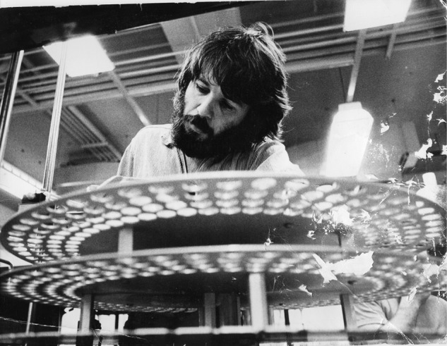
UCSC was an “experimental” campus and was in the third year of its existence. The theoretical chemist Terrell Hill, who recruited me, likened it to “an embryonic Cambridge or Oxford,” with its residential college system. A competing view was that it was an embryonic Haverford or Reed, focusing on undergraduate liberal-arts education. Fortunately, I was idealistic, fearless, and naïve. As an assistant professor, I taught five courses and served on eight faculty committees (and chaired five of them). I centered my life around the lab (Fig. 1), and when the timer rang, I went to class. When class was over, I headed back to the lab, where I asked students to wait for a break in my experiments to ask questions about my lectures. The ivory-tower atmosphere and relative isolation of UCSC were in a way liberating; there were not a lot of “experts” around to discourage you from going in unusual directions.
FIGURE 1.

The author in his laboratory at UCSC as an assistant professor in 1970.
My view of the ribosome in 1968 was a fairly conventional one for the time: the ribosome was believed to be sort of a multienzyme complex with lots of proteins, each of which contributed to a specific ribosome function. I decided to pick a couple of interesting proteins out of the fifty or so that had been identified and, like a good enzymologist, work on their structures and functions. The first question was which proteins to work on. Having a background in protein chemistry, I decided to bombard the ribosome with a wide range of chemical modification reagents known to react with the active sites of enzymes and test the ribosomes for loss of specific activities, such as tRNA binding or peptidyl transferase activity. When I found a loss of activity, I would then use Masayasu Nomura's in vitro reconstitution approach (1) to identify which protein had been functionally modified by the rescuing activity of the individual purified proteins. We tried all of the usual modification reagents and then some. One of the more memorable was the histidine-specific reagent diazonium-1H-tetrazole, which had to be synthesized on the spot because of its instability. I carried out a small-scale synthesis in a large test tube that was too tall for the length of my micropipette. In a display of youthful hubris to fix the problem, I etched a line halfway up the tube with my diamond pencil, touched a molten glass rod to the line, and struck the top of the tube sharply with the other end of the glass rod. I should have anticipated the potential chemical properties of a heterocyclic aromatic ring compound containing six nitrogen atoms and only a single carbon atom in its structure. Fortunately, the shards of glass unleashed by the explosion somehow missed my unshielded eyes. For the next weeks, anyone walking through that part of the lab could hear sharp popping noises coming from the floor as they stepped on the thousands of tiny crystals of diazonium-1H-tetrazole.
Strangely, the initial result of screening dozens of modification reagents was negative. None of the reagents knocked out ribosome activity, until my graduate student George Thomas tried photooxidation induced by the dye Rose Bengal, which had been used successfully to target active-site histidines in some enzymes. Rose Bengal finally caused inactivation of protein synthesis, including binding of tRNA to ribosomes. George's reconstitution experiments showed that modification of some ribosomal proteins was partly responsible for the loss of activity, but also that there had been functional modification of 16S rRNA. We were puzzled and troubled by this result until we found reports in the literature that guanine could also be targeted by Rose Bengal photooxidation. Around this time, in the spring of 1972, an undergraduate by the name of Brad Chaires came to my office to confess that he had made little headway on his senior thesis in my lab and needed to finish a project to graduate within the following couple of months. I suggested that he try to intentionally target the ribosomal RNA using kethoxal, a guanine-specific reagent. The following week, he came back to my office with an amazing result: kethoxal had completely inactivated the ribosome! Moreover, reconstitution experiments confirmed that the inactivation was indeed caused by modification of the ribosomal RNA; the proteins from the kethoxal-modified ribosome were 100% active. He also showed that binding tRNA to the ribosomes prior to modification protected them from inactivation and that inactivation was caused by modification of only a handful of guanines out of the more than 400 present in 16S rRNA. This was our first clear result, but not at all what we wanted to hear. No one would believe that RNA was involved in enzymatic function, much less in the function of such a complex and fundamental structure as the ribosome. Furthermore, the ribosomal RNAs were 2 orders of magnitude larger than any molecule that had been characterized by enzymologists. Worse, I knew next to nothing about RNA chemistry.
We next wondered which nucleotides were the ones whose kethoxal modification was causing loss of ribosome function. Unfortunately, the nucleotide sequence of 16S rRNA was incomplete, and there was no easy way to determine which of its more than 1500 bases might be functionally important. Our direction came from the observation that tRNA protected the ribosomes from inactivation by kethoxal: the critical guanines must be among the tRNA-protected bases. The first challenge was to figure out a way to identify the guanines that were being modified by kethoxal and then to identify the ones that were protected from modification by tRNA. We succeeded with the first part of the plan within a year or so, but the second part took more than an additional decade. The initial approach emerged during a conversation with Jim Dahlberg, who was visiting from Wisconsin. Jim had also been a postdoctoral fellow at the MRC, where Brian Hartley had introduced “diagonal thinking.” Whatever problem you were studying, there always seemed to be a two-dimensional paper electrophoresis “diagonal” solution. Jim asked me if the kethoxal reaction was reversible, and I immediately got it. If we cut the kethoxal-modified 16S rRNA with RNase T1, which cuts specifically at G, the modified guanines would be resistant to the nuclease. The result would be a mixture of all of the oligonucleotides ending in G, except for the kethoxal-modified sites, which would give a pair of two adjacent oligonucleotides connected by an internal kethoxal-modified G.
The first step of the diagonal method consisted of running the T1 digest of uniformly 32P-labeled RNA by paper electrophoresis on DEAE paper in the first dimension, separating the oligonucleotides according to their size and base composition. I then cut out the strip containing the first dimension and removed the kethoxal from the modified Gs by treatment with mild base while the oligonucleotides retained their positions bound to the DEAE paper. Next, I soaked the paper with RNase T1, which cut between the pairs of oligonucleotides at the sites that had been modified. The strip was then sewn onto a fresh sheet of DEAE paper at right angles to the direction of the first dimension, and the electrophoresis was rerun. When I turned on the darkroom right after developing the first autoradiogram, I was ecstatic. It showed a very dark diagonal line from all of the oligonucleotides that were unaffected by the redigestion step, but pairs of off-diagonal spots appeared, corresponding to the oligonucleotides surrounding the sites of kethoxal-modified guanines. I realized that we now had the means to identify the sites of kethoxal modification, which we could soon put into the context of the emerging complete sequence of 16S rRNA.
I then fell into a months-long slump, during which I could not reproduce my initial success, and my euphoria faded into depression and self-doubt. The problem was a dark streak running up from the origin of the second dimension, which had been absent in my first experiment. I began repeating the experiment, testing every possibility I could think of. This required growing cells in millicuries of [32P]orthophosphate, isolating blazingly hot ribosomes and ribosomal subunits and carrying out the two-dimensional diagonal electrophoresis procedure. Each trial took about a week and a half, and each time, the dreaded streak reappeared, even though I had been carefully using all of the same solutions that I had used in the successful trial. In desperation, I began remaking all of my reagents and buffers. When I made up a fresh bottle of water-saturated phenol that I was using to extract the RNA from the ribosomes, the streak vanished! Later that year, in the breakfast line at a meeting at Squaw Valley, I found myself standing next to Fred Sanger, whom I had not seen since my postdoctoral days at the MRC. He asked what I was up to and listened with rapt attention as I described my diagonal method, asking about experimental details such as pH, temperature, electrophoresis conditions, and so on. When I was finished, he smiled and said, “I'll bet you tried a lot of things that didn't work!” His remark, although at first seemingly telepathic to me, was a revealing look into the genius of one of the greatest experimental scientists of the twentieth century. I have since then repeated his words countless times to students and postdoctoral fellows and, of course, to myself during those moments of quiet desperation that we repeatedly face in our laboratories.
The ribosome teaches in silence.—Carl Woese
I began sequencing the off-diagonal oligonucleotides, which led to the identification of the sites of kethoxal modification of 16S rRNA. This was helped enormously by a methods chapter (2) written by Bart Barrell, who had been Sanger's technician during the development of RNA sequencing, and also by desperate phone calls to Howard Goodman, who had been a fellow postdoc scientist in Geneva and had taken a faculty position at University of California, San Francisco (UCSF). During this time, I was playing with the Randy Masters Group. I would start an electrophoresis run before dashing off to rehearse Randy's complex polyrhythmic arrangements and then return to the lab late at night to pull the racks out of the electrophoresis tanks. Some highlights were opening for the Duke Ellington Orchestra at the Del Mar Theater in Santa Cruz in 1974 and a concert at the Santa Cruz Civic Auditorium along with Bobby Hutcherson and Eddie Henderson and their bands. As my oligonucleotide sequences took shape, I began to doubt my skills as an RNA biochemist. Many of my sequences were in disagreement with the published ones. I was consoled by a phone conversation with Carl Woese at the University of Illinois at Urbana-Champaign; he had begun sequencing the RNase T1 oligonucleotides from different bacterial species to establish their phylogenetic relationships. This project led him to the discovery of the Archaea, the third domain of life. When I mentioned my discrepancies, Carl confirmed that my sequences were correct. Furthermore, he had found that over half of the published sequences of oligonucleotides of length greater than five were incorrect. His voice strained with barely contained anger. “These sequences are sacred scrolls. They should be entrusted only to those who appreciate what they mean,” he said. I was stunned. Everyone in the field used the partially finished 16S rRNA sequence as an important resource and assumed that it was correct and would soon be complete. Work had begun on attempting to fold it into a secondary structure, a major step toward an understanding of the ribosome structure. All of this was now cast in doubt. I gradually began to come to terms with the possibility that we might have to determine the sequence ourselves, which would entail a staggering amount of very challenging work.
An unexpected and exciting surprise from my conversation with Carl was that my oligonucleotide sequences were unusually rich in universally conserved sequences. At the time, conventional wisdom dictated that the most important sequences in ribosomal RNA would be the binding sites for ribosomal proteins (the putative “functional” molecules of the ribosome), yet the conserved kethoxal-reactive sites were exposed to the solvent, as would be expected if the ribosomal RNA itself were functionally important. Therefore, I was thrilled when I was invited to contribute a chapter to the Cold Spring Harbor volume Ribosomes (edited by Masayasu Nomura, Alfred Tissières, and Peter Lengyel) based on a meeting where I had presented my kethoxal sequence results for the first time. However, as I was nearing completion of my chapter, I received another letter from Nomura, uninviting me to contribute my chapter because of space limitations. This was surely the last and only time in my career that I would actually be disappointed not to have to write a review article.
Association of the two ribosomal subunits was also inactivated by the reaction of kethoxal with conserved sequences in 16S rRNA, as discovered by two undergraduates, Winship Herr and Nora Chapman. We also discovered that peptidyl transferase, the peptide bond-forming catalytic activity of the ribosome, was abolished by kethoxal modification of 50S ribosomal subunits. These experiments, carried out by another undergraduate, Rob Atchison, were never published because we had difficulty separating the 23S rRNA from the 50S proteins of the kethoxal-modified 50S subunits. Later, it seemed likely that this difficulty was caused by polymerization of our stock sample of kethoxal during storage. However, this result bolstered our convictions that ribosomal RNA was indeed functional, as did the finding by yet another undergraduate, Jim Breitmeyer, who found that a reactive electrophile attached to the α-amino group of an aminoacyl-tRNA became attached to 23S rRNA, not to a ribosomal protein.
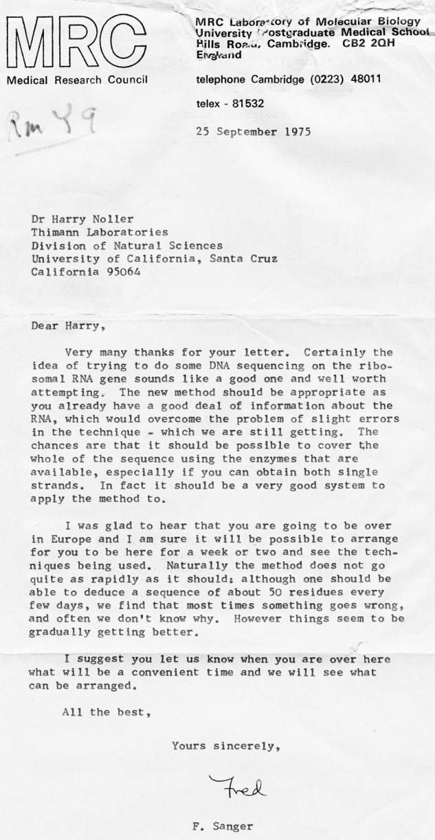
In the summer of 1975, coming up for my first sabbatical, I received a phone call from Christine Guthrie, who had just returned from the Gordon Research Conference on Nucleic Acids. She said, “Allan Maxam from Wally Gilbert's group and Bart Barrell from Fred Sanger's group announced that they are sequencing DNA on gels! Maybe you could use this approach to sequence ribosomal RNA.” Larry Soll at the University of Colorado in Boulder was known to have isolated a λ transducing phage that had picked up some ribosomal RNA genes from Escherichia coli, so there was a possible source of the DNA. I wrote to Fred Sanger, asking if he thought the new sequencing method might be applicable to doing the whole ribosomal RNA and if I might be able to visit his lab to learn it. His reply was encouraging (Fig. 2). As an aside, it is interesting to note that the state of the art of DNA sequencing in September 1975 was such that “ … one should be able to deduce a sequence of about 50 residues every few days … ”
FIGURE 2.

Letter from Fred Sanger with encouraging words for sequencing the ribosomal RNA genes by DNA sequencing.
I spent the first few months of my sabbatical in Berlin at the Max Planck Institute for Molecular Genetics in Heinz-Günter Wittmann's department, collaborating with Roger Garrett to apply the kethoxal diagonal method to the mapping of the binding sites for proteins L5, L18, and L25 on 5S rRNA. As one of the few people who worked in the lab after 5 p.m., one evening, I heard the unmistakable voice of Wolfman Jack as I wandered the halls. I traced its source to an Armed Forces Network program coming from a big German table radio sitting in a tiny closet of an office occupied by Jürgen Brosius, a graduate student who was sequencing ribosomal proteins under Brigitte Wittmann-Liebold for his doctoral research. Soon, we were going out for late-night dinners and beers, as Jürgen introduced me to the Hundekehle, the Zwiebelfisch, and other after-hours venues of the fascinating enclosed city of West Berlin.
Moreover, the experiments that Roger and I were doing were actually working, so Roger advised me to let the people in the business office know that I would be prolonging my stay for another month. However, Wittmann himself, a devoted protein chemist, was not thrilled by ribosomal RNA. One afternoon, he stormed into the lab while I was in the middle of an experiment, irately accusing me of manipulating his business office into giving me more money. I explained that I was happy to support myself from my own grant from the NIH, but that Roger had insisted that I simply inform the business office that I would be staying another month. Wittmann then asked, “And you really believe that ribosomal RNA is important for function? Even peptidyl transferase?” By now, he was becoming quite red in the face and stormed out of the laboratory. My thoughts began to turn to Geneva, the next stop on my sabbatical, where Alfred Tissières had offered me bench space for a few months.
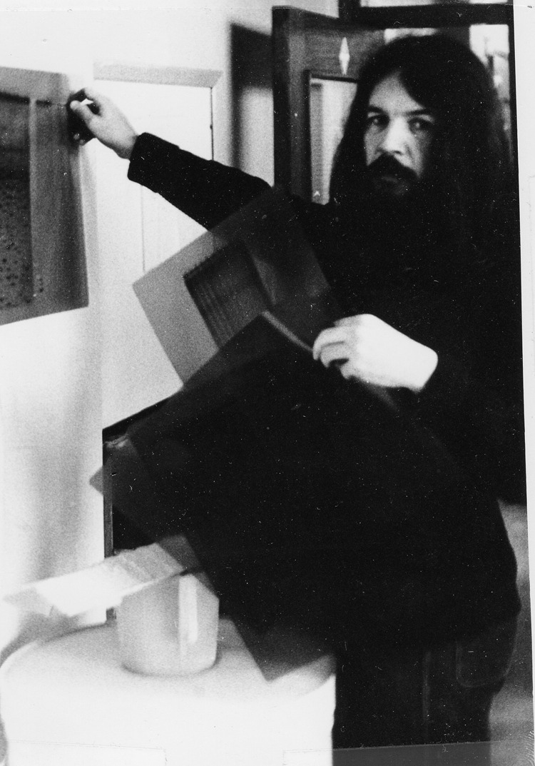
I had no idea a priori what I was going to do in Alfred's lab, especially because he no longer worked on ribosomes. This was resolved during my first day in the laboratory. I was describing to the postdoctoral fellow Jack Greenblatt that I was thinking about sequencing ribosomal RNA using DNA sequencing, and he said, “Oh, Joel Kirschbaum has a λ transducing phage with ribosomal RNA genes on it.” When I asked where Joel Kirschbaum worked, Jack replied, “He's right across the hall.” I was stunned. Next, Joel was handing me a sample of λdrifd18, which contained the entire rrnB operon, and explaining how to grow it and extract its DNA. Soon it was time to head to Cambridge to learn DNA sequencing. Fred Sanger had arranged for me to work alongside his right-hand man, Bart Barrell, who was now part of the group sequencing the complete genome of the phage φX174. Fred had reserved rooms for me in King's College. At the end of my first day in Fred's lab, I returned to my rooms to find an elderly gentleman fast asleep in my bed. Fred was greatly amused by this news, proudly reporting to everyone in the lab the “extras” that they had laid on for my stay at King's. Working with Bart was intense fun. At that time, micropipetting was done with Microcaps, which were small calibrated capillaries that you attached to a rubber adaptor and a long piece of rubber tubing for mouth pipetting. After addition of each 32P-labeled sample, Bart flicked the used Microcap into a wastebasket sitting in the middle of the bay about six feet away. The majority of the Microcaps hit their target; the rest formed a blazingly radioactive halo circling the base of the wastebasket. Bart was a superb teacher, and in a few days, I was admiring my first successful sequencing autoradiogram (Fig. 3).
FIGURE 3.

The author on sabbatical in Cambridge in April, 1976, holding the autoradiogram of his first successful DNA sequencing gel.
Among the postdoc fellows working on the φX174 project in Fred's lab was Wayne Barnes, who asked me if I had “clones” of the ribosomal RNA genes. When I mentioned Joel Kirschbaum's λdrifd18, he was astonished at my cluelessness. “No, I mean a clone! Where's the phage DNA?” he asked. I phoned Pia Malnoë, Alfred's Swedish technician in Geneva, and asked her to find a beaker on the second shelf in the cold room and to send the tube of DNA to me in Cambridge via air mail (FedEx had yet to come into existence). When the λdrifd18 DNA arrived, Wayne had me cutting it with restriction enzymes and cloning the pieces into the ColE1 plasmid (the naturally occurring ancestor of pBR322 and many other modern cloning vectors). Within a couple of days, I was making stabs of the clones and putting them into a package that I addressed to myself in Santa Cruz.
Everything you know is wrong.—The Firesign Theater
From the phone calls from Carl Woese and Chris Guthrie, Fred Sanger's letter, Joel Kirschbaum's phage, and Wayne Barnes's cloning expertise, I had the eerie sensation that everything was falling into place guided by a mysterious force. The final step was a crucial conversation with Jürgen Brosius at a sidewalk café in Geneva, when I explained to him where things were headed and persuaded him to come do postdoctoral research with me in Santa Cruz. Jürgen received a Fogarty Fellowship from the NIH for this, and he arrived in the lab the following year. We had only a tiny lab of about 600 square feet, but we managed to convince the department to give us an additional small room where we could set up DNA sequencing gel rigs. We had two gel boxes made and ran them off a single power supply, but Jürgen soon complained that we needed two more and then four more, until we ran out of supply money. Carl Woese stepped in and generously had eight more built for us at the University of Illinois, finally giving us a total of sixteen gel boxes. Jürgen was a force of nature, working day and night with an undergraduate, Tom Dull. They came to the lab in the morning, loaded sixteen warm pre-run sequencing gels, reloaded them a few hours later for short reads (to read the end-proximal sequences), and then stopped them and put on the x-ray film in the early afternoon. They then mounted fresh gels and pre-ran them while they ate and slept, coming in late afternoon to begin loading the next round of samples, finishing after midnight. Sequencing technology was far from perfect at that time. We had settled on the Maxam-Gilbert chemical sequencing method, a challenging technique that often failed to distinguish between Cs and Ts. There was readable sequence in about one out of four gel runs. However, with more experience, we started to be able to read 200 or more nucleotides at a time. We even had to make our own [γ-32P]ATP for end labeling DNA fragments and isolate our own restriction enzymes. Our first long sequence reads showed that the published partial sequence of 16S rRNA had mistakes not only in the sequences of the RNase T1 oligonucleotides, but also in their ordering within the sequence. In all, it contained more than 200 errors. We presented the sequence at the ribosome meeting in Salamanca in June 1978, hosted by the great Spanish antibiotic guru David Vázquez. Late at night after much partying, Vázquez was in fine form, on the loose in the bar with a dangerous-looking saber, demonstrating some kind of African sword dance. A well meaning attendee talked him out of the knife by trading in it for a pair of scissors. Vázquez then began cutting chunks of hair off the heads of scientists in the bar, leaving a stream of bald patches in his wake, urged on by a cheering crowd. When he spotted me (Fig. 3), a huge cheer went up as he cut off a two-foot-long fistful of hair, leaving me lopsided but otherwise mostly intact.
By the time we finished the sequence of E. coli 16S rRNA, we were, of course, well into the sequence of 23S rRNA and all of the rest of the DNA sequence of the rrnB operon. However, Carl Woese's paper on 5S rRNA had convinced me that the only way to figure out the secondary structures of the large ribosomal RNAs was to sequence several phylogenetically distinct versions of 16S and 23S rRNAs. Thus, in parallel with finishing the E. coli sequences, JoAnn Kop, Ramesh Gupta, Alexei Kopylov, Ginny Wheaton, and others in my group began sequencing the large ribosomal RNAs from Bacillus, Haloferax, and other species. In the meantime, other laboratories were beginning to sequence ribosomal RNA genes from many sources. Before long, it became difficult just to keep them straight because no computer software had been written to deal with alignment of multiple sequences. In fact, computer technology was barely able to deal with storage and retrieval of single sequences. Robin Gutell was entering sequences on a massive VAX750 (a “minicomputer” the size of a small refrigerator) using a remote terminal until we purchased our first lab computer, a Sun Microsystems workstation with 2-MB main memory ($15,000) and an 86-MB hard drive (another $15,000). When I informed a computer-savvy colleague that we had just bought an 86-MB hard drive, he laughed, “You'll never use all of that storage space!” The alignment problem was solved by a computer sciences undergrad, Ted Goldstein, who did his senior thesis project in our laboratory. After asking Robin a lot of questions, Ted disappeared for a couple of weeks and returned with a program he called STREAM, which was, quite possibly, the very first multiple-sequence alignment editor. Ted had ripped apart the source code for the UNIX operating system and tweaked the vi text editor to create his program, which we used successfully for many years.
Using STREAM and fistfuls of colored pens, we began dissecting the secondary structures of the large ribosomal RNAs in collaboration with Carl Woese, who had a wealth of phylogenetically diverse T1 oligonucleotide sequences to add to the rapidly developing database of complete sequences. We looked for compensatory base changes, i.e. complementary sequences in which mutational changes occurred in both strands such that Watson-Crick pairing was maintained. By the time we began on the secondary structure of 23S rRNA (2904 nucleotides), we waited until we had three complete sequences and then used a simple visual algorithm that we called “red dot-green dot.” Wherever there was a transversion, we put a red dot; transitions got a green dot. We then looked for complementary sequences that showed mirror-symmetric patterns of red and green dots. This approach remains the simplest and most powerful method for determining secondary structures of RNA, short of x-ray crystallography.
The next challenge was to develop a better tool to study ribosomal RNA (faster and more comprehensive) to be able to study the interactions of all of the bases (and possibly the riboses and phosphates) with the functional ligands and proteins of the ribosome. After hearing a talk in which primer extension was being used to identify breaks in DNA, I realized that we might be able to use this approach to map sites of modification by kethoxal and other reagents in the ribosomal RNA using reverse transcriptase. A similar method was being developed in parallel in Tom Cech's lab at the University of Colorado in Boulder unbeknownst to us. After getting advice on the use of reverse transcriptase from Norm Pace at Indiana University, I tried it on some 16S rRNA that I had modified with several reagents and bingo! There were the bands at the modification sites. This caught the attention of three graduate students, Danesh Moazed, Seth Stern, and Ted Powers, whom divine providence had sent to our lab for this purpose. After optimizing the method, Danesh set out to map functional ligands, and Seth and Ted began to map the binding sites for the ribosomal proteins. A few days later, I was working at my bench when I was confronted by the three of them. “What are you doing?” they asked. Taken aback, I started to explain that I was making some ribosomal protein stock solutions. “No. We are doing that,” they said. “You are writing the papers.” I started to argue but stopped when I realized I would not win that argument. These three students exploded with exciting results for the next several years. They mapped the binding sites for the tRNAs, protein synthesis factors, ribosomal proteins, and antibiotics. Out of this came the hybrid-states mechanism for translocation, the placement of antibiotics in functional sites in the ribosomal RNA, and an initial model for the three-dimensional folding of 16S rRNA. The universal conservation of the ribosomal RNA sequences protected by the tRNAs and factors provided overwhelming evidence to convince us that ribosomal RNA was at the heart of ribosome function.
However, Wittmann's protein-centric point of view continued to represent thinking in the field at that time. Indeed, it was representative of the convictions of biochemists in general. After giving a talk on the functional role of ribosomal RNA at Berkeley, a colleague confided to me that a distinguished biochemist had remarked, “What a crackpot idea!” I was reminded of the Firesign Theater's landmark album Everything You Know Is Wrong, an anthem for the state of the ribosome field. Of course, there was an upside to this: we had very little competition studying the functional role of ribosomal RNA for about a decade. However, once self-splicing introns were discovered by Tom Cech, Norm Pace, and Yale University's Sidney Altman in the early 1980s, it quickly became respectable to work on ribosomal RNA function. The final straw occurred after my talk at the 1987 Cold Spring Harbor Symposium, when Phil Sharp, who is at the Massachusetts Institute of Technology (MIT), said to me, “So, Harry, why don't you nail it?” I stared at him blankly. “What do you mean, Phil?” I was so immersed in my own belief in the functional role of ribosomal RNA that I was unable to see the question from the outside. There was so much circumstantial evidence that I had long since stopped questioning it.
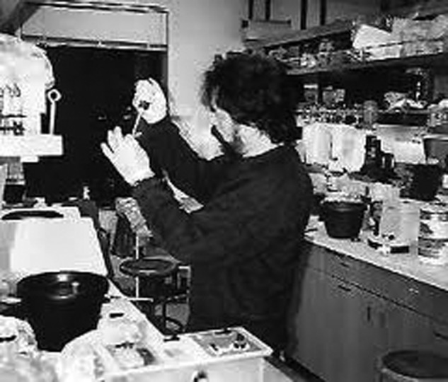
I then remembered an experiment that had been done by a postdoctoral fellow, Ludwika Zagorska (later Zimniac). As a control in a peptidyl transferase assay she was doing, she had included the strong ionic detergent SDS in one of her reactions. In place of the fMet-puromycin product, a smear appeared in that lane. It occurred to me that the smear might have actually been the product, whose migration on paper electrophoresis was distorted by the presence of the detergent. When I got back to the lab from Cold Spring Harbor, I treated some ribosomes with SDS but then removed the SDS by ethanol precipitation from the extracted ribosomes before carrying out the reaction. As I had guessed, a beautiful product spot appeared: the SDS-treated ribosomes were fully active in peptide bond formation! Next, I upped the ante by adding some proteinase K, a standard method for deproteinizing nucleoprotein samples. Again, there was full activity. Finally, I added an equal volume of phenol to the SDS/proteinase K-treated ribosomes and vortexed them continuously for half an hour. The E. coli ribosomes finally lost activity with this treatment, but the Thermus aquaticus ribosomes retained their full activity. Although not all of the ribosomal protein was removed by this treatment, I decided that it was interesting enough to publish. I had intended to submit it as a letter to the Journal of Molecular Biology, but Christine Guthrie, who was on sabbatical at UCSC from UCSF, was so excited about the result that she convinced me to send it to Science. I was surprised at the RNA community's reaction to this result, but it was a relief not to have to justify my passion for ribosomal RNA after that (Fig. 4).
FIGURE 4.

The author in his laboratory in 1991 doing the peptidyl transferase experiments.
Just get the spots.—Brian Matthews
At each stage, our desire to know the three-dimensional structure of the ribosome grew more intense. Modeling exercises in our lab and in Richard Brimacombe's lab at the Max Planck Institute for Molecular Genetics in Berlin gave us a general sense of where the interesting things were in three dimensions (in fact, we fairly accurately predicted the arrangement of the three tRNAs in the ribosome). However, these attempts always came up short of what we really wanted: a crystal structure of the whole ribosome, with its mRNA and tRNAs in place. Alex Rich had a notorious policy of requiring his students at MIT to spend their first year attempting to crystallize ribosomes. I began thinking that everyone in the field should spend, say, 5% of their effort on ribosome crystallization. In the late 1980s, Ted Powers and I began an effort to crystallize T. aquaticus 70S ribosomes without much success until one day when Ted found a beautiful crystal in one of his drops. I phoned Steve Harrison, who had solved the structures of viruses in his lab at Harvard, and he flew to Santa Cruz to retrieve the crystal for analysis. A few days later, Steve phoned with the sad news that it was a salt crystal, most likely magnesium ammonium phosphate. I was embarrassed but puzzled. I had carefully avoided having any phosphate present in our drops for this very reason. It turned out that the polyethylene glycol we were using as precipitant was contaminated with phosphate.
A few years later, I heard from Marat Yusupov, who was then working in Strasbourg at the CNRS lab. He had noticed our paper on in vitro reconstitution of the head of the 30S subunit, done by Ray Samaha, and suggested coming to Santa Cruz to crystallize it for structure determination. I recognized Marat's name from the paper from Russia in which crystallization of the Thermus thermophilus 70S ribosome had been reported (3). I replied, “Why don't we try to solve the whole thing?” A few months later, Marat and his wife, Gulnara, arrived in Santa Cruz to begin the project. Unfortunately, the crystal form that had been obtained in Russia diffracted to no better than about 20 Å. However, after a few months in Santa Cruz, the Yusupovs found a new crystal form of the T. thermophilus 70S ribosome in the I422 space group, and before long, it was diffracting to around 12 Å. On a seminar visit to Eugene, I told Brian Matthews about what we were up to and expressed to him my worries about the next steps. “Brian, if we get these crystals to diffract well, how on earth will we be able to solve the structure? The ribosome is much too big to use heavy atoms for phasing.” Brian's reply was reassuring. “Just get the spots,” he said. “You'll figure out how to phase them.”
Around this time, I met Jamie Cate, a young crystallographer who had just solved the structure of the P4–P6 domain of the group I intron as a graduate student in Jennifer Doudna's lab at Yale. When I asked him whether he might be interested in solving the ribosome, Jamie answered me with raised eyebrows. He joined our small group, which we called Mission:Impossible. We knew that we were up against some of the best crystallographers in the world, but among ourselves, we had an almost perfectly complementary set of researchers. We spent many long nights at the Advanced Light Source, running on coffee, donuts, and barbecued ribs from the legendary Everett & Jones Barbecue in West Berkeley. The Yusupovs continued to improve the diffraction properties of the crystals, and Jamie began to work on phasing the ribosome at low resolution. He started with the molecular envelope of the E. coli 70S ribosome from Joachim Frank's cryo-EM reconstruction, packing it with Gaussian spheres that were matched to the average scattering power of the ribosome, essentially filling the ribosomal envelope with BBs. One day, he discovered a molecular replacement solution that provided phases to about 20 Å resolution, allowing him to calculate an electron density map that looked like a ribosome. The proof that we had independent phases came from a difference map from ribosomes that had a tRNA bound to the A site versus ones with a vacant A site. We crowded around the SGI monitor to look at the result. A chubby L-shaped density appeared in exactly the position that we had predicted for the A site from our biochemical work! This was the aha moment. With these low-resolution phases, Jamie could then calculate higher resolution phases using the heavy-atom cluster tantalum bromide. From those phases, he could find individual iridium hexammine heavy atoms for phasing to the limits of our diffraction data, which the Yusupovs improved first to 7.8 Å and then to 5.5 Å. Meanwhile, the Steitz and Ramakrishnan groups at Yale and Cambridge, respectively, and the Yonath group in Israel were solving the structures of the ribosomal subunits at all-atom resolution. Although at lower resolution, we could see the whole thing: how the subunits fitted together with their dozen intersubunit bridges; how the tRNAs bound to the A, P, and E sites of the ribosome; and the path of the mRNA through the ribosome. As we anticipated, all of the functional sites were made almost exclusively of ribosomal RNA; the ribosomal proteins were scattered mainly around the periphery of the ribosome. However, just as the structure was unfolding, Jamie was recruited to a faculty position at the Whitehead Institute for Medical Research and the Yusupovs to positions in Strasbourg. Eventually, we rebuilt our crystallography group at Santa Cruz with the arrival of Andrei Korostelev, Sergei Trakhanov, Martin Laurberg, Jie Zhou, and others, and we began solving all-atom structures of the 70S ribosome trapped in different functional states.
In parallel, we began to complement the static snapshots of the crystal structures of the ribosome with studies of its structural dynamics in solution using FRET methods, including single-molecule FRET, pioneered by the postdoctoral fellows Robyn Hickerson and Dmitri Ermolenko, in collaborations with Bob Clegg and Taekjip Ha at the University of Illinois and their co-workers. A collaboration with Nacho Tinoco and Carlos Bustamante at the University of California, Berkeley, has provided yet another view of the behavior of single ribosomes using optical tweezers. Paradoxically, as methods have become more and more powerful, with higher and higher resolution, the problem of understanding the molecular mechanism of action of the ribosome has become increasingly challenging and ever more complex. This has proved to be a happy situation, making it possible to spend my entire career on a single problem. Fortunately, the pursuit of this problem continues to be a rewarding one, illuminating one of the deepest and most central mechanisms in all of biology. Or, as the headline writers for the Oakland Tribune would have put it, “Scientists Continue Search for Secret of Life.”
REFERENCES
- 1. Nomura M., Mizushima S., Ozaki M., Traub P., Lowry C. V. (1969) Structure and function of ribosomes and their molecular components. Cold Spring Harbor Symp. Quant. Biol. 34, 49–61 [DOI] [PubMed] [Google Scholar]
- 2. Barrell B. G. (1971) in Procedures in Nucleic Acids Research (Cantoni G. L., Davies D. R., eds) Vol. 2, pp. 751–779, Harper & Row, New York [Google Scholar]
- 3. Trakhanov S., Yusupov M., Shirokov V., Garber M., Mitschler A., Ruff M., Thierry J. C., Moras D. (1989) Preliminary X-ray investigation of 70S ribosome crystals from Thermus thermophiles. J. Mol. Biol. 209, 327–328 [DOI] [PubMed] [Google Scholar]


