Abstract
Since superior mesenteric vein thrombosis (SMVT) is a relatively rare disease and shows no specific symptom, its diagnosis tends to be delayed. In this report, we present a patient in whom acute appendicitis was complicated by SMVT and portal vein thrombosis (PVT). A definitive diagnosis could be made by abdominal contrast-enhanced CT, and acute appendicitis was surgically treated. Anticoagulant therapy was continued for about half a year after surgery. Abdominal contrast-enhanced CT after discharge showed no recurrence of SMVT or PVT. We consider that acute appendicitis induced SMVT or PVT caused by the effect of inflammation. There is the possibility that these conditions lead to intestinal congestion or necrosis and liver dysfunction; appropriate diagnosis and treatment are necessary.
Keywords: Acute appendicitis, Superior mesenteric vein thrombosis, Portal vein thrombosis, Liver dysfunction
Introduction
Since superior mesenteric vein thrombosis (SMVT) is a relatively rare disease and shows no specific symptom, its early diagnosis is reported to be difficult.
In this report, we present a patient with acute appendicitis complicated by SMVT and portal vein thrombosis (PVT) who followed an uneventful course after surgical treatment of the primary disease with a review of the literature.
Case report
A 45-year-old man whose BMI was 23.2 kg/m2 visited our outpatient clinic primarily due to fever and abdominal pain. Although mild pain was noted in the right lower abdominal region, there was no other symptom including rebound tenderness, and the patient was prescribed drugs and allowed to go home. After 2 days, pain of the right lower abdomen was slightly alleviated, but a fever above 39 °C persisted. The patient consulted the emergency outpatient clinic of our hospital. Blood tests showed high values indicating inflammatory reaction, and abdominal ultrasonography and contrast-enhanced CT showed enlargement of the appendix. The patient was immediately admitted.
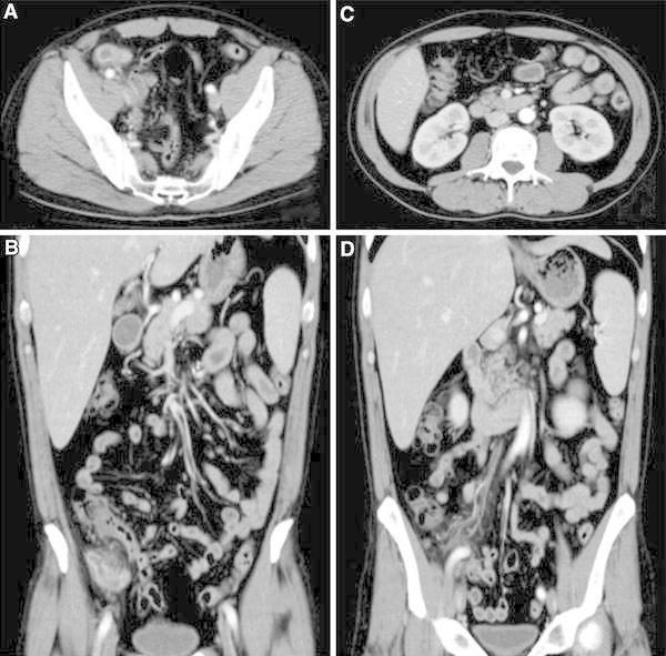
On admission, the body temperature was 39.0 °C, the abdomen was flat, pain in the right lower abdominal region was mild, and no rebound tenderness was noted. On the blood tests at the outpatient visit 2 days before, the WBC was 8,300/μl, and CRP was 7.5 mg/dl, but they were exacerbated to 11,000/μl and 34.13 mg/dl, respectively. Also, the platelet count was reduced to 75,000/μl, and liver dysfunction was suggested by increases in the AST 71 IU/l, ALT 58 IU/l, T-Bil 2.5 mg/dl, and LDH 303 IU/l. Abdominal contrast-enhanced CT showed increased radiodensity of the enlarged appendix and surrounding adipose tissue (Fig. 1).
Fig. 1.

Abdominal contrast-enhanced CT on admission. a, b An enlarged appendix is observed with increased radiodensity in the surrounding adipose tissue. c, d There is no finding suggesting the presence of thrombi in the SMV or ICV
After admission, as abdominal findings were unremarkable despite of fever and blood test results. Decreases in the platelet count, mild increases in the transaminase levels, and increases in the total bilirubin level were noted. We suspected other diseases, for example, viral infection and blood disease as a cause of inflammation. We initially planned to manage the acute appendicitis conservatively with the possibility of taking into consideration of emergency surgery.
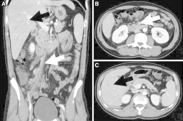
Detailed examinations were performed. Blood cultures were negative. Tests for viral infections such as cytomegalovirus and EB virus infections were negative, and for fungal infection was negative too. As an empiric treatment, meropenem (MEPM) was administered at 0.5 g × 3/day. The fever, high inflammatory reaction, and liver dysfunction were temporarily alleviated, but the WBC and fever elevated again on the 5th hospital day. Abdominal contrast-enhanced CT was performed again, and the result showed no remarkable changes in appendicitis. However, a vine-like low-density area along the ileocolic vein (ICV) and defects in the lumen of the SMV and dendritic low-density area of the portal vein in S5 of the liver were recognized (Fig. 2). We suspected the occurrence of SMVT and PVT induced by inflammation extending from protracted appendicitis. We carried out appendectomy by laparotomy on emergency.
Fig. 2.

Abdominal contrast-enhanced CT on the 5th hospital day. a, b A vine-like low-density area along the ICV and defects in the lumen of SMV were noted (white arrows). a, c A dendritic low-density area was noted in the portal region in S5 of the liver (black arrows)
Intraoperatively, the appendix adhered to the surrounding tissues but could be detached. The mesoappendix was markedly thickened, and there were findings suggestive of abscess formation in the mesoappendix. No sign of edema, congestion, or necrosis was noted in the large intestine, small intestine, and mesentery. Therefore, only an appendectomy was performed.
Histopathological examinations indicated the diagnosis of phlegmonous or gangrenous appendicitis. Abscesses were noted in the mesoappendix, and fibrinoid necrosis was observed in the vascular wall, particularly the venous wall, in the mesoappendix. No clear thrombus, suppurating thrombus, or biofilm was found in the blood vessels.
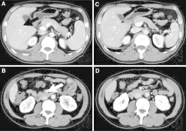
The postoperative course was uneventful, and improvements were observed in inflammatory reaction, platelet count, transaminase levels, and total bilirubin level. Postoperatively, heparin administration was started at 16,000 U/day, and heparin was substituted for warfarin with the resumption of oral nutrition. On the 7th postoperative day, regression and alleviation of SMVT and PVT were confirmed by abdominal contrast-enhanced CT.
On abdominal contrast-enhanced CT on the 48th postoperative day, no sign of recurrence of SMVT or PVT was noted, but the SMV was atrophied (Fig. 3). Oral warfarin administration was discontinued half a year after surgery. The blood AT III activity, protein C antigen and activity levels, and antinuclear antibody level were measured after surgery. As these parameters were all within the normal range, abnormality of the congealing fibrinogenolysis system was excluded.
Fig. 3.

Abdominal contrast-enhanced CT on the 7th and 48th postoperative days. a, b On the 7th postoperative day, SMVT was reduced in size (white arrows), and the images of PVT became unclear (black arrow). c, d On the 48th postoperative day, no sign of recurrence of SMVT or PVT was noted
Discussion
SMVT is a relatively rare disease first reported by Warren et al. in 1935 [1]. Its diagnosis tends to be delayed because of its nonspecific symptoms [2]. The presence of secondary causes has been reported in about 80 % of the cases, and abnormalities of the congealing fibrinogenolysis system (a decrease in anticoagulant factor, antiphospholipid antibody syndrome), portal hypertension, inflammatory diseases of the abdomen, laparotomy, malignant tumor, and trauma have been reported as such secondary causes [3]. If SMVT is caused by an inflammatory disease of the abdomen, appendicitis, diverticulitis, cholecystitis, pancreatitis, intra-abdominal infection, and intrapelvic infection are considered. Blood cultures are often positive, and Escherichia coli, B. fragilis, Proteus mirabilis, Klebsiella pneumoniae, Enterobacter species, etc., are detected as causative microorganisms [4].
When SMVT is caused by appendicitis, as in our patient, the following mechanisms of thrombogenesis are considered possible: (1) local sepsis due to abscesses in the mesoappendix causes a hypercoagulability state, and thrombi are formed locally. (2) Bacteria infiltrate into the SMV, cause angiitis such as portal vein phlebitis, and induce thrombus formation. (3) Bacteria invade into tissues around the veins, cause periphlebitis along the vessels, and as inflammation extends into the vascular lumens, blood clotting is promoted, leading to thrombus formation [3]. In our patient, histopathological examination was performed concerning the mesoappendix alone, and details of SMVT or PVT were unclear. However, as no thrombus was noted in the blood vessels of the mesoappendix, as fibrinoid necrosis was observed in the vascular wall, and as abdominal contrast-enhanced CT showed a vine-like low-density area along the ICV, the third mechanism is considered the most likely.
Contrast-enhanced CT is an effective diagnostic examination. Filling defect in the lumen of SMV caused by thrombus is recognized in 90 % or more of cases [5]. In some cases, the organ blood flow and collaterals can also be evaluated by changing the phase. It was also effective in our patient for their evaluation.
SMVT is treated conservatively if abdominal symptoms are mild, but surgical treatment is the first choice when intestinal necrosis is strongly suspected. If the progression of thromboembolism is slow, the intestinal blood flow remains relatively intact because of the development of collaterals, and necrosis is avoided. Therefore, conservative treatment using anticoagulants is attempted first. However, if SMVT is caused by inflammation, such as appendicitis as in our patient, thrombi are formed rapidly with exacerbation of inflammation, possibly leading to intestinal edema, congestion, and necrosis. If inflammation cannot be controlled sufficiently by conservative treatment, aggressive choice of surgical removal of the primary lesion is necessary even without signs of intestinal necrosis [6].
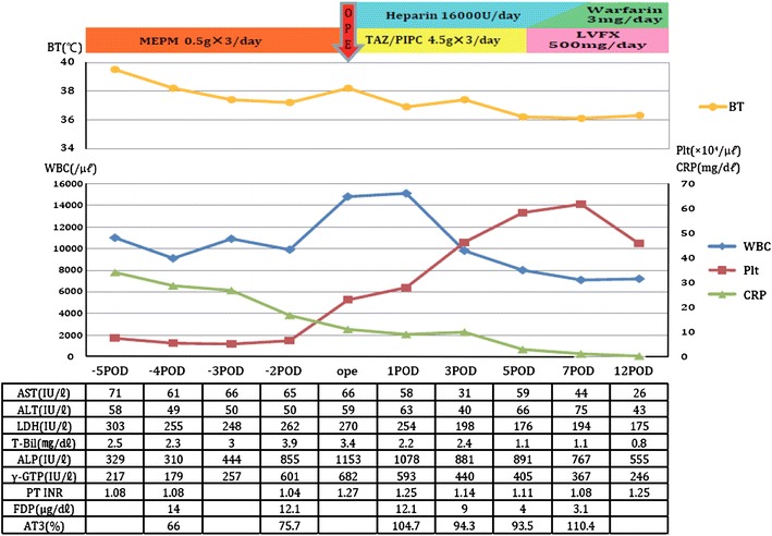
In our patient, thrombus formation was observed from the ICV to the SMV, but physical and imaging examinations suggested that the intestinal blood flow was maintained by the collateral blood flows. However, thrombogenesis extended to the intrahepatic portal system caused by the progression of phlebitis. At the diagnosis of PVT, the control of inflammation by conservative treatment is considered to have become insufficient, and thrombosis was likely to spread further. With a delay of the diagnosis and further progression of PVT, the patient may have developed severe liver dysfunction. Intestinal necrosis caused by the progression of SMVT and obstruction of collaterals and exacerbation of liver dysfunction caused by the progression of PVT were considered possible, and since a recovery of the platelet count was observed around this time, we decided to perform appendectomy. As a result, the alleviation of inflammatory reaction, liver and coagulation dysfunction were recognized (Fig. 4).
Fig. 4.

Clinical course of the patient. Circles body temperature (BT), Diamonds white blood cells (WBC), Squares platelet (Plt), Triangles C-reactive protein (CRP). We recognized improvements of inflammatory reaction, platelet count, and liver dysfunction. MEPM meropenem, TAZ/PIPC, Zosyn; LVFX levofloxacin, POD post operative day, ope operation
No clear guidelines have been proposed concerning postoperative anticoagulant therapy [7, 8]. Some authors report that the recurrence rate of, and mortality rate caused by SMVT was lower in patients who underwent postoperative anticoagulant therapy than in those who did not [9]. On the other hand, some authors report that anticoagulant therapy is unnecessary if the primary disease has been controlled adequately even when it is complicated by SMVT [10]. In our patient, heparin was administered from immediately after surgery, and the administration of warfarin, with which heparin was substituted, was continued for about half a year. No recurrence of thrombosis was noted even after the discontinuation of warfarin administration.
In this case, we took the wrong timing of the operation because we suspected other diseases and SMVT and PVT developed caused by appendicitis. From this experience, we consider that SMVT or PVT induced by intraperitoneal inflammation such as appendicitis, the primary disease should be aggressively treated by surgery if the control of inflammation by conservative treatment is not effective. In recent years, a concept of interval appendectomy is spreading in appendicitis treatment, and the cases where appendectomy is performed before managed conservatively are increasing. Some treatments may not be able to control inflammation enough, like in this case, and develop SMVT, PVT. As a cause of liver dysfunction under conservative treatment of appendicitis, one should consider the complication of SMVT and PVT as a differential disease.
In summary, SMVT and PVT are relatively rare as a complication of appendicitis. If acute appendicitis is complicated with liver dysfunction, early diagnosis and sufficient control of inflammation of the primary disease are important while taking into consideration SMVT and PVT.
Conflict of interest
The authors declare that they have no conflict of interest.
References
- 1.Warren S, Eberhard TP. Mesenteric venous thrombosis. Surg Gynecol Obstet. 1935;61:102–121. [Google Scholar]
- 2.Singh P, Yabav N, Visvalingam V, Indaram A, Bank S. Pylephlebitis: diagnosis and management. Am J Gastroenterol. 2001;32:262–265. doi: 10.1111/j.1572-0241.2001.03736.x. [DOI] [PubMed] [Google Scholar]
- 3.Fukutomi S, Yasutomi J, Kusashio K, Suzuki M, Fukao K, Miyazaki M. A case of acute appendicitis complicated with superior mesenteric vein thrombosis (in Japanese with English abstract) Jpn J Surg Assoc. 2008;69(3):581–585. doi: 10.3919/jjsa.69.581. [DOI] [Google Scholar]
- 4.Chang YS, Min SY, Joo SH, Lee SH. Septic thrombophlebitis of the porto-mesenteric veins as a complication of acute appendicitis. World J Gastroenterol. 2008;14(28):4580–4582. doi: 10.3748/wjg.14.4580. [DOI] [PMC free article] [PubMed] [Google Scholar]
- 5.Hassan HA, Ranfman JP. Mesenteric venous thrombosis. South Med J. 1999;92:558–562. doi: 10.1097/00007611-199906000-00002. [DOI] [PubMed] [Google Scholar]
- 6.Nitta H, Kato T, Shibata Y, Suzuki M, Onoue S, Nagasawa K, et al. A case of superior mesenteric venous thrombosis successfully treated in two-stage operation. Jpn J Gastroenterol Surg. 2005;38:1602–1606. doi: 10.5833/jjgs.38.1602. [DOI] [Google Scholar]
- 7.Nishimori H, Ezoe E, Ura H, Imaizumi H, Meguro M, Furuhata T, et al. Septic thrombophlebitis of the portal and superior mesenteric veins as a complication of appendicitis: report of a case. Surg Today. 2004;34:173–176. doi: 10.1007/s00595-003-2654-8. [DOI] [PubMed] [Google Scholar]
- 8.Condat B, Pessione F, Helene Denninger M, Hillaire S, Valla D. Recent portal or mesenteric venous thrombosis: increased recognition and frequent recanalization on anticoagulant therapy. Hepatology. 2000;32:466–470. doi: 10.1053/jhep.2000.16597. [DOI] [PubMed] [Google Scholar]
- 9.Abdu RA, Zakhour BJ, Dallia DJ. Mesenteric venous thrombosis: 1911 to 1984. Surgery. 1987;101:383–388. [PubMed] [Google Scholar]
- 10.Crowe PM, Sager G. Reversible superior mesenteric vein thrombosis in acute pancreatitis. The CT appearances. Clinic Radiol. 1995;50:628–633. doi: 10.1016/S0009-9260(05)83293-3. [DOI] [PubMed] [Google Scholar]


