Abstract
Background
Imprinting is an important epigenetic regulator of gene expression that is often disrupted in cancer. While loss of imprinting (LOI) has been reported for two genes in prostate cancer (IGF2 and TFPI2), disease-related changes in methylation across all imprinted gene regions has not been investigated.
Methods
Using an Illumina Infinium Methylation Assay, we analyzed methylation of 396 CpG sites in the promoter regions of 56 genes in a pooled sample of 12 pairs of prostate tumor and adjacent normal tissue. Selected LOI identified from the array was validated using the Sequenom EpiTYPER assay for individual samples and further confirmed by expression data from publicly available datasets.
Results
Methylation significantly increased in 52 sites and significantly decreased in 17 sites across 28 unique genes (P < 0.05), and the strongest evidence for loss of imprinting was demonstrated in tumor suppressor genes DLK1, PLAGL1, SLC22A18, TP73, and WT1. Differential expression of these five genes in prostate tumor versus normal tissue using array data from a publicly available database were consistent with the observed LOI patterns, and WT1 hypermethylation was confirmed using quantitative DNA methylation analysis.
Conclusions
Together, these findings suggest a more widespread dysregulation of genetic imprinting in prostate cancer than previously reported and warrant further investigation.
Keywords: Imprinting, Prostate cancer, Differential methylation, DLK1, PLAGL1, SLC22A18, TP73, WT1
Background
Genomic imprinting is the epigenetic phenomenon by which alleles of select genes are differentially expressed according to the parent of origin [1]. In humans, approximately 65 genes have been validated as imprinted [2]. It has been suggested that imprinting is regulated primarily by DNA methylation of imprinting control regions (ICRs), which is established in the germ line and maintained throughout subsequent development [3].
Loss of monoallelic expression at imprinted genes, known as loss of imprinting (LOI), has been associated with many cancer types including leukemia, colorectal, liver, and lung cancer [4] and may play a role as an early driver in tumorigenesis [5]. Abnormal methylation of imprinted genes can be detrimental given frequent roles in promoting and restricting cellular growth. For example, loss of methylation of the maternal allele of insulin-like growth factor-II (IGF2) has been associated with increased expression of the growth-promoting gene in Wilms’ tumor [6].
While studies have suggested a role for IGF2 and tissue factor pathway inhibitor-2 (TFPI2) LOI in prostate cancer [7-9], the literature is restricted largely to these two genes. Here, we present a comprehensive investigation of methylation patterns at imprinted genes in prostate cancer. Our results indicate an overall dysregulation of imprinted gene methylation levels in prostate tumor tissue as compared to adjacent normal tissue, with pronounced gain of methylation at five tumor suppressor genes.
Methods
Study subjects
Procedures for participant recruitment have been described previously [10]. Briefly, study subjects were identified via the Yale Cancer Center Rapid Case Ascertainment system and all patients consented to the donation of tissue to the Yale-New Haven Hospital (YNHH) tissue bank. Samples were obtained according to protocols approved by the Research Ethics Board from YNHH, New Haven County, Connecticut and the Connecticut Department of Public Health Human Investigations Committee. Tissue sections from seventeen pairs of formalin-fixed paraffin-embedded (FFPE) prostate cancers and corresponding adjacent normal tissue specimens, obtained from patients who had undergone surgery between 2005 and 2009 at YNHH, were mounted on slides and examined by an expert pathologist. Gleason grades varied between specimens, with a composite score ranging from 6 to 9. No subjects who had received either chemo- or radio-therapy were included in the study.
Isolation of genomic DNA
Sections of tumor and adjacent normal tissue were pathologically reviewed, manually microdissected, and collected into 1.5 ml microtubes. The DNeasy Blood & Tissue Kit (Qiagen, Valencia, CA) was used to isolate genomic DNA according to the manufacturer’s protocols.
Methylation assay
Equal amounts of DNA from tumor and matching adjacent normal tissue from twelve subjects (a sufficient amount of DNA was unavailable for five of the seventeen subjects) were combined by tissue type for CpG methylation microarray analysis. Methylation of imprinted genes was assessed using the Illumina Infinium HumanMethylation27 Array (Illumina, Inc., San Diego, CA). The CpG sites of the imprinted genes were located within promoter regions, ranging from 3 to 1,495 bp upstream of the transcription start site (average distance: 426 ± 373 bp). A methylation index (β) was obtained for each site, which is a continuous variable ranging between 0 and 1 representing the ratio of the intensity of the methylated-probe signal to the total locus signal intensity (a β value of 0 corresponds to no methylation while a value of 1 corresponds to 100% methylation at the specific CpG locus measured). Complete array data have been uploaded to the Gene Expression Omnibus (GEO) database (http://www.ncbi.nlm.nih.gov/geo/; accession number GSE26319).
Validation by quantitative DNA methylation analysis
In order to confirm methylation microarray results we carried out quantitative DNA methylation analysis using Sequenom’s EpiTYPER assay (Sequenom, Inc., San Diego, CA). Methylation levels at WT1 were analyzed using tumor and adjacent normal tissue DNA from five subjects (non-pooled) for which DNA was of sufficient concentration. Analysis was conducted using pre-validated primers from the Sequenom’s Imprinting EpiPanel designed to target imprinting control regions of known imprinted genes (Amplicon WT1.ALT.TRANSCRIPT_05; Forward primer: GTAGGGGTTAGGGGAGGTAAAGT; Reverse primer: CCCAATCACAATACAACTACAATCA). Average methylation levels in tumor versus normal tissue were compared for individual CpG sites and for overall WT1 methylation.
Expression confirmation in publicly available datasets
We used the Oncomine expression profiling database version 4.4 (http://www.oncomine.org; accessed March 3rd, 2012) to search for expression array comparisons between prostate cancer tissue and normal tissue (either from adjacent normal tissue or healthy controls) in five genes with the strongest evidence of LOI in our dataset. We searched the database for expression differences in human prostate cancer using the gene symbol as the keyword (e.g. “DLK1”).
Statistical analysis
The Infinium methylation data were analyzed using Illumina's GenomeStudio software, which employs a custom model to yield a DiffScore and p-value for each CpG site based on a comparison of the mean methylation level in tumor tissue versus that of adjacent normal tissue. To control for multiple comparisons, adjustments were made in order to obtain an adjusted P value (designed as the false discovery rate, or Q value) for each observation using the method originally proposed by Benjamini-Hochberg [11]. CpG sites were defined as differentially methylated if the Q values obtained were < 0.05. Average methylation levels derived from SEQUENOM EpiTYPER analysis were compared between tumor and adjacent normal samples using a two sample t-test.
Results
Global disruption of methylation at imprinted genes
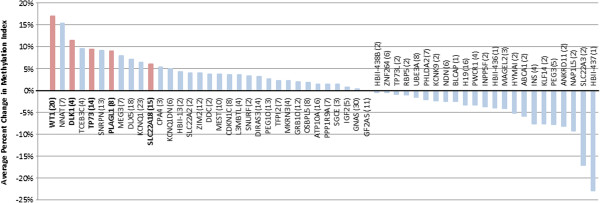
Based on our analysis of a pooled sample of 12 pairs of prostate tumor and adjacent normal tissues, our results demonstrate an overall disruption of methylation patterns of imprinted genes in tumor tissue. Among 397 CpG sites analyzed in 56 imprinted genes that were covered by the Illumina Infinium HumanMethylation27 array, the methylation index (β) in adjacent normal tissues was 0.453 on average, and in prostate cancer tissue methylation was significantly higher at 52 sites (13.1%) and significantly lower at 17 (4.3%) sites (P < 0.05), jointly spanning 28 unique genes (Table 1). Average percent changes in the methylation index were calculated for each of the 56 imprinted genes, in which percent changes were averaged across the set of corresponding CpG sites for each gene (mean number of CpG sites/gene: 7.1 ± 6.2). Of the 56 genes, 13 demonstrated a higher average methylation of at least 5%, and 9 demonstrated a lower average methylation of at least 5% (Figure 1).
Table 1.
Methylation indices (β) for CpG sites with significant methylation changes (P < 0.05)
| Gene | CpG site | Normal tissue(β1) | Tumor tissue(β2) | Change in methylation index(β2-β1) | Fold change | Gene | CpG site | Normal tissue(β1) | Tumor tissue(β2) | Change in methylation index(β2-β1) | Fold change |
|---|---|---|---|---|---|---|---|---|---|---|---|
| ATP10A |
cg10734665 |
0.717 |
0.394 |
−0.323 |
−1.820 |
IGF2 |
cg20339650 |
0.060 |
0.254 |
0.194 |
4.233 |
| ATP10A |
cg11015241 |
0.453 |
0.714 |
0.261 |
1.576 |
IGF2AS |
cg13791131 |
0.050 |
0.253 |
0.203 |
5.060 |
| CDKN1C |
cg05559445 |
0.360 |
0.567 |
0.207 |
1.575 |
INS |
cg25336198 |
0.732 |
0.530 |
−0.202 |
−1.381 |
| DIRAS3 |
cg09118625 |
0.657 |
0.847 |
0.190 |
1.289 |
KCNQ1 |
cg06719391 |
0.329 |
0.092 |
−0.237 |
−3.576 |
| DLK1 |
cg17412258 |
0.088 |
0.380 |
0.291 |
4.318 |
KCNQ1 |
cg20751395 |
0.847 |
0.690 |
−0.157 |
−1.228 |
| DLX5 |
cg11500797 |
0.451 |
0.652 |
0.201 |
1.446 |
KCNQ1 |
cg17820828 |
0.087 |
0.263 |
0.176 |
3.023 |
| DLX5 |
cg24115040 |
0.368 |
0.581 |
0.212 |
1.579 |
KCNQ1 |
cg12578166 |
0.552 |
0.813 |
0.261 |
1.473 |
| DLX5 |
cg20080624 |
0.549 |
0.767 |
0.219 |
1.397 |
KCNQ1 |
cg01734338 |
0.530 |
0.900 |
0.369 |
1.698 |
| DLX5 |
cg06537230 |
0.057 |
0.380 |
0.323 |
6.667 |
KCNQ1DN |
cg05656180 |
0.507 |
0.707 |
0.200 |
1.394 |
| DLX5 |
cg06911084 |
0.320 |
0.709 |
0.389 |
2.216 |
KCNQ1DN |
cg13081704 |
0.557 |
0.759 |
0.202 |
1.363 |
| GNAS |
cg01355739 |
0.882 |
0.640 |
−0.243 |
−1.378 |
KLF14 |
cg06533629 |
0.548 |
0.319 |
−0.229 |
−1.718 |
| GNAS |
cg17414107 |
0.360 |
0.125 |
−0.235 |
−2.880 |
MEG3 |
cg25836301 |
0.558 |
0.745 |
0.187 |
1.335 |
| GNAS |
cg06044900 |
0.284 |
0.123 |
−0.161 |
−2.309 |
MEG3 |
cg09280976 |
0.643 |
0.870 |
0.226 |
1.353 |
| GNAS |
cg07284407 |
0.485 |
0.682 |
0.197 |
1.406 |
MEST |
cg09872616 |
0.138 |
0.309 |
0.171 |
2.239 |
| GNAS |
cg01565918 |
0.457 |
0.720 |
0.263 |
1.575 |
MEST |
cg17347253 |
0.338 |
0.585 |
0.247 |
1.731 |
| GNAS |
cg09437522 |
0.299 |
0.565 |
0.266 |
1.890 |
NAP1L5 |
cg12759554 |
0.714 |
0.407 |
−0.307 |
−1.754 |
| H19 |
cg25852472 |
0.801 |
0.513 |
−0.288 |
−1.561 |
NNAT |
cg23566503 |
0.645 |
0.823 |
0.178 |
1.276 |
| H19 |
cg10602543 |
0.790 |
0.560 |
−0.229 |
−1.411 |
NNAT |
cg12862537 |
0.712 |
0.903 |
0.191 |
1.268 |
| H19 |
cg11492040 |
0.626 |
0.414 |
−0.212 |
−1.512 |
NNAT |
cg18433380 |
0.401 |
0.625 |
0.224 |
1.559 |
| H19 |
cg23977670 |
0.888 |
0.721 |
−0.167 |
−1.232 |
NNAT |
cg10642330 |
0.567 |
0.865 |
0.299 |
1.526 |
| HBII-437 |
cg08993557 |
0.686 |
0.458 |
−0.228 |
−1.498 |
PEG10 |
cg25524350 |
0.141 |
0.581 |
0.440 |
4.121 |
| PLAGL1 |
cg08263357 |
0.429 |
0.678 |
0.249 |
1.580 |
TP73 |
cg25115460 |
0.378 |
0.745 |
0.367 |
1.971 |
| PLAGL1 |
cg17895149 |
0.128 |
0.418 |
0.290 |
3.266 |
TP73 |
cg16607065 |
0.311 |
0.708 |
0.397 |
2.277 |
| PLAGL1 |
cg25350411 |
0.298 |
0.611 |
0.313 |
2.050 |
WT1 |
cg04096767 |
0.463 |
0.670 |
0.207 |
1.447 |
| PPP1R9A |
cg11164400 |
0.435 |
0.725 |
0.291 |
1.667 |
WT1 |
cg22511262 |
0.265 |
0.500 |
0.235 |
1.887 |
| SLC22A18 |
cg18655584 |
0.677 |
0.860 |
0.182 |
1.270 |
WT1 |
cg15446391 |
0.322 |
0.571 |
0.249 |
1.773 |
| SLC22A18 |
cg03336167 |
0.234 |
0.878 |
0.643 |
3.752 |
WT1 |
cg13301003 |
0.320 |
0.580 |
0.260 |
1.813 |
| SLC22A3 |
cg25313204 |
0.956 |
0.626 |
−0.330 |
−1.527 |
WT1 |
cg01693350 |
0.505 |
0.781 |
0.276 |
1.547 |
| SNRPN |
cg24993443 |
0.549 |
0.793 |
0.244 |
1.444 |
WT1 |
cg05222924 |
0.336 |
0.617 |
0.281 |
1.836 |
| SNRPN |
cg11265941 |
0.297 |
0.595 |
0.298 |
2.003 |
WT1 |
cg12006284 |
0.315 |
0.724 |
0.410 |
2.298 |
| SNRPN |
cg22678136 |
0.422 |
0.827 |
0.405 |
1.960 |
WT1 |
cg22533573 |
0.075 |
0.575 |
0.500 |
7.667 |
| TCEB3C |
cg02432101 |
0.644 |
0.861 |
0.218 |
1.337 |
ZIM2 |
cg06244906 |
0.796 |
0.553 |
−0.243 |
−1.439 |
| TP73 |
cg00565688 |
0.580 |
0.230 |
−0.351 |
−2.522 |
ZIM2 |
cg02162069 |
0.561 |
0.751 |
0.190 |
1.339 |
| TP73 |
cg26208930 |
0.601 |
0.866 |
0.265 |
1.441 |
ZIM2 |
cg16519742 |
0.635 |
0.867 |
0.232 |
1.365 |
| TP73 | cg03846767 | 0.430 | 0.749 | 0.319 | 1.742 |
Figure 1.
Average percent change in methylation index (β) for tumor tissue (relative to normal) across all measured CpG sites for all imprinted genes. Numbers in parentheses indicate the number of CpG sites measured, and red bars indicate genes selected for further analysis. Of the 56 genes, 13 demonstrated higher average methylation indices of at least 5%, and 9 demonstrated lower average methylation indices of at least 5%.
Strongest evidence for methylation dysregulation at five imprinted genes
Results from five genes were particularly notable based on the magnitude of methylation change from adjacent normal tissue and the consistency in the direction of these changes across multiple CpG sites (Figure 2). CpG site cg17412258, 212 bp upstream of the DLK1 transcription start site, showed 4.32 fold higher methylation in the tumor samples relative to the adjacent normal tissue samples, with a 5% higher average methylation index for the other CpG sites. SLC22A18 demonstrated similar methylation changes, where 3.75 fold higher methylation was observed at CpG site cg03336167 with a tendency toward higher methylation at remaining sites. Three CpG sites in the promoter region of PLAGL1 showed statistically significant higher methylation (P < 0.05), including 3.27 fold higher methylation at cg17895149. Four CpG sites in the proximal TP73 promoter region demonstrated statistically significant higher methylation indices (1.44, 1.74, 1.97, and 2.28 fold higher methylation) (P < 0.05), while one site further upstream from the transcription start site (917 bp) showed statistically significantly lower methylation (P < 0.05). WT1 demonstrated the greatest consistency in methylation aberration, with increased methylation at 16 of 20 CpG sites. This increased methylation was statistically significant (P < 0.05) at eight CpG sites including 7.67 fold higher methylation at cg22533573. All five of the identified genes (DLK1, PLAGL1, SLC22A18, TP73, and WT1) have presumed tumor suppressing functions and taken together demonstrate a strong tendency towards higher methylation at the CpG sites assessed.
Figure 2.
Changes in methylation in tumor tissue by CpG Site in DLK1, PLAGL1, SLC22A18, TP73, and WT1. Bars represent the percentage change in the methylation index (β) in pooled tumor samples relative to pooled adjacent tissue samples. All measured CpG sites for the five genes are presented.
Validation of WT1 hypermethylation by quantitative DNA methylation analysis
We sought to confirm the hypermethylation observed at WT1 using SEQUENOM’s EpiTYPER quantitative methylation assay. Results were consistent with the prior analysis by microarray, demonstrating roughly 2–4 fold hypermethylation across all eight CpG sites analyzed in the WT1 imprinting control region (including cg22533573, as discussed above) in comparing individual tumor and adjacent normal tissue pairs. Averaged across the eight analyzed CpG sites and five tumor-normal tissue pairs with adequate DNA for this confirmation, we observed a 2.04-fold increase in methylation in tumor tissue relative to adjacent normal tissue (average normal methylation level: 25.2%; average tumor methylation level: 51.4%; P = 0.0104).
Expression confirmation of differentially methylated genes using public data
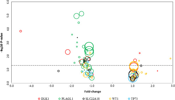
We used the Oncomine expression profiling database to search for expression array comparisons between prostate cancer tissue and prostate tissue from healthy controls in DLK1, PLAGL1, SLC22A18, TP73, and WT1; expression data for all available studies are presented in Figure 3. Six of fourteen studies reported statistically significant (P < 0.05) expression changes for DLK1, demonstrating lower expression of the gene in prostate cancer tissues relative to healthy control tissues in four studies and higher expression in two. These studies reported expression fold changes of −4.547 (n = 34) [12], -2.216 (n = 89) [13], -1.496 (n = 102) [14], -1.242 (n = 88) [15], 1.124 (n = 57) [16], and 1.350 (n = 21) [17], respectively. Of 38 studies, 27 reported significantly lower expression of PLAGL1 in prostate cancer tissue relative to healthy controls. These studies reported lower expression fold changes ranging from 1.143 [18] to 1.995 [17], and no study reported higher expression of the gene in tumor tissue. Five of thirteen studies were identified that showed significantly lower expression for SLC22A18, with fold changes of −1.734 (n = 13) [19], -1.411 (n = 21) [17], -1.400 (n = 26) [20], -1.341 (n = 52) [21], and −1.157 (n = 57) [16]. Four out of 22 studies showed significant WT1 expression changes, including two studies demonstrating lower expression fold changes of −1.315 (n = 102) [14], and −1.106 (n = 35) [22], and two studies showing significantly higher expression with a fold change of 1.327 (n = 21) [17] and 2.878 (n = 21) [17]. Of nine studies, one (n = 35) [22] demonstrated a significant change in expression of TP73, with a −1.218 fold change.
Figure 3.
Expression of five selected genes (DLK1, PLAGL1, SLC22A18, TP73, and WT1) in prostate cancer tissue relative to normal tissue in all studies of prostate cancer identified from the publicly available Oncomine expression database. Study results are plotted according to the -log(P-value) and expression fold-change in tumor versus normal tissue, and sample size is represented by the size of the circles (range: n = 12 to n = 160). The dotted line represents statistical significance at P = 0.05.
Discussion
This work represents the first comprehensive investigation of methylation changes in prostate cancer. Our results demonstrate an overall disruption of methylation at imprinted genes in prostate cancer tissue with a greater tendency toward hypermethylation than hypomethylation. Based on the magnitude and consistency of hypermethylation across multiple CpG sites, the strongest evidence for disrupted methylation patterns at imprinted genes was demonstrated at five tumor suppressor genes: DLK1, PLAGL1, SLC22A18, TP73, and WT1. Of the five genes, LOI has been reported for WT1 in Wilms’ tumor development, [23] and has not been reported for the other four genes in the context of cancer development. Statistically significant hypermethylation across eight CpG sites in the WT1 imprinting control region was confirmed using quantitative DNA methylation analysis (P = 0.0104).
All five of the identified genes are presumed tumor suppressors and have been reported to play roles in cancer development. DLK1, which encodes a transmembrane protein and is involved in cell differentiation, has been linked to liver cancer development and progression [24,25]. PLAGL1 is thought to be a transcriptional regulator and has been associated with pheochromocytoma, a tumor of the adrenal grand [26]. SLC22A18 is a transporter of organic cations, and has been associated with glioma and breast cancer progression and survival [27,28]. WT1 plays an important role in normal development of the urogenital system, and is named after its association with Wilms’ tumor development [29]. It has also been associated with breast cancer [30], colorectal cancer [31], and thyroid cancer [32]. TP73 is an important member of the p53 family of cell cycle regulatory proteins, which is likely disrupted in the majority of cancers [33,34]. Under normal imprinting control, DLK1, PLAGL1, and WT1 are paternally expressed, while SLC22A18 and TP73 are maternally expressed.
We hypothesized that the increased methylation observed would be associated with lower expression of these five genes, as previous research has established the role that DNA methylation plays in regulating gene expression [35,36]. Methylation impacts gene expression by altering chromatin structure and modifying interactions between proteins and DNA [37]. Specifically, methylated promoter CpG islands attract methyl-CpG binding proteins and transcriptional repressors, thereby interfering with transcription factor binding and reducing transcription of the associated gene [38,39]. Accordingly, we queried the Oncomine database to determine if expression of identified genes significantly differed between prostate cancer and normal prostate tissue in publicly available datasets. Evidence was provided to support a tendency towards reduced expression in prostate tumor tissue at DLK1, PLAGL1, SLC22A18, with less conclusive expression data for WT1 and TP73.
Taken together, our results suggest that observed promoter hypermethylation may be involved in the downregulation of these normally imprinted tumor suppressor genes, which may have important functional consequences for the development and progression of prostate cancer. For example, the transmembrane protein DLK1 has been shown to negatively regulate NOTCH1 [40], which is overexpressed in prostate cancer and is associated with human prostate cancer cell invasion [41]; this suggests that DLK1 downregulation may promote cell invasion via increased NOTCH1 expression. Loss of PLAGL1 expression has been associated with progression from benign to metastatic prostate tumors via the acquisition of androgen-independence, which enables prostate cancers to grow in the absence of androgens [42]. Expression of SLC22A18 (also known as TSSC5) has been observed in adult human prostate tissue and may be involved in growth regulation and small molecule transport, including the export of potentially genotoxic substances [43]; underexpression of this protein may consequently increase the risk of tumor formation. Finally, reduced expression of WT1 and TP73 proteins may have major implications for tumorigenesis: WT1 is a transcription factor that has been shown to regulate growth and induce apoptosis when overexpressed in prostate cancer cells [44], and p73 is a p53-family protein that is key to apoptosis and growth arrest in human prostate cancer cells [45].
Previous studies of loss of imprinting in prostate cancer have focused on TFPI2 and IGF2, with one study reporting lower methylation in the former [8] and three studies providing inconsistent reports of methylation changes in the latter [7,9,46]. No significant changes in methylation were identified in our data at TFPI2 CpG sites, and IGF2 methylation was higher at one CpG site and lower at the other four that were assessed. The resulting effects on expression of these two genes were variable across studies identified in the Oncomine expression database.
Conclusions
This study was limited by the inability to assess expression changes for our samples, as we were unable to extract sufficient RNA from our tissue samples to conduct qPCR analyses. It should also be noted that loss of imprinting has been previously observed in normal tissue from cancer patients [47], which suggests that some differential methylation events at these genes would not be detectable in comparing tumor tissue to adjacent normal tissue. Despite these limitations, this study represents the first comprehensive assessment of methylation changes in prostate cancer. Our results suggest an overall tendency towards disruption of methylation at imprinted loci in prostate cancer tissue, and our data provide the first suggestion of disrupted imprinting patterns in cancer for four imprinted genes (DLK1, PLAGL1, SLC22A18, and TP73). Although our results need to be further confirmed by larger studies, these findings suggest a more widespread dysregulation of genomic imprinting in prostate cancer than previously reported. Future investigations such as studying the biological significance of dysregulated imprinting genes are also warranted.
Competing interests
The authors declare that they have no competing interests.
Authors’ contributions
DIJ analyzed the data and prepared the manuscript. YM and AF were involved in data analysis. WKK and YZ designed the study and were involved in data analysis, interpretation, and manuscript preparation. All authors read and approved the final manuscript.
Pre-publication history
The pre-publication history for this paper can be accessed here:
Contributor Information
Daniel I Jacobs, Email: daniel.jacobs@yale.edu.
Yingying Mao, Email: bearmmyy@gmail.com.
Alan Fu, Email: alan.fu@yale.edu.
William Kevin Kelly, Email: William.Kelly@Jefferson.edu.
Yong Zhu, Email: yong.zhu@yale.edu.
Acknowledgments
This work was supported by the Yale Cancer Center Pilot Grant.
References
- Robertson KD. DNA methylation and human disease. Nat Rev Genet. 2005;6(8):597–610. doi: 10.1038/nrg1655. [DOI] [PubMed] [Google Scholar]
- Fang F, Hodges E, Molaro A, Dean M, Hannon GJ, Smith AD. Genomic landscape of human allele-specific DNA methylation. P Natl Acad Sci. 2012;109(19):7332–7337. doi: 10.1073/pnas.1201310109. [DOI] [PMC free article] [PubMed] [Google Scholar]
- Kacem S, Feil R. Chromatin mechanisms in genomic imprinting. Mamm Genome. 2009;20(9):544–556. doi: 10.1007/s00335-009-9223-4. [DOI] [PubMed] [Google Scholar]
- Feinberg AP, Cui H, Ohlsson R. DNA methylation and genomic imprinting: insights from cancer into epigenetic mechanisms. Semin Cancer Biol. 2002;12(5):389–398. doi: 10.1016/S1044-579X(02)00059-7. [DOI] [PubMed] [Google Scholar]
- Sawan C, Vaissière T, Murr R, Herceg Z. Epigenetic drivers and genetic passengers on the road to cancer. Mutat Res Fundam Mol Mech Mutagen. 2008;642(1–2):1–13. doi: 10.1016/j.mrfmmm.2008.03.002. [DOI] [PubMed] [Google Scholar]
- Ravenel JD, Broman KW, Perlman EJ, Niemitz EL, Jayawardena TM, Bell DW, Haber DA, Uejima H, Feinberg AP. Loss of Imprinting of Insulin-Like Growth Factor-II (IGF2) Gene in Distinguishing Specific Biologic Subtypes of Wilms Tumor. J Natl Cancer Inst. 2001;93(22):1698–1703. doi: 10.1093/jnci/93.22.1698. [DOI] [PubMed] [Google Scholar]
- Jarrard DF, Bussemakers MJ, Bova GS, Isaacs WB. Regional loss of imprinting of the insulin-like growth factor II gene occurs in human prostate tissues. Clin Cancer Res. 1995;1(12):1471–1478. [PubMed] [Google Scholar]
- Ribarska T, Ingenwerth M, Goering W, Engers R, Schulz WA. Epigenetic Inactivation of the Placentally Imprinted Tumor Suppressor Gene TFPI2 in Prostate Carcinoma. Cancer Genomics Proteomics. 2010;7(2):51–60. [PubMed] [Google Scholar]
- Bhusari S, Yang B, Kueck J, Huang W, Jarrard DF. Insulin-like growth factor-2 (IGF2) loss of imprinting marks a field defect within human prostates containing cancer. Prostate. 2011;71(15):1621–1630. doi: 10.1002/pros.21379. [DOI] [PMC free article] [PubMed] [Google Scholar]
- Kim SJ, Kelly WK, Fu A, Haines K, Hoffman A, Zheng T, Zhu Y. Genome-wide methylation analysis identifies involvement of TNF-α mediated cancer pathways in prostate cancer. Cancer Lett. 2011;302(1):47–53. doi: 10.1016/j.canlet.2010.12.010. [DOI] [PubMed] [Google Scholar]
- Benjamini Y, Hochberg Y. Controlling the False Discovery Rate: A Practical and Powerful Approach to Multiple Testing. J Roy Stat Soc B Met. 1995;57(1):289–300. [Google Scholar]
- Welsh JB, Sapinoso LM, Su AI, Kern SG, Wang-Rodriguez J, Moskaluk CA, Frierson HF, Hampton GM. Analysis of Gene Expression Identifies Candidate Markers and Pharmacological Targets in Prostate Cancer. Cancer Res. 2001;61(16):5974–5978. [PubMed] [Google Scholar]
- Wallace TA, Prueitt RL, Yi M, Howe TM, Gillespie JW, Yfantis HG, Stephens RM, Caporaso NE, Loffredo CA, Ambs S. Tumor Immunobiological Differences in Prostate Cancer between African-American and European-American Men. Cancer Res. 2008;68(3):927–936. doi: 10.1158/0008-5472.CAN-07-2608. [DOI] [PubMed] [Google Scholar]
- Singh D, Febbo PG, Ross K, Jackson DG, Manola J, Ladd C, Tamayo P, Renshaw AA, D'Amico AV, Richie JP. et al. Gene expression correlates of clinical prostate cancer behavior. Cancer Cell. 2002;1(2):203–209. doi: 10.1016/S1535-6108(02)00030-2. [DOI] [PubMed] [Google Scholar]
- Yu YP, Landsittel D, Jing L, Nelson J, Ren B, Liu L, McDonald C, Thomas R, Dhir R, Finkelstein S. et al. Gene Expression Alterations in Prostate Cancer Predicting Tumor Aggression and Preceding Development of Malignancy. J Clin Oncol. 2004;22(14):2790–2799. doi: 10.1200/JCO.2004.05.158. [DOI] [PubMed] [Google Scholar]
- Liu P, Ramachandran S, Ali Seyed M, Scharer CD, Laycock N, Dalton WB, Williams H, Karanam S, Datta MW, Jaye DL. et al. Sex-Determining Region Y Box 4 Is a Transforming Oncogene in Human Prostate Cancer Cells. Cancer Res. 2006;66(8):4011–4019. doi: 10.1158/0008-5472.CAN-05-3055. [DOI] [PubMed] [Google Scholar]
- Arredouani MS, Lu B, Bhasin M, Eljanne M, Yue W, Mosquera J-M, Bubley GJ, Li V, Rubin MA, Libermann TA. et al. Identification of the Transcription Factor Single-Minded Homologue 2 as a Potential Biomarker and Immunotherapy Target in Prostate Cancer. Clin Cancer Res. 2009;15(18):5794–5802. doi: 10.1158/1078-0432.CCR-09-0911. [DOI] [PMC free article] [PubMed] [Google Scholar]
- Taylor BS, Schultz N, Hieronymus H, Gopalan A, Xiao Y, Carver BS, Arora VK, Kaushik P, Cerami E, Reva B. et al. Integrative Genomic Profiling of Human Prostate Cancer. Cancer Cell. 2010;18(1):11–22. doi: 10.1016/j.ccr.2010.05.026. [DOI] [PMC free article] [PubMed] [Google Scholar]
- Varambally S, Yu J, Laxman B, Rhodes DR, Mehra R, Tomlins SA, Shah RB, Chandran U, Monzon FA, Becich MJ. et al. Integrative genomic and proteomic analysis of prostate cancer reveals signatures of metastatic progression. Cancer Cell. 2005;8(5):393–406. doi: 10.1016/j.ccr.2005.10.001. [DOI] [PubMed] [Google Scholar]
- LaTulippe E, Satagopan J, Smith A, Scher H, Scardino P, Reuter V, Gerald WL. Comprehensive Gene Expression Analysis of Prostate Cancer Reveals Distinct Transcriptional Programs Associated with Metastatic Disease. Cancer Res. 2002;62(15):4499–4506. [PubMed] [Google Scholar]
- Tomlins SA, Mehra R, Rhodes DR, Cao X, Wang L, Dhanasekaran SM, Kalyana-Sundaram S, Wei JT, Rubin MA, Pienta KJ. et al. Integrative molecular concept modeling of prostate cancer progression. Nat Genet. 2007;39(1):41–51. doi: 10.1038/ng1935. [DOI] [PubMed] [Google Scholar]
- Vanaja DK, Cheville JC, Iturria SJ, Young CYF. Transcriptional Silencing of Zinc Finger Protein 185 Identified by Expression Profiling Is Associated with Prostate Cancer Progression. Cancer Res. 2003;63(14):3877–3882. [PubMed] [Google Scholar]
- Brown KW, Power F, Moore B, Charles AK, Malik KTA. Frequency and Timing of Loss of Imprinting at 11p13 and 11p15 in Wilms' Tumor Development. Mol Cancer Res. 2008;6(7):1114–1123. doi: 10.1158/1541-7786.MCR-08-0002. [DOI] [PubMed] [Google Scholar]
- Z-h J, R-j Y, Dong B, Xing B-c. Progenitor gene DLK1 might be an independent prognostic factor of liver cancer. Expert Opin Biol Ther. 2008;8(4):371–377. doi: 10.1517/14712598.8.4.371. [DOI] [PubMed] [Google Scholar]
- Xu X, Liu R-F, Zhang X, Huang L-Y, Chen F, Fei Q-L, Han Z-G. DLK1 as a Potential Target against Cancer Stem/Progenitor Cells of Hepatocellular Carcinoma. Mol Cancer Ther. 2012;11(3):629–638. doi: 10.1158/1535-7163.MCT-11-0531. [DOI] [PubMed] [Google Scholar]
- Jarmalaite S, Laurinaviciene A, Tverkuviene J, Kalinauskaite N, Petroska D, Böhling T, Husgafvel-Pursiainen K. Tumor suppressor gene ZAC/PLAGL1: altered expression and loss of the nonimprinted allele in pheochromocytomas. Cancer genet. 2011;204(7):398–404. doi: 10.1016/j.cancergen.2011.07.002. [DOI] [PubMed] [Google Scholar]
- Chu S-H, Feng D-F, Ma Y-B, Zhang H, Zhu Z-A, Li Z-Q, Jiang P-C. Promoter methylation and downregulation of SLC22A18 are associated with the development and progression of human glioma. J Transl Med. 2011;9(1):156. doi: 10.1186/1479-5876-9-156. [DOI] [PMC free article] [PubMed] [Google Scholar]
- He H, Xu C, Zhao Z, Qin X, Xu H, Zhang H. Low expression of SLC22A18 predicts poor survival outcome in patients with breast cancer after surgery. Cancer Epidemiol. 2011;35(3):279–285. doi: 10.1016/j.canep.2010.09.006. [DOI] [PubMed] [Google Scholar]
- Lee SB, Haber DA. Wilms Tumor and the WT1 Gene. Exp Cell Res. 2001;264(1):74–99. doi: 10.1006/excr.2000.5131. [DOI] [PubMed] [Google Scholar]
- Silberstein GB, Van Horn K, Strickland P, Roberts CT, Daniel CW. Altered expression of the WT1 Wilms tumor suppressor gene in human breast cancer. Proc Natl Acad Sci. 1997;94(15):8132–8137. doi: 10.1073/pnas.94.15.8132. [DOI] [PMC free article] [PubMed] [Google Scholar]
- Cawkwell L, Lewis FA, Quirke P. Frequency of allele loss of DCC, p53, RBI, WT1, NF1, NM23 and APC/MCC in colorectal cancer assayed by fluorescent multiplex polymerase chain reaction. Br J Cancer. 1994;70(5):813–818. doi: 10.1038/bjc.1994.404. [DOI] [PMC free article] [PubMed] [Google Scholar]
- Oji Y, Miyoshi Y, Koga S, Nakano Y, Ando A, Nakatsuka S-i, Ikeba A, Takahashi E, Sakaguchi N, Yokota A. et al. Overexpression of the Wilms' tumor gene WT1 in primary thyroid cancer. Cancer Science. 2003;94(7):606–611. doi: 10.1111/j.1349-7006.2003.tb01490.x. [DOI] [PMC free article] [PubMed] [Google Scholar]
- Sigal A, Rotter V. Oncogenic Mutations of the p53 Tumor Suppressor: The Demons of the Guardian of the Genome. Cancer Res. 2000;60(24):6788–6793. [PubMed] [Google Scholar]
- Hollstein M, Sidransky D, Vogelstein B, Harris CC. p53 Mutations in human cancers. Science. 1991;253(5015):49–53. doi: 10.1126/science.1905840. [DOI] [PubMed] [Google Scholar]
- Bird AP. Gene expression: DNA methylation– how important in gene control? Nature. 1984;307(5951):503–504. doi: 10.1038/307503a0. [DOI] [PubMed] [Google Scholar]
- Robertson KD, Jones PA. DNA methylation: past, present and future directions. Carcinogenesis. 2000;21(3):461–467. doi: 10.1093/carcin/21.3.461. [DOI] [PubMed] [Google Scholar]
- Ng H-H, Adrian B. DNA methylation and chromatin modification. Curr Opin Genet Dev. 1999;9(2):158–163. doi: 10.1016/S0959-437X(99)80024-0. [DOI] [PubMed] [Google Scholar]
- Jones PA, Takai D. The Role of DNA Methylation in Mammalian Epigenetics. Science. 2001;293(5532):1068–1070. doi: 10.1126/science.1063852. [DOI] [PubMed] [Google Scholar]
- Robertson KD, Wolffe AP. DNA methylation in health and disease. Nat Rev Genet. 2000;1(1):11–19. doi: 10.1038/35049533. [DOI] [PubMed] [Google Scholar]
- Baladrón V, Ruiz-Hidalgo MJ, Nueda ML, Díaz-Guerra MJM, García-Ramírez JJ, Bonvini E, Gubina E, Laborda J. dlk acts as a negative regulator of Notch1 activation through interactions with specific EGF-like repeats. Exp Cell Res. 2005;303(2):343–359. doi: 10.1016/j.yexcr.2004.10.001. [DOI] [PubMed] [Google Scholar]
- Hafeez BB, Adhami VM, Asim M, Siddiqui IA, Bhat KM, Zhong W, Saleem M, Din M, Setaluri V, Mukhtar H. Targeted knockdown of Notch1 inhibits invasion of human prostate cancer cells concomitant with inhibition of matrix metalloproteinase-9 and urokinase plasminogen activator. Clin Cancer Res. 2009;15(2):452–459. doi: 10.1158/1078-0432.CCR-08-1631. [DOI] [PMC free article] [PubMed] [Google Scholar]
- Murillo H, Schmidt LJ, Karter M, Hafner KA, Kondo Y, Ballman KV, Vasmatzis G, Jenkins RB, Tindall DJ. Prostate cancer cells use genetic and epigenetic mechanisms for progression to androgen independence. Gene Chromosome Canc. 2006;45(7):702–716. doi: 10.1002/gcc.20333. [DOI] [PubMed] [Google Scholar]
- Yamada HY, Gorbsky GJ. Tumor suppressor candidate TSSC5 is regulated by UbcH6 and a novel ubiquitin ligase RING105. Oncogene. 2005;25(9):1330–1339. doi: 10.1038/sj.onc.1209167. [DOI] [PMC free article] [PubMed] [Google Scholar]
- Fraizer G, Leahy R, Priyadarshini S, Graham K, Delacerda J, Diaz M. Suppression of prostate tumor cell growth in vivo by WT1, the Wilms' tumor suppressor gene. Int J Oncol. 2004;24(3):461–471. [PubMed] [Google Scholar]
- Yu J, Baron V, Mercola D, Mustelin T, Adamson E. A network of p73, p53 and Egr1 is required for efficient apoptosis in tumor cells. Cell Death Differ. 2006;14(3):436–446. doi: 10.1038/sj.cdd.4402029. [DOI] [PubMed] [Google Scholar]
- Fu VX, Dobosy JR, Desotelle JA, Almassi N, Ewald JA, Srinivasan R, Berres M, Svaren J, Weindruch R, Jarrard DF. Aging and Cancer-Related Loss of Insulin-like Growth Factor 2 Imprinting in the Mouse and Human Prostate. Cancer Res. 2008;68(16):6797–6802. doi: 10.1158/0008-5472.CAN-08-1714. [DOI] [PMC free article] [PubMed] [Google Scholar]
- Cui H, Horon IL, Ohlsson R, Hamilton SR, Feinberg AP. Loss of imprinting in normal tissue of colorectal cancer patients with microsatellite instability. Nat Med. 1998;4(11):1276–1280. doi: 10.1038/3260. [DOI] [PubMed] [Google Scholar]


